
UNIVERSITÀ DEGLI STUDI DI TRIESTE - core.ac.uk · universitÀ degli studi di trieste xxviii ciclo...
163
UNIVERSITÀ DEGLI STUDI DI TRIESTE XXVIII CICLO DEL DOTTORATO DI RICERCA IN SCIENZE E TECNOLOGIE CHIMICHE E FARMACEUTICHE CONVENIENT COVALENT FUNCTIONALIZATION OF CARBON NANOTUBES FOR RADIOACTIVITY DELIVERY Settore scientifico-disciplinare: CHIM/08 DOTTORANDA AGNIESZKA GAJEWSKA COORDINATORE PROF. MAURO STENER SUPERVISORE DI TESI PROF. TATIANA DA ROS ANNO ACCADEMICO 2014 / 2015
Transcript of UNIVERSITÀ DEGLI STUDI DI TRIESTE - core.ac.uk · universitÀ degli studi di trieste xxviii ciclo...
UNIVERSITÀ DEGLI STUDI DI TRIESTE XXVIII CICLO DEL DOTTORATO DI RICERCA
IN SCIENZE E TECNOLOGIE CHIMICHE E FARMACEUTICHE
CONVENIENT COVALENT
FUNCTIONALIZATION OF CARBON
NANOTUBES FOR RADIOACTIVITY
DELIVERY
Settore scientifico-disciplinare: CHIM/08
DOTTORANDA AGNIESZKA GAJEWSKA COORDINATORE PROF. MAURO STENER SUPERVISORE DI TESI PROF. TATIANA DA ROS
ANNO ACCADEMICO 2014 / 2015


Acknowledgements Luckily, after three years of the unconditional support I would like to
thank to:
My supervisor Prof. Tatiana Da Ros for giving me this great chance to
develop my Ph.D. thesis at University of Trieste. Also for the patience, for
many essential advices and having door open in a case of any doubts.
Prof. Maurizio Prato for the opportunity to work in his group in Trieste. For
collaboration in the frame of the project.
I would like to thank the jury members Prof. Ester Vazquez Fernandez-
Pacheco, Prof. Sonia Merino, Prof. Enzo Menna and Prof. Silvia Marchesan
for accepting to read and judge my work.
Special word of thanks goes to Claudio Gamboz for his useful help with
TEM.
Prof. Benjamin G. Davis, for having accepted me in his wonderful group in
Oxford. To Sonia, Reida and Christopher for such a good care and
hospitality, and to all, canal punting team for great time and kind
treatment.
Dr. Khuloud Al-Jamal for the opportunity of all biological test. For the
great help and assistants, as well for high climbing goals to Rebecca,
Julie, Cinzia, Maxime and Houmam and all group who was so helpful to
the chemist in biolab.
All Raddel group: Anne, Ana, Sandra, Adem, Aritz, Martin, Robert for
such a lovely integration and multicultural experience which we could
expand in our network.
Cinzia, for being the best hotelmate and tripadvisor in one.
Magdalena, Elzbieta and Markus - almost Polish team - thank you for
your Radler support and collaboration almost on every level.
Of course, I cannot forget the financial support of the RADDEL project
from European Commission under the FP7 People programme (Grant
agreement number: 290023).

I've been fortunate to work with great colleagues from Prato Group. In
particular I would like to thank you to:
Adrian, for help at the beginning of my PhD adventure.
Caroline, “mother of all doctoral students” for helping, listening and
making things possible.
Michela, Sussana, Nuria, Davide, Angela, Dani, Eleonora, Francesco,
Cristina, Maribel, Andrea, Mimmo, Michele, Akcan, Anirban. “So far and
yet so near” - for Pharmacy - C11 integration.
Maria, Ana and Arturito for many nice moments, chats, Spanish lessons,
and other entertaining contributions in our daily coffee/chocolate
brioche/tee time.
Jenni and Manu, you are the best Galician ever, hay parfavar don’t grow
up!
The new and old batches of master students: Lor enzo, Alexa, Jacopo,
Silvia, Federico for a pleasant fresh wind in the long PhD’s live, for
excellent atmosphere in the lab and the sunny lunches.
Zois, my big, Greek friend, thank you for all advices, chemistry
discussions and smart suggestions, also in the life!
Valentina, for infinite, Roman energy which turns the Earth and lights the
Sun.
Giuly, for understanding, simplifying all complicated world to few
eloquent emoticons and being always in the right time and
place when I needed you.
Tanja, fortunately, there were also the moments of relaxation, long
conversation, kebeb, Lasko beer and future planning... Thank you!
All my friends from Poland Sylwia, Wiola, Tetyana, Justyna, Iza
for being, knowing my complicated character and loving me as I am.
Dziękuję moim rodzicom, Jakubowi i Ewelinie, Karolowi i
Martynie, Joannie i Tadeuszowi, którzy przez te trzy lata
dzielnie znosili rozłąkę i zawsze pomagali w trudnych chwilach.
Finally, a thousand thanks for all this constant support and
understanding during these last years I would like to dedicate to my
husband Piotr. Thank you for always believing in me!
LIST OF ABBREVIATIONS
Boc t-butyloxycarbonyl Boc2O Di-t-butyl dicarbonate CNTs Carbon Nanotubes DCM Dichloromethane DIEA Diisopropylethiylamine DMF Dimethylformamide EI Electron impact mass spectrometer ESI Electrospray ionization mass spectrometer Et2O Diethyl ether HiPCO High-pressure carbon monoxide HR-TEM High Resolution Transmission Electron Microscopy HOMO Highest occupied molecular orbital LUMO Lowest unoccupied molecular orbital MW Microwave MWCNTs Multi-Wall Carbon Nanotubes NMP N-Methyl-2-pyrrolidone NMR Nuclear Magnetic Resonance o-DCB 1,2-Dichlorobenzene oxSWCNTs Oxidized SWCNTs PEG Polyethyleneglycol Pht Phthalimide group
RBM Radial breathing mode RGD Arginine-glycine-aspartic acid peptide ROS Reactive Oxygen Species SPECT Single-photon emission computed tomography SWCNTs Single Wall Carbon Nanotube TEA Triethylamine TEG Triethylene glycol TEM Transmission Electronic Microscopy TFA Trifluoroacetic Acid TGA Thermogravimetric Analysis THF Tetrahydrofuran TLC Thin Layer Chromatography tR Retention time UV-Vis Ultra Violet - Visible X@CNTs Carbon nanotubes filled with X
XPS X-Ray Photoemission Spectroscopy

ABSTRACT
The attachment of radioactive metal ions on the external surface of
carbon nanotubes allows the labeling and tracking of CNTs with good sensitivity
the same result can be achieved by encapsulating emitting elements into the
CNT cavities. This feature could be perfect to explore in biomedical applications
including imaging and therapy, depending on radionuclide.
Both methods have their advantages but in this thesis the direct labeling
by creation of nanocapsules was chosen as easier to control and more suitable
for the external attachment of biologically active molecules.
The first part of the presented work, described in Chapter II, has been
dedicated to new functionalization pathway of CNTs. The main attention has
been given, to design reaction which 1) will work on SWCNTs and MWCNTs with
a high degree of functionalization; 2) will not cause damages of the tubes
leading to leakage of filled materials; 3) can be applied in a large scale and 4)
will be fast enough for functionalization of CNTs filled with short life-time
isotopes. The cycloaddition of 1,3-dipoles on CNTs was performed using various
approaches but none of them was enough efficient to provide an easy setup for
further exploration on filled CNTs. To accomplish the goal we have used
arylation reaction, already described in literature. To fit to the requirements for
biological application we have synthesized a linker which would provide high
solubility and amine functions for further binding with biological molecules. The
direct functionalization by arylation method of several filled nanotubes was the
subject of Chapter III and on some of those compounds biological tests have
been performed as reported in Chapter IV. The cytotoxicity of filled and
functionalized SWCNTs and MWCNTs was examined. The biodistribution study

was performed on mice using MWCNTs filled with the radioisotope of
samarium-153 (153Sm) and functionalized with previously synthesized linker.
Our results suggest that application of diazonium-based arylation of
filled CNTs can be generally adopt as an efficient and convenient technique for
functionalization, without breaking and opening the tubes.
The functionalized nanocapsules filled with 153Sm can be used for in vivo
delivery coupled with long-term tracing studies.

RIASSUNTO
L’introduzione di elementi radioattivi sulla superficie esterna di nanotubi
di carbonio (CNTs) permette di tracciare i CNTs con una buona sensibilità e lo
stesso risultato può essere ottenuto incapsulando i radioisotopi nella cavità
interna dei nanotubi stessi. Questa metodologia potrebbe essere perfetta per
diverse applicazioni biomediche, tra cui l'imaging e la terapia radiativa, a
seconda del radionuclide presente.
Entrambe le metodologie presentano dei vantaggi, ma nell’ambito di
questo lavoro di ricerca si è scelto di lavorare con sistemi che presentavano
derivati all’interno della loro struttura, con la creazione di nanocapsule essendo
queste più adatte a legare sulla superficie esterna molecole biologicamente
attive.
La prima parte del presente lavoro, descritta nel Capitolo II, è stata
dedicata allo studio di nuove metodologie di funzionalizzazione dei CNTs, con
particolare attenzione ad approcci:1) in grado di funzionalizzare riccamente sia
SWCNTs che MWCNTs; 2) che non danneggino la superficie dei nanotubicon
possibilità di fuoriuscite dei materiali incapsulati; 3) che possano essere applicati
su scala relativamente larga e 4) che siano abbastanza rapidi da consentire
un’efficiente funzionalizzazione di CNTs con isotopi con breve tempo di emivita.
La cicloaddizione di diversi 1,3-dipoli su nanotubi di carbonio è stata effettuata
utilizzando vari approcci, ma nessuno di essi ha dato risultati così soddisfacenti
da impiegare le nuove procedure studiate per la funzionalizzazione delle
nanocapsule.
Per raggiungere l'obiettivo abbiamo utilizzato un'altra reaziona già
descritta in letteratura e a tale scopo è stato sintetizzato un linker che fornisse
una buona solubilità al sistema e presentasse gruppi amminici terminali da
poter sfruttare per la successiva introduzione di molecole biologicamente attive.
La funzionalizzazione mediante arilazione di diversi nanotubi riempiti con sali di
Gadolinio, Samario, Iodio e Lutezio è stata oggetto di studio (Capitolo III). La
citotossicità di SWCNTs e MWCNTs con incapsulato il samario cloruro è stata
studiata,come riportato nel Capitolo IV, e grazie alla presenza di materiale
radiativo (153Sm) è stato possibile effettuare un'analisi di biodistribuzione nei
topi.
I nostri risultati suggeriscono che l'applicazione della reazione di
arilazione può essere generalmente adottata quale tecnica efficace e
conveniente per funzionalizzazione di CNTs che presentano materiale
incapsulato al loro interno, senza che la struttura esterna venga danneggiata e
vi sia rilascio del materiale radiativo. Le nanocapsule a base di 153Sm potrebbero
quindi trovare utilizzo in teranostica.
TABLE OF CONTENTS
I. Introduction 1
I.1 Structure of carbon nanotubes 3
I.2 Properties of carbon nanotubes 5
I.3 Synthesis of Carbon Nanotubes 8
I.4 Reactivity of Carbon Nanotubes 9
I.5 Functionalization of Carbon Nanotubes 11
I.5.1 Non-covalent functionalization 11
I.5.2 Defect groups functionalization 13
I.5.3 Endohedral functionalization 15
I.5.4 Covalent Sidewall functionalization 20
I.6 Biomedical application of carbon nanotubes 25
I.6.1 Carbon nanotubes as cell substrates 25
I.6.2 Carbon nanotube for delivery of drugs 27
I.6.3 Carbon nanotubes for imaging 30
I.7 Toxicity - physical determinants 31
I.7.1 Effect of purity on toxicity 31
I.7.2 Effect of CNTs functionalization 32
I.7.3 The length of carbon nanotubes 33
Bibliography 34
II. Covalent functionalization of carbon nanotubes 41
II.1 Introduction 41
II.2 Cycloaddition 43
II.2.1 1,3-Dipolar cycloaddition of azomethine ylide 44
II.2.2 Results of cycloaddition of azomethine ylide 49
II.2.3 Nitrile oxides 60
II.2.4 Results of nitrile oxides cycloaddition 63
II.2.5 Conclusions 71
II.3 Diazonium-Based Functionalization 72
II.3.1 Results of Diazonium-Based Functionalization 73
II.3.2 Conclusions 82
II.4 Experimental part 83
II.4.1 Synthesis of the organic precursors 83
II.4.2 Functionalization 90
Bibliography 100
III. Functionalization of filled CNTs for radioactivity delivery 103
III.1 Introduction 103
III.2 Result and discussion 106
III.2.1 Functionalization of filled SWCNTs 106
III.2.2 Functionalization of filled MWCNTs 114
III.2.3 Microwave accelerated functionalization of filled CNTs 117
III.3 Conclusions 121
III.4 Experimental Part 122
III.4.1 Chemical Synthesis 122
III.4.2 Preparation of filled CNTs (X@CNTs) 122
III.4.3 Functionalization of CNTs 125
Bibliography 127
IV. Biomedical application of functionalized, filled CNTs 129
IV.1 Introduction 129
IV.2 Results of in vitro study 133
IV.3 Results of in vivo study 137
IV.4 Conclusions 140
IV.5 Experimental Part 141
IV.5.1 Preparation of SmCl3@CNTs 141
IV.5.2 Preparation of 153SmCl3@MWCNTs 141
IV.5.3 Functionalization of SmCl3@CNTs 141
IV.5.4 Functionalization of 153SmCl3@MWCNTs 141
IV.5.5 Cell toxicity assays 142
IV.5.6 Biodistribution study 143
Bibliography 145
V. Appendix 147
1
I. Introduction
The vision of tiny, active robots which manufacture various substances to
eliminate damages in the body after injection to the bloodstream was proposed
in 1959 by physicist, future Nobel Prize laureate, Richard Feynman in his work
“There's Plenty of Room at the Bottom”. Nowadays, nanotechnology, a relatively
young scientific discipline, gives the possibility to manipulate the matter on
atomic, molecular, and supramolecular scale and it is applied across all fields of
science, such as chemistry, biology, physics, materials science and engineering.
Medical application of nanoscale materials, which by definition are in
range of 1-100 nm, is a topic of interest for many researches around the world.
Nowadays, different nanotherapeutics have been already used for early
diagnosis of cancer, accurate cancer imaging, cancer therapy and targeted drug
delivery. Furthermore, it is generally unquestionable that these materials will
significantly contribute to the next generation of health care technologies in
treating various diseases.
Carbon nanotubes (CNTs) are particularly interesting nanomaterial, as
they exhibit an amazing model of a one-dimensional system with fascinating
physical and chemical properties. Since their discovery by Iijima in 1991
2
nanotubes were an object of intense research all around the world.1 At first,
carbon nanotubes were applied to different purposes, i.e. for electronics, optics,
plastics, and other materials of the nanotechnology fields. Since the beginning
of XXI century, they started to be studied in pharmacy and medicine for
theranostic uses. Due to their high surface area, excellent chemical stability and
rich electronic structure, CNTs are able to conjugate and interact with numerous
therapeutic molecules i.e. drugs, proteins, antibodies, DNA, enzymes. It was
demonstrated that carbon nanotubes are an excellent transporter medium,
which penetrate into cells, without damaging them.2,3 Many studies have shown
that, when bonded to CNTs, therapeutic molecules were delivered more
effectively and safely into cells compared to traditional methods.4 This excellent
breakthrough had a positive input on traditional concepts of pharmacology.5,6
Recently also other biomolecules (i.e. genes, radioisotopes, vaccines) were
successfully transported by means of CNTs to achieve gene therapy,
radiotherapy, immunotherapy, tissue regeneration and diagnosis of various
diseases.7 For this reason, despites novelty of those findings, in a very short
time, many scientists from a wide variety of disciplines, have focused their
attention towards carbon nanotubes.8
The research described within this PhD thesis is focused on the
modification of carbon nanotubes for biomedical applications. The main aim
was the sidewall functionalization of the CNTs already filled with radioactive
isotopes, which can be applied for cancer imaging and/or treatment. The main
part of the research activity was devoted to improve dispersibility of CNTs in
aqueous media by their direct, covalent functionalization and to shorten the
reaction time of this transformation. Pharmacological studies on the new
functionalized CNTs were performed and the perspectives of this promising
material are commented in the conclusions.
3
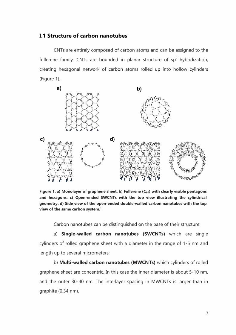
I.1 Structure of carbon nanotubes
CNTs are entirely composed of carbon atoms and can be assigned to the
fullerene family. CNTs are bounded in planar structure of sp2 hybridization,
creating hexagonal network of carbon atoms rolled up into hollow cylinders
(Figure 1).
Carbon nanotubes can be distinguished on the base of their structure:
a) Single-walled carbon nanotubes (SWCNTs) which are single
cylinders of rolled graphene sheet with a diameter in the range of 1-5 nm and
length up to several micrometers;
b) Multi-walled carbon nanotubes (MWCNTs) which cylinders of rolled
graphene sheet are concentric. In this case the inner diameter is about 5-10 nm,
and the outer 30-40 nm. The interlayer spacing in MWCNTs is larger than in
graphite (0.34 nm).
d)c)
b)a)
Figure 1. a) Monolayer of graphene sheet. b) Fullerene (C60) with clearly visible pentagons
and hexagons. c) Open-ended SWCNTs with the top view illustrating the cylindrical
geometry. d) Side view of the open-ended double-walled carbon nanotubes with the top view of the same carbon system.1
4
It was discovered that both types of CNTs can spontaneously close their
ends by forming a graphitic dome, at high temperature or during synthesis
experiments.9
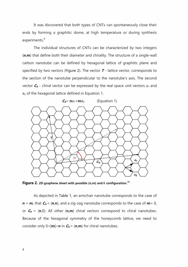
The individual structures of CNTs can be characterized by two integers
(n,m) that define both their diameter and chirality. The structure of a single-wall
carbon nanotube can be defined by hexagonal lattice of graphitic plane and
specified by two vectors (Figure 2). The vector T - lattice vector, corresponds to
the section of the nanotube perpendicular to the nanotube’s axis. The second
vector Ch - chiral vector can be expressed by the real space unit vectors a1 and
a2 of the hexagonal lattice defined in Equation 1.
Ch= na1+ma2 (Equation 1)
Figure 2. 2D graphene sheet with possible (n,m) and configuration.10
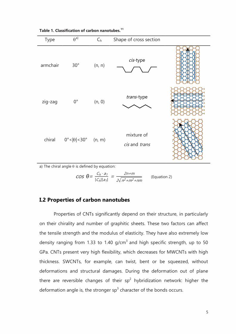
As depicted in Table 1, an armchair nanotube corresponds to the case of
n = m, that Ch = (n,n), and a zig-zag nanotube corresponds to the case of m= 0,
or Ch = (n,0). All other (n,m) chiral vectors correspond to chiral nanotubes.
Because of the hexagonal symmetry of the honeycomb lattice, we need to
consider only 0<|m|<n in Ch = (n,m) for chiral nanotubes.
5
Table 1. Classification of carbon nanotubes.11
Type a) Ch Shape of cross section
armchair 30° (n, n) cis-type
zig-zag 0° (n, 0) trans-type
chiral 0°<||<30° (n, m) mixture of
cis and trans
a) The chiral angle is defined by equation:
cos θ= Ch · a1
|Ch||a1| = 2n+m
2 n2+m2+nm (Equation 2)
I.2 Properties of carbon nanotubes
Properties of CNTs significantly depend on their structure, in particularly
on their chirality and number of graphitic sheets. These two factors can affect
the tensile strength and the modulus of elasticity. They have also extremely low
density ranging from 1.33 to 1.40 g/cm3 and high specific strength, up to 50
GPa. CNTs present very high flexibility, which decreases for MWCNTs with high
thickness. SWCNTs, for example, can twist, bent or be squeezed, without
deformations and structural damages. During the deformation out of plane
there are reversible changes of their sp2 hybridization network: higher the
deformation angle is, the stronger sp3 character of the bonds occurs.
6
CNTs have also very interesting electronic properties. It is very fascinating
that these structures, made entirely from only one element, may exhibit many
different electronic behaviors. CNT’s electronic conductivity, for example,
sensitively depends on the tube diameter and the helicity of the tube lattice.
Changes in those parameters cause a shift from a metallic to a semiconducting
state. This relation is preserved for SWCNTs and also MWCNTs.12 There are two
estimated electronic band gaps: one has a defined value around 0.5–0.6 eV for
semiconducting tubes, the other has significantly larger values of 1.7–2.0 eV for
a metallic tubes.13
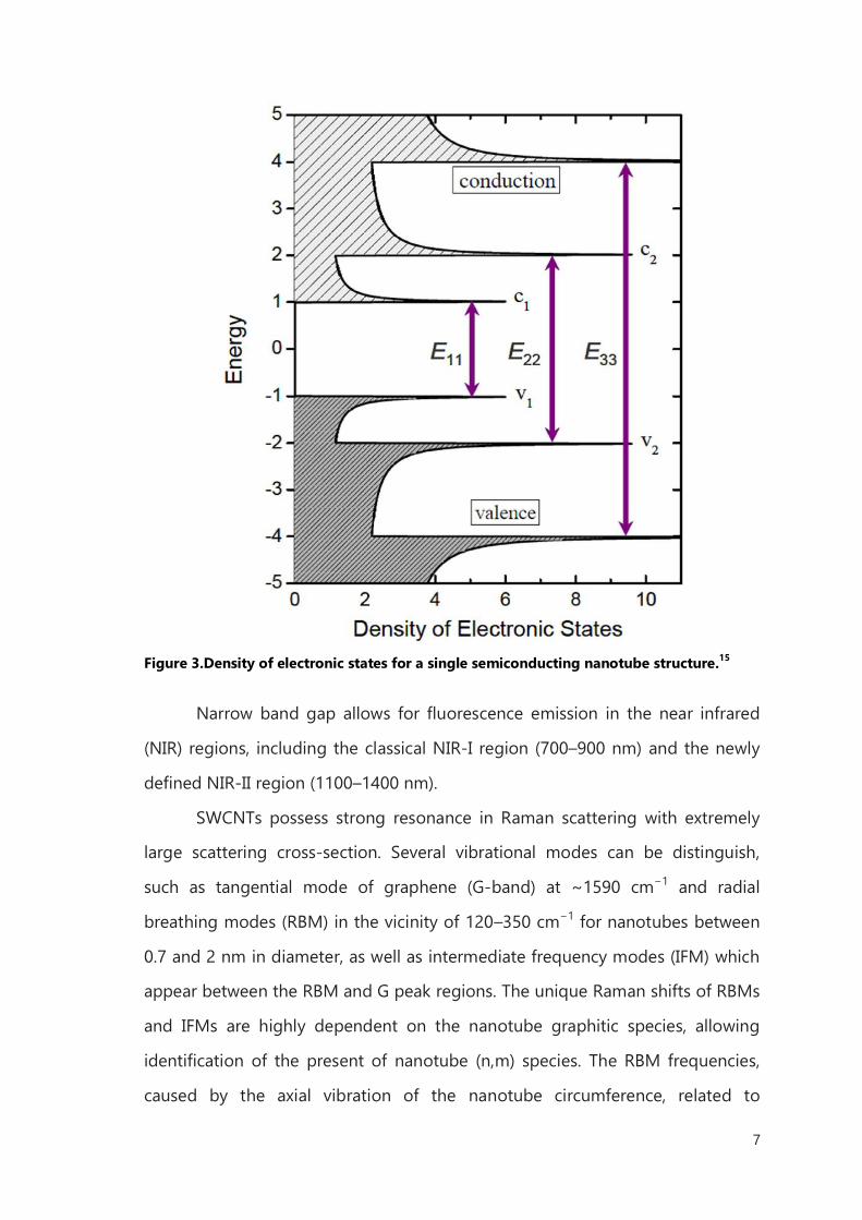
In the individual semiconducting SWCNT, the density of electronic states
has a series of sharp peaks, named van Hove singularities (HVSs), with energies
depending mainly on the tube diameter. The energy of the Fermi level is
defined as zero, and energy levels above and below the Fermi level are
conduction and valence bands, respectively. The optical transition occurs when
an electron or a hole is excited from one energy level to another, denoted Epq,
with p and q representing the order of conduction and valence bands,
respectively. Transition is allowed only when p = q (e.g., E11, E22, and E33). The
emission and absorption spectra in the first van Hove transition (E11) are
strikingly correspondent, so the fluorescence can be assigned to the E11
emission of semiconducting SWCNTs. It is proposed that the excitation of the
semiconducting SWCNT in the second van Hove transition E22 is followed by fast
electronic relaxation before fluorescence emission in the first van Hove
transition, E11 (Figure 3).14
7
Figure 3.Density of electronic states for a single semiconducting nanotube structure.15
Narrow band gap allows for fluorescence emission in the near infrared
(NIR) regions, including the classical NIR-I region (700–900 nm) and the newly
defined NIR-II region (1100–1400 nm).
SWCNTs possess strong resonance in Raman scattering with extremely
large scattering cross-section. Several vibrational modes can be distinguish,
such as tangential mode of graphene (G-band) at ~1590 cm−1 and radial
breathing modes (RBM) in the vicinity of 120–350 cm−1 for nanotubes between
0.7 and 2 nm in diameter, as well as intermediate frequency modes (IFM) which
appear between the RBM and G peak regions. The unique Raman shifts of RBMs
and IFMs are highly dependent on the nanotube graphitic species, allowing
identification of the present of nanotube (n,m) species. The RBM frequencies,
caused by the axial vibration of the nanotube circumference, related to
8
nanotube diameter with the relation ωRBM=228/dt+16 cm−1.16 The IFMs have not
been studied as extensively as the other modes, but certain modes with
frequencies between 380 and 650 cm−1 have been assigned to nanotube (n,m)
species or families of species. Some IFMs exhibit stepwise dispersion, changing
in frequency upon excitation at different laser energies, while other modes are
non-dispersive. Other information gained from carbon nanotube samples by
Raman spectra includes the presence, in metallic nanotubes, the fano
resonances. They are characterized by asymmetric line broadening expressed by
the Breit-Wigner-Fano (BWF) line shape of the tangential mode. This
phenomenon can be interpreted as the inference between the phonon and a
continuum spectrum of electronic transitions near the band gap. The Raman
spectra present also the sp3 carbon defects via the 1297 cm−1 disorder mode (D-
peak), and the presence of van der Waals contact in the RBMs. For instance, at
785 nm excitation, the (10,2) nanotube at 267 cm−1 comes into resonance upon
such contact, denoting aggregation or “bundling”.17
However, manipulation of CNTs is limited by several problems, such as:
1) formation of bundles due to π-π stacking and van der Waals interactions;
2) very low solubility in organic solvents and water;
3) low reactivity of pristine CNTs under many chemical reaction conditions, etc.
I.3 Synthesis of Carbon Nanotubes
Today’s fabrication methods are focused on three main techniques:
1. The arc-discharge method, also used by Iijima, where two graphite
electrodes are arced together in argon atmosphere, forming free carbon
atoms which then condense as nanotubes on the container walls;
2. Laser ablation, where graphite is evaporated by an intense laser pulse and
the carbon atoms condense as nanotubes.1 These methods are well
established in producing high-quality and nearly perfect nanotube
9
structures, despite large amounts of byproducts associated during the
process;
3. Chemical Vapor Deposition (CVD), where the flow of carbon rich gas (e.g.
methane or acetylene) is heated to nearly 900 °C, which results in nanotube
formation on metal catalyst nanoparticles, on a supporting substrate.
For SWCNTs, any of the three synthetic methods has yielded homogeneous
diameters and chirality. Nonetheless, arc-discharge and laser ablation
techniques are known for their impressively narrow diameter distributions,
approximately 1.4 nm.18
I.4 Reactivity of Carbon Nanotubes
In principle, the chemical reactivity of carbon nanotubes can be
compared with the reactivity of the other allotropes of carbon - graphite and
fullerene. However, due to the unique structure of CNTs, there are differences
with respect to what occur in other poly-aromatic carbon compounds.
Compared to graphite, in the atomic structure of single layer of CNTs, there is a
curvature of bonds between the sp2 carbon atoms that leads to local bond
stress.19 Therefore, CNTs in general exhibit higher chemical reactivity than a flat
layer of graphene. Due to the shape of closed ends of CNTs, the reactivity of
these portions should be comparable to the fullerenes. The reactivity of a
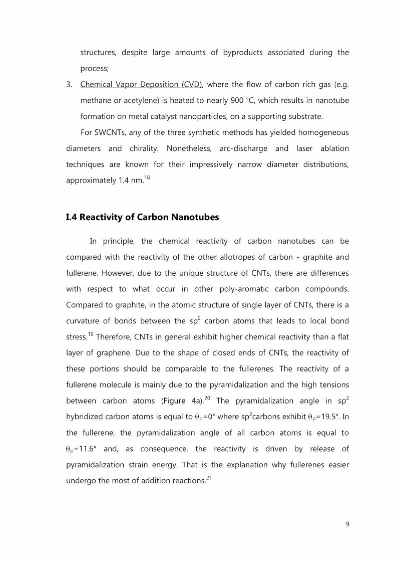
fullerene molecule is mainly due to the pyramidalization and the high tensions
between carbon atoms (Figure 4a).20 The pyramidalization angle in sp2
hybridized carbon atoms is equal to p=0° where sp3carbons exhibit p=19.5°. In
the fullerene, the pyramidalization angle of all carbon atoms is equal to
p=11.6° and, as consequence, the reactivity is driven by release of
pyramidalization strain energy. That is the explanation why fullerenes easier
undergo the most of addition reactions.21
10
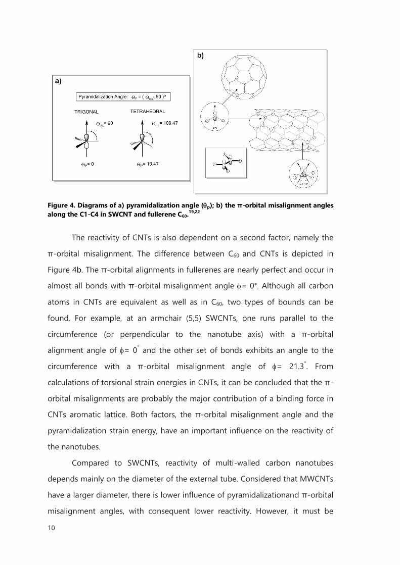
Figure 4. Diagrams of a) pyramidalization angle (p); b) the π-orbital misalignment angles along the C1-C4 in SWCNT and fullerene C60.
19,22
The reactivity of CNTs is also dependent on a second factor, namely the
π-orbital misalignment. The difference between C60 and CNTs is depicted in
Figure 4b. The π-orbital alignments in fullerenes are nearly perfect and occur in
almost all bonds with π-orbital misalignment angle = 0°. Although all carbon
atoms in CNTs are equivalent as well as in C60, two types of bounds can be
found. For example, at an armchair (5,5) SWCNTs, one runs parallel to the
circumference (or perpendicular to the nanotube axis) with a π-orbital
alignment angle of = 0° and the other set of bonds exhibits an angle to the
circumference with a π-orbital misalignment angle of = 21.3°. From
calculations of torsional strain energies in CNTs, it can be concluded that the π-
orbital misalignments are probably the major contribution of a binding force in
CNTs aromatic lattice. Both factors, the π-orbital misalignment angle and the
pyramidalization strain energy, have an important influence on the reactivity of
the nanotubes.
Compared to SWCNTs, reactivity of multi-walled carbon nanotubes
depends mainly on the diameter of the external tube. Considered that MWCNTs
have a larger diameter, there is lower influence of pyramidalizationand π-orbital
misalignment angles, with consequent lower reactivity. However, it must be
11
highlighted that there are many factors for the CNTs chemical reactivity and
they are strongly connected with their structural cohesion. Since defects are
formed directly during the manufacture it is hardly to say how they will affect
the general reactivity of the tubes.
I.5 Functionalization of Carbon Nanotubes
The application of carbon nanotubes in any branch of nanotechnology
strongly depends on their treatment and processability. The very low solubility
in all organic solvents and water, strongly limits their applicability.23 CNTs can be
briefly held in solution only by sonication process. Another method to maintain
CNTs in solution is the surface modification. In this way, depending on the
attached functional groups, solubility in certain solvents can be increased. There
are several methods for CNTs modification which allows applications of this
material for hydrogen storage, biosensors, in medicine.24,25,26
There are many different approaches for the functionalization of carbon
nanotubes, however they can be roughly divided into four classes:
1. Non-covalent functionalization;
2. Defect functionalization;
3. Endohedral functionalization;
4. Covalent sidewall functionalization.
I.5.1 Non-covalent functionalization
The non-covalent functionalization of carbon nanotubes is based on van
der Waals, π-π stacking and current exchange effects between the extended π-
system along the CNTs and the bounding molecules. These interactions are
strong enough to break bundles of CNTs, but also debundling process has to be
carried out under ultrasound irradiation. If the selected molecules are also
12
carrying charges, individualized CNTs are prevented by electrostatic repulsions
from re-bundling. This approach is preferred for non-oxidative purification of
CNTs. The big advantage over other methods is that the electronic structure of
the nanotubes will not be changed and their outstanding electrical and physical
properties remained unaffected.
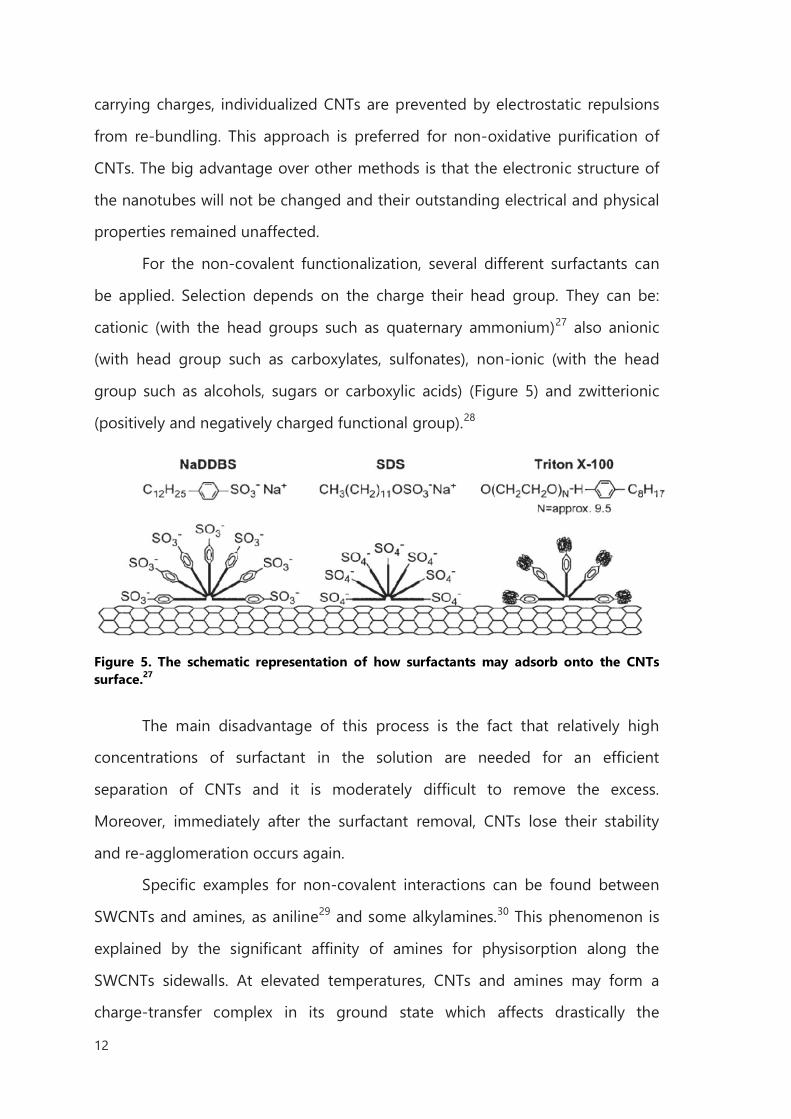
For the non-covalent functionalization, several different surfactants can
be applied. Selection depends on the charge their head group. They can be:
cationic (with the head groups such as quaternary ammonium)27 also anionic
(with head group such as carboxylates, sulfonates), non-ionic (with the head
group such as alcohols, sugars or carboxylic acids) (Figure 5) and zwitterionic
(positively and negatively charged functional group).28
Figure 5. The schematic representation of how surfactants may adsorb onto the CNTs surface.27
The main disadvantage of this process is the fact that relatively high
concentrations of surfactant in the solution are needed for an efficient
separation of CNTs and it is moderately difficult to remove the excess.
Moreover, immediately after the surfactant removal, CNTs lose their stability
and re-agglomeration occurs again.
Specific examples for non-covalent interactions can be found between
SWCNTs and amines, as aniline29 and some alkylamines.30 This phenomenon is
explained by the significant affinity of amines for physisorption along the
SWCNTs sidewalls. At elevated temperatures, CNTs and amines may form a
charge-transfer complex in its ground state which affects drastically the
13
electrical properties of individual SWCNTs. Complex can be simply removed by
washing with acetone.
An efficient coating of CNTs can be achieved with organic polymers31,
where the polymer wraps individual CNTs within a bundle and leads to their
separation.32 Polymers such as poly(p-phenylenevinylene-co-2,5-dioctyloxy-m-
phenylenevinylene) (PmPV-co-DOctOPV) have a typical selectivity for SWCNTs
within a certain diameter range.33 Thanks to the presence of polar portions of
the polymer chains, derivatives as polyvinylpyrrolidone (PVP) or polystyrene
sulfonate (PSS) are able to stabilize CNT-polymer complexes and prevent their
agglomeration even in water.34 Other non-covalent functionalizations are
established in bio-medical field: a large number of biomolecules, such as
sugars,31 proteins and peptides, can be adsorbed onto CNTs surface.35 A non-
covalent binding of SWCNTs with DNA is also possible and leads to
individualization of the CNTs in an aqueous medium. Different strong
interactions between single-stranded DNA and CNTs were observed to be
dependent on the diameter and the electronic properties of the CNTs.36,37
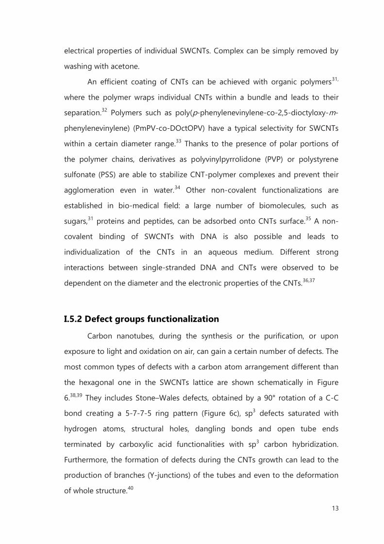
I.5.2 Defect groups functionalization
Carbon nanotubes, during the synthesis or the purification, or upon
exposure to light and oxidation on air, can gain a certain number of defects. The
most common types of defects with a carbon atom arrangement different than
the hexagonal one in the SWCNTs lattice are shown schematically in Figure
6.38,39 They includes Stone–Wales defects, obtained by a 90° rotation of a C-C
bond creating a 5-7-7-5 ring pattern (Figure 6c), sp3 defects saturated with
hydrogen atoms, structural holes, dangling bonds and open tube ends
terminated by carboxylic acid functionalities with sp3 carbon hybridization.
Furthermore, the formation of defects during the CNTs growth can lead to the
production of branches (Y-junctions) of the tubes and even to the deformation
of whole structure.40
14
Figure 6. Defects on SWCNTs. (a) Infinite tube (periodic boundary conditions);(b) H-saturated open tube on both sides (used to characterize the saturation energy); (c)pentagon and heptagon defects on the sidewall; (d) closed tube; (e) oxidized hole obtained by removing a hexagon at the cap; (f) the same as at the wall.
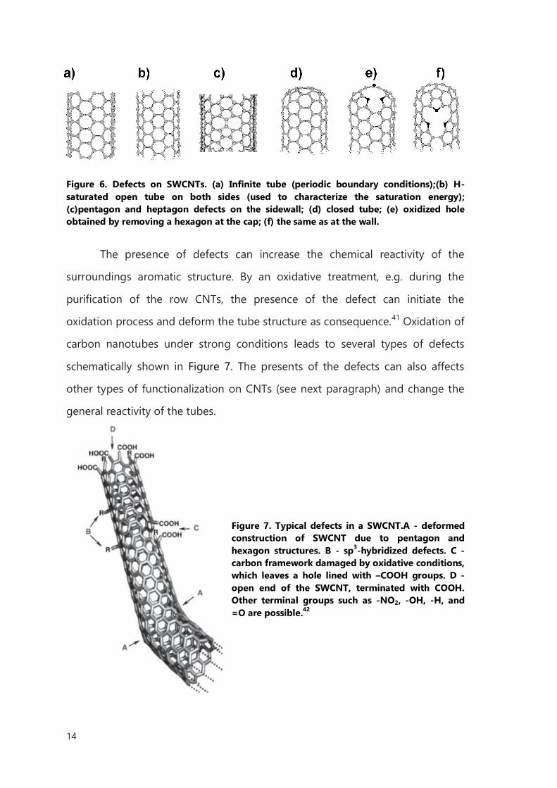
The presence of defects can increase the chemical reactivity of the
surroundings aromatic structure. By an oxidative treatment, e.g. during the
purification of the row CNTs, the presence of the defect can initiate the
oxidation process and deform the tube structure as consequence.41 Oxidation of
carbon nanotubes under strong conditions leads to several types of defects
schematically shown in Figure 7. The presents of the defects can also affects
other types of functionalization on CNTs (see next paragraph) and change the
general reactivity of the tubes.
Figure 7. Typical defects in a SWCNT.A - deformed construction of SWCNT due to pentagon and hexagon structures. B - sp3-hybridized defects. C - carbon framework damaged by oxidative conditions, which leaves a hole lined with –COOH groups. D - open end of the SWCNT, terminated with COOH. Other terminal groups such as -NO2, -OH, -H, and =O are possible.42
15
I.5.3 Endohedral functionalization
Endohedral functionalization of CNTs means that the inner cavity of the
tube is filled with single atoms or small molecules. The new structure is
described as X@CNT [hybrid nanotubes in which the inner cavity has been filled
with foreign atoms] where X is the chemical symbol of the content of the tube.
Endohedral modification is usually performed to create a new organization of
several confined materials with controlled size, structure and shape, where the
internal cavity of the tube serves as a template. Secondly, the new material can
gain new physical, chemical and electronic properties depending on the type of
CNTs and foreign material.
CNTs can be filled in one step, by in situ method, during the CNTs
growth.43 However in most of cases, filling is separated from synthesis, occurring
ex situ, through the open tips. Several gases, molten or sublimated materials can
be introduced,44 as well as metal nanoparticles,43 oxides,45 salts,46 organic
compounds among which dyes such as beta-carotene47 or porphyrines48 and
other carbon nanostructures such carbon nanoribbons49 and C6050 (Figure 8).
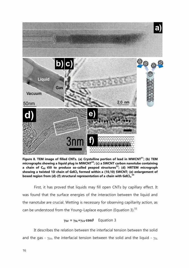
16
Figure 8. TEM image of filled CNTs. (a) Crystalline portion of lead in MWCNT51; (b) TEM micrographs showing a liquid plug in MWCNT52; (c) a SWCNT carbon nanotube containing a chain of C60 t50 to produce so-called peapod structures53; (d) HRTEM micrograph showing a twisted 1D chain of GdCl3 formed within a (10,10) SWCNT; (e) enlargement of boxed region from (d)-(f) structural representation of a chain with GdCl3.
54
First, it has proved that liquids may fill open CNTs by capillary effect. It
was found that the surface energies of the interaction between the liquid and
the nanotube are crucial. Wetting is necessary for observing capillarity action, as
can be understood from the Young–Laplace equation (Equation 3).55
SV = SL+LV cos Equation 3
It describes the relation between the interfacial tension between the solid
and the gas - SV, the interfacial tension between the solid and the liquid - SL
a)
2.0 nm
c)b)
d) e)
f).
17
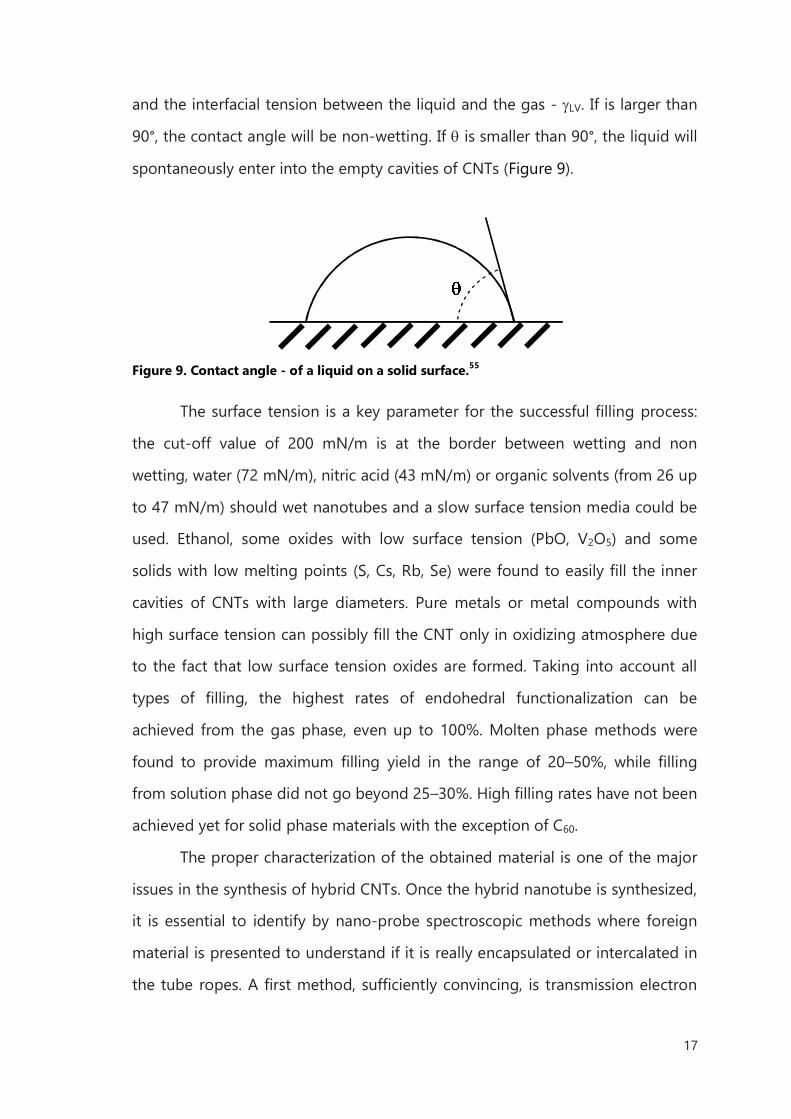
and the interfacial tension between the liquid and the gas - LV. If is larger than
90°, the contact angle will be non-wetting. If is smaller than 90°, the liquid will
spontaneously enter into the empty cavities of CNTs (Figure 9).
Figure 9. Contact angle - of a liquid on a solid surface.55
The surface tension is a key parameter for the successful filling process:
the cut-off value of 200 mN/m is at the border between wetting and non
wetting, water (72 mN/m), nitric acid (43 mN/m) or organic solvents (from 26 up
to 47 mN/m) should wet nanotubes and a slow surface tension media could be
used. Ethanol, some oxides with low surface tension (PbO, V2O5) and some
solids with low melting points (S, Cs, Rb, Se) were found to easily fill the inner
cavities of CNTs with large diameters. Pure metals or metal compounds with
high surface tension can possibly fill the CNT only in oxidizing atmosphere due
to the fact that low surface tension oxides are formed. Taking into account all
types of filling, the highest rates of endohedral functionalization can be
achieved from the gas phase, even up to 100%. Molten phase methods were
found to provide maximum filling yield in the range of 20–50%, while filling
from solution phase did not go beyond 25–30%. High filling rates have not been
achieved yet for solid phase materials with the exception of C60.
The proper characterization of the obtained material is one of the major
issues in the synthesis of hybrid CNTs. Once the hybrid nanotube is synthesized,
it is essential to identify by nano-probe spectroscopic methods where foreign
material is presented to understand if it is really encapsulated or intercalated in
the tube ropes. A first method, sufficiently convincing, is transmission electron
18
microscopy (TEM). On TEM images it is possible to observe separated or
bundled tubes where the foreign material is visible by differences in contrast.
The crucial problem of this technique is that it is based on local observations,
where the hybrid nanotubes have to be well separated. For a full
characterization, numerous images are necessary to have representative image
of the sample. Where the filling is crystallized inside the inert cavities, the high-
resolution microscope can revealed specific lattice distances of the inserted
material. It also might not be sufficient to use nano-probe energy-dispersive X-
ray spectroscopy (X-EDS) for exact identification of the filling materials due to
intrinsic spectroscopy limitations. X-EDS is hardly quantitative, and provides
chemical information on which elements are present in a chosen area of the
sample. Electron energy loss spectroscopy (EELS) might be a better choice, since
it is potentially able to provide both qualitative and quantitative chemical data
about atom identification, oxidation state, band structure, etc., but fine structure
EELS work is necessary, which is not a routine-based procedure. Anyway, if such
accurate methods are not used, it is quite risky to deduce the chemical
identification of the filling materials from just considering the expected chemical
reactions involved during the filling process.
Another method of chemical analysis is atomic emission spectroscopy
(AES). This technique employs the intensity of light emitted from a flame,
plasma, arc, or spark at a particular wavelength to determine the quantity of an
element in a sample. The wavelength of the atomic spectral line gives the
identity of the element, while the intensity of the emitted light is proportional to
the number of atoms of the element itself. The limitation of this method is that
there is no distinction between the inside and the outside of CNT so it is not
possible to differentiate among filled material and compound deposited on the
external surface of the tubes. Values reported in the literature should thus often
be considered only as rough evaluation.
19
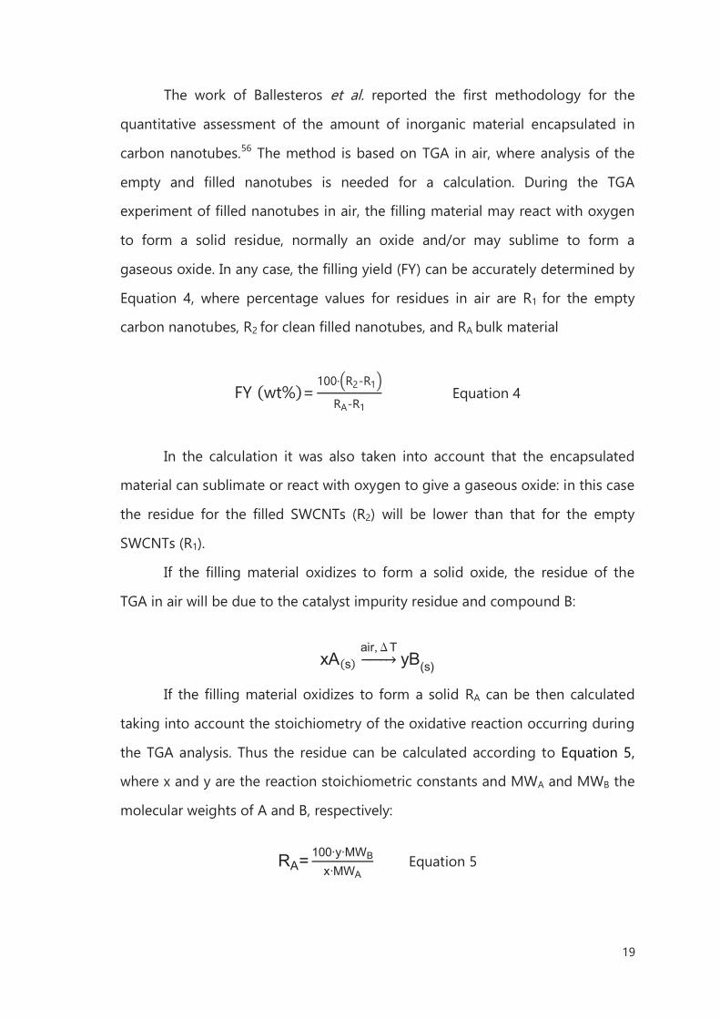
The work of Ballesteros et al. reported the first methodology for the
quantitative assessment of the amount of inorganic material encapsulated in
carbon nanotubes.56 The method is based on TGA in air, where analysis of the
empty and filled nanotubes is needed for a calculation. During the TGA
experiment of filled nanotubes in air, the filling material may react with oxygen
to form a solid residue, normally an oxide and/or may sublime to form a
gaseous oxide. In any case, the filling yield (FY) can be accurately determined by
Equation 4, where percentage values for residues in air are R1 for the empty
carbon nanotubes, R2 for clean filled nanotubes, and RA bulk material
FY (wt%)=100· R2-R1
RA-R1 Equation 4
In the calculation it was also taken into account that the encapsulated
material can sublimate or react with oxygen to give a gaseous oxide: in this case
the residue for the filled SWCNTs (R2) will be lower than that for the empty
SWCNTs (R1).
If the filling material oxidizes to form a solid oxide, the residue of the
TGA in air will be due to the catalyst impurity residue and compound B:
xA(s)air,ΔT⎯⎯⎯ yB(s)
If the filling material oxidizes to form a solid RA can be then calculated
taking into account the stoichiometry of the oxidative reaction occurring during
the TGA analysis. Thus the residue can be calculated according to Equation 5,
where x and y are the reaction stoichiometric constants and MWA and MWB the
molecular weights of A and B, respectively:
RA= 100∙y∙MWBx∙MWA
Equation 5
20
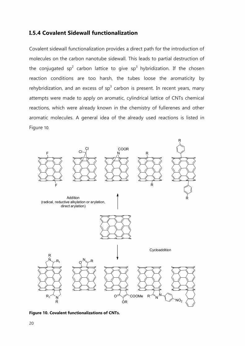
I.5.4 Covalent Sidewall functionalization
Covalent sidewall functionalization provides a direct path for the introduction of
molecules on the carbon nanotube sidewall. This leads to partial destruction of
the conjugated sp2 carbon lattice to give sp3 hybridization. If the chosen
reaction conditions are too harsh, the tubes loose the aromaticity by
rehybridization, and an excess of sp3 carbon is present. In recent years, many
attempts were made to apply on aromatic, cylindrical lattice of CNTs chemical
reactions, which were already known in the chemistry of fullerenes and other
aromatic molecules. A general idea of the already used reactions is listed in
Figure 10.
Addition (radical, reductive alkylation or arylation,
direct arylation)
Cycloaddition
F
F
ClCl N
COORR
R
R
R
N
N
RR1
R1
R
NO R
OOR
COOMe NNR
NO2
Figure 10. Covalent functionalizations of CNTs.
21
One of the first successful implementation of covalent reactions on
carbon nanotubes was applied in 1996: the fluorination.57,58 STM studies have
shown that the reaction occurs where the fluorine atoms are adsorbed
perpendicular to the axis. This can occur in two different addition patterns,
namely 1,2-addition and 1,4-addition. Calculations have shown that isomer 1,4-
should be more stable, but, due to the low energy barrier between the two
substitution pathways, both isomers likely occur at the same time. After this
discovery, a variety of different pathways were found for efficient fluorination of
carbon nanotubes, including plasma-based functionalization.59 Fluorinated
nanotubes have been widely used as starting material for further chemical
modifications, based on the nucleophilic substitution of fluorine atoms. For
examples, the reactions of alkyl lithium and Grignard reagents lead to alkyl
functionalization of sidewalls.60 They can be de-halogenated under treatment
with hydrazine or strong nucleophiles and functionalized in a second step.61
Zhang et al. showed that fluorinated CNTs are even capable to react in Diels-
Alder [4 + 2] reaction.62 It is also possible that, by thermal oxidation, fluorinated
or alkylated CNTs loose the side appendix and regenerate the original
nanotubes.
A reaction of alkyl lithium compounds with non-fluorinated SWCNTs was
reported in 2003 by Viswana et al.63 The approach was developed to in situ
composite synthesis by attachment of polystyrene (PS) chains to full-length
pristine SWCNTs without disrupting the original structure, based on an
established anionic polymerization. The reaction was performed by carbanion
formation with sec-butyl lithium as an initial step for polymerization of styrene
at the CNT sidewall. The reaction was performed also on MWCNTs with n-butyl
lithium and chlorinated polypropylene.64 A successful alkylation of CNTs was
performed with sec-butyl lithium, and subsequent carboxylation with CO2.65 In
the first reaction step, the nucleophilic addition of alkylcarbanionis used and
then the second step is the addition of CO2 as electrophile.
22
Another method is the reductive alkylation under the Birch conditions.
The reaction is performed in liquid ammonia with alkali metals such as sodium,
lithium or potassium to reduce SWCNTs and generate soluble CNT anions. It
was shown that negatively charged single-wall and multiwall nanotubes can be
derivatized by addition of alkyl halides.66 The Birch method was extended to
arylation, and besides the use of NH3 as solvent, the reactions was performed
with the radical anion salt of lithium naphthalenide in THF.67 In 2007
Chattopadhyay et al. found that SWCNTs treated with lithium in liquid ammonia
react also with aryl and alkyl sulfides by single electron transfer (SET).68
In 2001 Tour proposed an electrochemical reduction of aryl diazonium
salts to provide reactive radicals that can covalently react with CNTs.61 It was
postulated that an electron from the CNTs is transfer on to diazonium salt, and
then, after elimination of nitrogen, a reactive aryl radical is formed. By varying
the aryl diazonium salt, SWCNTs can be functionalized to obtain well-soluble
products. Soon the reaction was extended to a solvent-based thermally induced
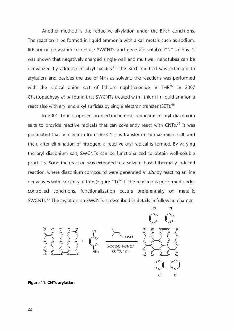
reaction, where diazonium compound were generated in situ by reacting aniline
derivatives with isopentyl nitrite (Figure 11).69 If the reaction is performed under
controlled conditions, functionalization occurs preferentially on metallic
SWCNTs.70 The arylation on SWCNTs is described in details in following chapter.
Cl
NH2
+
Cl Cl
Cl Cl
ONO
o-DCB/CH3CN 2:160 oC, 12 h
Figure 11. CNTs arylation.
23
Following the idea of radical additions to CNTs, organic xanthates and
peroxides are also suitable for covalent functionalization.71 Pennetreau et al.
stated that the reactivity of dilauroyl peroxide moieties onto the CNT surface
along with xanthate-derived radicals are inversely proportional to the stability of
the radical that is formed during the reaction.
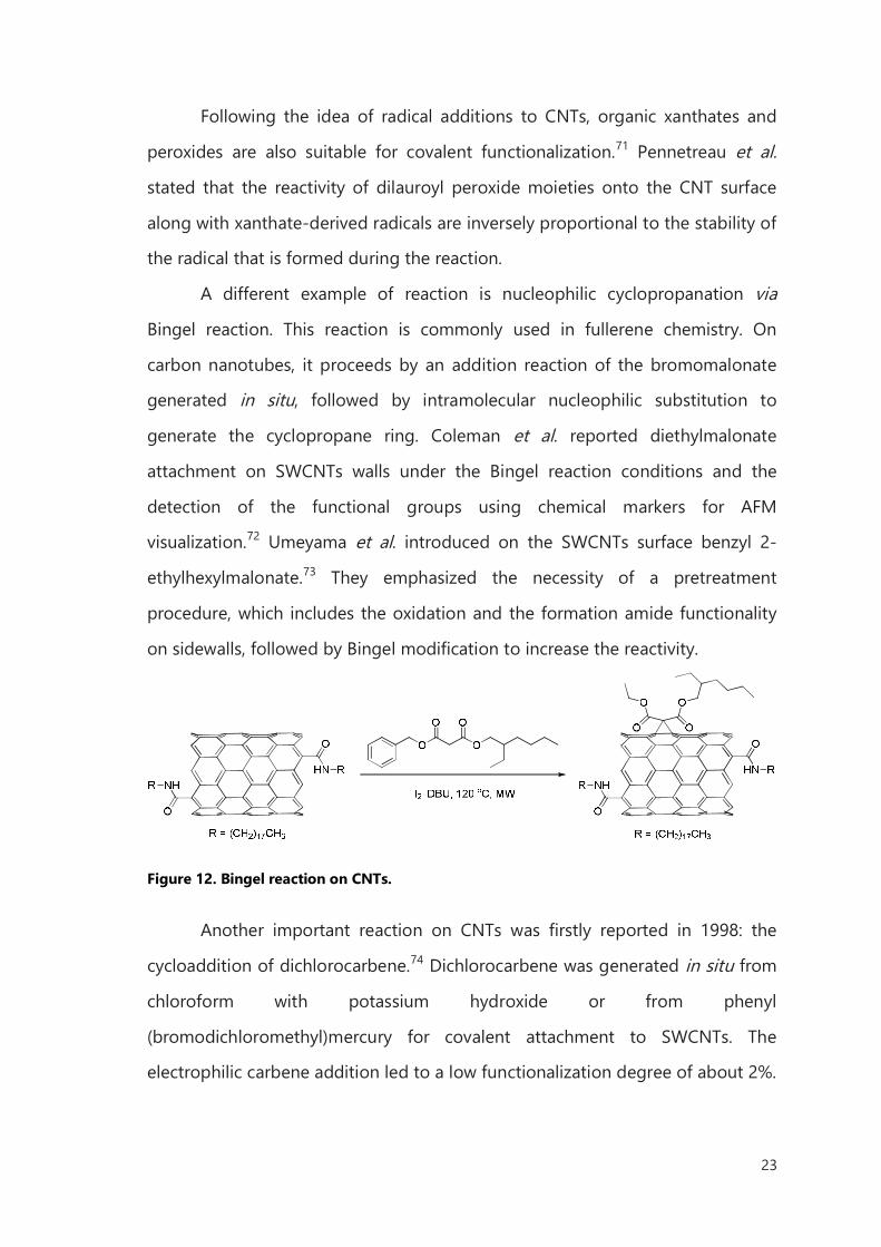
A different example of reaction is nucleophilic cyclopropanation via
Bingel reaction. This reaction is commonly used in fullerene chemistry. On
carbon nanotubes, it proceeds by an addition reaction of the bromomalonate
generated in situ, followed by intramolecular nucleophilic substitution to
generate the cyclopropane ring. Coleman et al. reported diethylmalonate
attachment on SWCNTs walls under the Bingel reaction conditions and the
detection of the functional groups using chemical markers for AFM
visualization.72 Umeyama et al. introduced on the SWCNTs surface benzyl 2-
ethylhexylmalonate.73 They emphasized the necessity of a pretreatment
procedure, which includes the oxidation and the formation amide functionality
on sidewalls, followed by Bingel modification to increase the reactivity.
Figure 12. Bingel reaction on CNTs.
Another important reaction on CNTs was firstly reported in 1998: the
cycloaddition of dichlorocarbene.74 Dichlorocarbene was generated in situ from
chloroform with potassium hydroxide or from phenyl
(bromodichloromethyl)mercury for covalent attachment to SWCNTs. The
electrophilic carbene addition led to a low functionalization degree of about 2%.
24
The functionalization was more efficient for small diameter tubes and caused
great enhancement of charge transfer on semiconducting tubes.
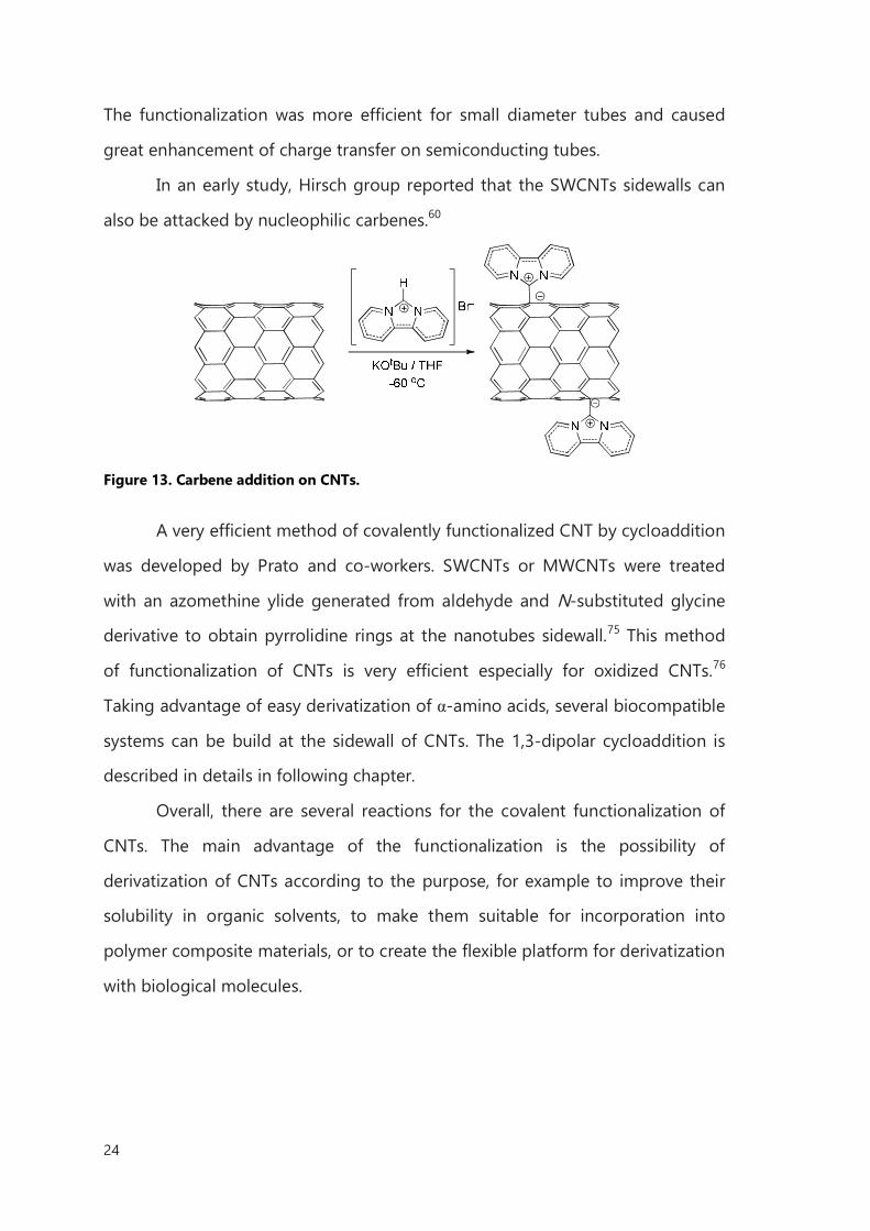
In an early study, Hirsch group reported that the SWCNTs sidewalls can
also be attacked by nucleophilic carbenes.60
Figure 13. Carbene addition on CNTs.
A very efficient method of covalently functionalized CNT by cycloaddition
was developed by Prato and co-workers. SWCNTs or MWCNTs were treated
with an azomethine ylide generated from aldehyde and N-substituted glycine
derivative to obtain pyrrolidine rings at the nanotubes sidewall.75 This method
of functionalization of CNTs is very efficient especially for oxidized CNTs.76
Taking advantage of easy derivatization of α-amino acids, several biocompatible
systems can be build at the sidewall of CNTs. The 1,3-dipolar cycloaddition is
described in details in following chapter.
Overall, there are several reactions for the covalent functionalization of
CNTs. The main advantage of the functionalization is the possibility of
derivatization of CNTs according to the purpose, for example to improve their
solubility in organic solvents, to make them suitable for incorporation into
polymer composite materials, or to create the flexible platform for derivatization
with biological molecules.
25
I.6 Biomedical application of carbon nanotubes
The properties of CNTs allow to envision application in many biomedical
areas by offering unique alternatives to present technologies, from tissue
scaffolds to drug delivery vehicles.
I.6.1 Carbon nanotubes as cell substrates
One of the very promising and important implementation of CNTs is
connected with their topological and chemical structure. It is well established
that topographies and patterns can influence the cellular behaviors. By
controlling the nanoscale topography of cellular substrates, the implantation of
medical devices facilitate new biological processes, including embryogenesis,
angiogenesis, and pathological conditions.
CNTs as nanoparticles are inherently appropriate for surface
modifications by simple incorporation or deposition on their surface. They have
a fibrillar shape and versatile optical, electrical, and mechanical characteristics
for applications as a cellular substrate. Many groups have successfully utilized
CNTs for cellular growth surfaces to provide structural support or present novel
properties.77 For instance, a range of cell phenotypes were presented to have
high binding affinities for CNTs surfaces, demonstrating the CNTs may be used
for a variety of tissue implantation devices or novel substrates.78,79
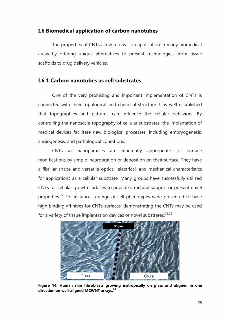
Figure 14. Human skin fibroblasts growing isotropically on glass and aligned in one direction on well-aligned MCWNT arrays.80
50 µm
Glass CNTs
26
Several papers have presented CNTs as conducive to neuronal adhesion
and safe for neural processes outgrowth, suggesting that CNTs are
biocompatible with neurons. Cellot at al. reported CNTs to provide shortcut for
the electrical signaling connecting tight junctions adhered onto the nanotube
surface at proximal and distal portions of the neuron.81 Mature neuronal cells
have also been derived directly from human embryonic stem cells (hESCs) using
polymer-grafted CNTs thin film scaffolds.82 Malarkey and co-workers, by
modulating the thickness and the conductivity of a CNTs film, were able to
change neuron morphology, neurite outgrowth, and the number of growth
cones.83 Even if till now neuronal interfacing has no direct clinical benefits,
research developments in this area may help to explain biological mechanisms
and neural interactions relevant to injury and disease. Preferential interactions of
neural cells and CNTs permit the study of axonal outgrowth and connection
between neural clusters and patterned CNTs. Also the directed growth and
migration along CNT surface architectures allowed the study to prevent and
repair of nerve injuries, such as spinal cord injury or stroke.84
Lee et al. pretreated rats with amine-modified SWCNTs to protect neurons and
to improve the recovery of behavioral functions in rats with induced stroke.
Authors suggest that CNTs with positive charges may have contributed to a
favorable environment for neurons.85 Roman et al. investigated the
administration of PEGylated SWCNTs after traumatic neural cord injury, which
could promote regeneration of axons into the lesion cavity and functional
recovery of the hindlimbs.86 They found that, after a spinal cord injury (SPI),
neurofilament-positive fibers of SWCNT-PEG induced a modest improvement in
hindlimb locomotor recovery without inducing hyper algesia. These data
suggest that SWCNT-PEG may be an effective material to promote axonal repair
and regeneration after SPI.
27
I.6.2 Carbon nanotube for delivery of drugs
The high biocompatibility of CNTs and their possibility to penetrate the
cells provide the excellent intracellular vehicle to interact with mammalian cells.
In 2004 Pantarotto et al. demonstrated the translocation of CNTs in cellular
membranes and proposed uptake mechanism, intracellular distribution,
elimination from cells and possible adverse effects.87 Two types of CNTs
functionalization were reported: SWCNTs FITC-labeled and peptide–SWCNTs
conjugated. Generally, these compounds were able to pass the cell membrane
and the peptide-SWCNTs were found to accumulate in the nuclei of the cells,
whereas the FITC-labeled SWCNTs were distributed in the cytoplasm. The
internalization was not affected by the temperature or the presence of
endocytosis inhibitors. So it was possible to claim that the uptake mechanism
was endocytosis-independent and it was hypothesized that the cylindrical shape
and high aspect ratio of functionalized CNTs allowed their penetration through
the plasma membrane in a needle-like uptake. Kam et al. also studied the
cellular uptake of fluorescent-labeled SWCNTs and fluorescent peptide–
SWCNTs conjugate.88 Their results suggested an uptake by endocytosis because
it was inhibited at low temperatures. Nanotubes were localized in the
endosomes and in the cytoplasm, but not into the nucleus.
These contrast results are ascribed to the different physical and chemical
properties of used nanotubes, as well the type of functionalization.
In particularly the uptake mechanism is very important for delivery application.
In the case of endocytic uptake, CNTs are initially covered by intracellular
vesicles, called endosomes, with mild acidic pH. Next there is the fusion of the
endosomes with lysosomes and pH reaches 5.5 for hydrolytic enzymes.
Unfortunately in such conditions drugs and nucleic acids can degrade. Thus, in
same case it is preferable that CNTs escape lysosomes. The needle-like uptake
pathway transports the nanotube directly into the cytoplasm and there are more
chances to avoid the lysosomal degradation.
28
CNTs can be combined with the drug in two ways: linked to the outer
walls or by internal filling. In the case of external functionalization, this can be
non-covalent or covalent. The non-covalent way is simply based on the
interaction of CNTs and drugs after mixing. However, the stability of the
produced system depends on environmental factors. This approach can find a
convenient application when the drug can be released from complex in slightly
acidic microenvironment of endosomes and lysosomes, as for CNT-doxorubicin
couple, which is bound via π-π interactions. At physiological pH the amino
group of the doxorubicin sugar moiety is deprotonated, promoting strong
hydrophobic interactions with the nanotubes sidewalls and conferring low
solubility in water. In contrast, at lower pH as in cancer cells or lysosomes, the
amino group becomes protonated, increasing the solubility in water and
resulting in the release of the drug from the nanotubes.89 Other examples of
non-covalent binding to CNTs include paclitaxel90 and camptothecin.91
The covalent binding between CNTs and drugs is usually based on
cleavable bonds like disulfides92, esters93, carbamates94. In these cases, the drugs
are bound not directly to the nanotube but to the linkers which have been
introduced to increase the solubility and biocompatibility of the carbon scaffold.
The most common linkers are so far poly(ethylene glycol) (PEG) and linear
polymers, such aspoly(vinyl alcohol)95. In recent years, branched polymers have
become more popular, among these dendrimers96 and dendrons.97
When a drug is delivered inside the cavities of the tubes, it can be
transferred with high protection and released in a controlled way. Hampel et al.
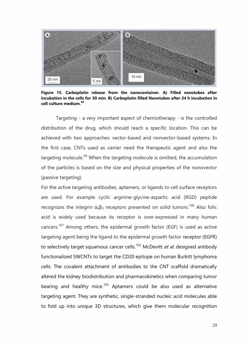
have filled MWCNTs with carboplatin; they used wet chemistry approach to
reach approximately 30 wt% of filling yield.98 The complex displays a cytotoxic
effect depending on the concentration reached inhuman bladder cancer cells.
The study was performed also with empty tubes, showing strong inhibition of
the cells viability only in the case of carboplatin filled CNTs (Figure 15).
29
Figure 15. Carboplatin release from the nanocontainer. A) Filled nanotubes after incubation in the cells for 30 min. B) Carboplatin filled Nanotubes after 24 h incubation in cell culture medium.98
Targeting - a very important aspect of chemiotherapy - is the controlled
distribution of the drug, which should reach a specific location. This can be
achieved with two approaches: vector-based and nonvector-based systems. In
the first case, CNTs used as carrier need the therapeutic agent and also the
targeting molecule.99 When the targeting molecule is omitted, the accumulation
of the particles is based on the size and physical properties of the nonovector
(passive targeting).
For the active targeting antibodies, aptamers, or ligands to cell surface receptors
are used. For example cyclic arginine-glycine-aspartic acid (RGD) peptide
recognizes the integrin αvβ3 receptors presented on solid tumors.100 Also folic
acid is widely used because its receptor is over-expressed in many human
cancers.101 Among others, the epidermal growth factor (EGF) is used as active
targeting agent being the ligand to the epidermal growth factor receptor (EGFR)
to selectively target squamous cancer cells.102 McDevitt et al. designed antibody
functionalized SWCNTs to target the CD20 epitope on human Burkitt lymphoma
cells. The covalent attachment of antibodies to the CNT scaffold dramatically
altered the kidney biodistribution and pharmacokinetics when comparing tumor
bearing and healthy mice.103 Aptamers could be also used as alternative
targeting agent. They are synthetic, single-stranded nucleic acid molecules able
to fold up into unique 3D structures, which give them molecular recognition
30
properties similar to antibodies. They are easily producible for variety of targets
with little or no immunogenicity in therapeutic submissions. However, they
usually have an inefficient cell uptake but Van der Bossche et al. have
demonstrated that aptamers bound onto CNTs are translocated into the cytosol
of different cell types independently of receptor-mediated uptake.104
I.6.3 Carbon nanotubes for imaging
By functionalization, CNTs can be conjugated with imaging agents, such as
metallic nanoparticles, quantum dots, or isotopes to make possible imaging via
conventional techniques. Chen et al. studied the conjugation of CNTs with super
paramagnetic iron oxide nanoparticles (SPIO) and NIR fluorescent quantum dots
CdTe.105 The CNTs-SPIO-CdTe nanohybrids exhibited the superparamagnetic
behavior of SPIO with a saturation magnetization of about 65 emug-1 at room
temperature and strong emission band located at a near-infrared wavelength of
734 nm. Al-Faraj et al. and Vittorio et al. used magnetic resonance imaging
(MRI) for visualizing CNTs in cells106 or living organisms107 taking advantage of
the presence of iron oxide impurities.
The recent studies about application of CNTs for radiotherapy presented several
examples of CNTs as a radioisotope carrier. In the case of encapsulation of the
radionuclides, Hong et al., presented single-walled CNTs filled with sodium
iodide-125 (Auger and γ-emitter) for in vitro and in vivo study.108 The material
had a specific tissue accumulation (lung) and the leakage of the radionuclide
was not observed.
31
I.7 Toxicity - physical determinants influencing the toxicity of
carbon nanotubes
The biological application of CNTs strongly depends on all health hazards
that the new material can cause. Since several years, in vivo and in vitro
toxicological studies begun to determine the effect of this material in cells and
tissues. However, it turned out that many parameters of CNTs, (as contaminants,
surface chemistry, processing method, agglomerate state, length, diameter, and
more others) can have various toxic effects.
I.7.1 Effect of purity on toxicity
As it was reported, the toxic effect of the CNTs can arise not directly from
CNTs, but from the residues produced during the synthetic process as nickel,
cobalt or iron nanoparticles, which can persevere in the CNTs and generate
reactive oxygen species (ROS) in biological environment. It turns out that ROS
cause inflammatory symptoms and induce mitochondrial membrane
degradation, depletion of antioxidant agents, rise in inflammatory biomarkers,
and decreases the cell viability. It has been demonstrated that 30% of iron in
SWCNTs is able to generate free radicals within 15 min of exposure to
epidermal keratinocytes in the presence of DMPO (5,5-dimethyl-1-pyrroline-1-
oxide).109 In a later study it was shown that higher amounts of catalyst generate
higher concentrations of free radicals and increase inflammatory responses.110
Additionally nickel alters the expression of the gene encoding the protein
HIF1A, a factor of transcript involved in the regulation of inflammatory genes
and apoptosis.111 All studies on the toxicity of CNTs must than take into account
the nature of the metallic catalyst and its percentage/quantity. It is difficult to
obtain pure CNTs by removing all traces of catalysts and several methods can
be employed to decrease residual catalysts including centrifugation, high-
temperature annealing112 and oxidation treatment by acid reflux.113
32
I.7.2 Effect of CNTs functionalization
Toxicity can be also influenced by the modification on CNTs surface. Only
with the acid-treatment on CNTs surface, it is possible to introduce number of
defect sites along the CNT surface (see above). Muller et al. changed the
number of defect sites on MWCNTs by mechanical grinding and annealing at
high temperature and demonstrated that acute pulmonary toxicity and
genotoxicity increased after intratracheally administration of MWCNTs with a
larger number of defect sites.114 However, another study by Kagan et al. showed
that oxidized SWCNTs can be biodegraded easier by myeloperoxidase enzyme,
found in neutrophils and macrophages. The enzyme interacts with carboxylic
sites on the nanotubes surface115 and oxidized CNTs may be more
biocompatible than pristine CNTs from this point of view. Following this
pathway Sayes and co-workers examined cell viability in presence of oxidized
and phenylated tubes. They discovered that the phenylated tubes exhibit lower
toxicity, and this can be due to the hindering of the defect sides of the tubes.116
Dumortier at al. have examined the toxicity of CNTs functionalized by 1,3-
dipolar cycloaddition. They concluded that CNTs, fully soluble in aqueous
culture media, did not modified primary immune cells viability in vitro.117
Also the CNTs surface area and their hydrophobic nature impact on the
toxicity. The tubes, in fact, can potentially interact with numerous molecules like
proteins, RNA, DNA, enzymes with toxic effects on the biological
environment.118 Dutta and co-workers found that the bovine or human serum
albumin adsorption onto the CNT surface resulted in inflammatory responses
after uptake by macrophage cells. Normally that effect occurs only when
albumin adopts structurally changes or gets damaged.119
The effect of functionalization of the tubes highlights the importance of
assessing toxicity profile for every type of new CNTs modification.
33
I.7.3 The length of carbon nanotubes
Finally, the CNTs length cannot be ignored. CNT sizes have an important
effect on clearance. The length can range from nanometers up to millimeters.
The exposure to long fiber-like material can induce dangerous damages in DNA
and genetic mutations over a period of exposure, causing an extremely
malignant form of cancer, mesothelioma. Symptoms of these bio-persistent
fibers are the granulomas, which are the signs of oxidative stress, causing
excessive fibrous tissue.120
Macrophage are the cells responsible for the removal of foreign material
like CNTs from living organism. Fibers with a length exceeding 20 µm are
extremely resistant to phagocytosis121 and Poland et al. shown that CNTs as
spherical or stellate shaped agglomerates with less than 20 µm have no
significant damaging reactions compared to samples with individualized
MWCNTs, agglomerates, and ropes of MWCNTs with lengths exceeding 20
µm.120
In conclusion, CNTs should be used without the presence of metal
catalysts, should have an appropriate functionalization for the planned purpose
with low surface oxidation, should present covered surface to effectively escape
biointeractions, and should be short to avoid long retention times.
34
Bibliography
1 S. Iijima; Nature 1991, 354, 56−58. 2 D. Pantarotto, J. P. Briand, M. Prato, A. Bianco; Chem. Commun. 2004, 7, 16−17. 3 Y. Zhang, Y. Bai, B. Yan; Drug Discovery Today 2010, 15, 428−435. 4 N. W. Kam, H. Dai; J. Am. Chem. Soc. 2005 127, 6021–6026.
5 B. G. P. Singh, C. Baburao, V. Pispati, H. Pathipati, N. Muthy, S. R. V. Prassana, B. Ganesh
Rathode; International Journal of Research in Pharmacy and Chemistry 2012, 2, 523–532. 6 Z. Liu, S. Tabakman, K. Welsher, H. Dai; Nano Research 2009, 2, 85–120. 7 G. Seeta Rama Raju, Leah Benton, E. Pavitra, Jae Su Yu; Chem. Commun. 2015, 51,
13248−13259. 8 H. Gong, R. Peng, and Z. Liu; Advanced Drug Delivery Reviews 2013, 65, 1951–1963. 9 J.−C. Charlier, X. Blase, A. De Vita, R. Car; Appl. Phys. A 1999, 68, 267–273.
10 M. S. Dresselhaus, G. Dresselhaus, R. Saito, A. Jorio; Physics Reports 2005, 409, 47–99. 11 L. Ortolani, F. Houdellier, M. Monthioux, V. Morandi; Carbon 2010, 48, 3050–3056. 12 A. Thess, P. Nikolaev, H. Dai, C. Xu, A. G. Rinzler, D. T. Colbert, G. E. Scuseria, R. E. Smalley;
Science 1996, 273, 483−487. 13 J. W. G. Wildoer, L. C. Venema, A. G. Rinzler, R. E. Smalley, C. Dekker; Nature 1998, 39, 59−62. 14 H. Huang, M. Zou, X. Xu, F. Liu, N. Li; Trends in Analytical Chemistry 2011, 30, 1109−1119. 15 S. M. Bachilo, M. S. Strano, C. Kittrell, R. H. Hauge, R. E. Smalley, R. B. Weisman; Science 2002,
298, 2361−2366.
16 Z. Luo, F. Papadimitrakopoulos, S. K. Doorn; Phys. Rev. B. 2007, 75, 205438. 17 J. W. Kang, F. T. Nguyen, N. Lue, R. R. Dasari, D. A. Heller; Nano Lett. 2012, 12, 6170–6174. 18 R. Andrews, D. Jacques, D. Qian, T. Rantell; Acc. Chem. Res. 2002, 35, 1008−1017. 19 S. Niyogi, M. A. Hamon, H. Hu , B. Zhao, P. Bhowmik, R. Sen, M. E. Itkis, R. C. Haddon; Acc.
Chem. Res. 2002, 35, 1105−1113. 20 Mi. Sola, J. Mestres, M. Duran; J. Phys. Chem. 1995, 99, 10752−10758. 21 A.V. Narlikar, Y.Y. Fu, Oxford Handbook of Nanoscience and Technology: Vol. 1: Basic Aspects,
Oxford University Press, 2010.
22 A. Hirsch, O. Vostrowsky; Top Curr. Chem. 2005, 245, 193– 237. 23 J. L. Bahr, E. T. Mickelson, M. J. Bronikowski, R. E. Smalley J. M. Tour; Chem. Commun. 2001,
193–194. 24 R. H.Baughman, A.A. Zakhidov, W. A. De Heer; Science 2002, 297, 787−792. 25 K. Cho, X. Wang, S. Nie, Z. G. Chen, D. M. Shin; Clinical Cancer Research 2008, 14, 1310−1316.
35
26 T. Dvir, B. P. Timko, D. S. Kohane, R. Langer; Nature Nanotechnology 2011, 6, 13−22. 27 M. F. Islam, E. Rojas, D. M. Bergey, A. T. Johnson, A. G. Yodh; Nano Lett. 2003, 3, 269−273. 28 H. Dodziuk, A. Ejchart, W. Anczewski, H. Ueda, E. Krinichnaya, Grygoriy Dolgonosa, W. Kutner;
Chem. Commun. 2003, 986–987. 29 Y. Sun, S. R. Wilson, D. I. Schuster; J. Am. Chem. Soc. 2001, 123, 5348.
30 D. Chattopadhyay, I. Galeska, F. Papadimitrakopoulos; J. Am. Chem. Soc. 2003, 125, 3370. 31 T. Fujigaya, N. Nakashima; Sci. Technol. Adv. Mater. 2015, 16, 024802. 32 A. Star, Y. Liu, K. Grant, L. Ridvan, J. F. Stoddart, D. W. Steuerman, M. R. Diehl, A. Boukai, J. R.
Heath; Macromolecules 2003, 36, 553. 33 A. B. Dalton, C. Stephan, J. N. Coleman, B. McCarthy, P. M. Ajayan, S. Lefrant, P. Bernier, W. J.
Blau, H. J. Byrne; J. Phys. Chem. B 2000, 104, 10012. 34 S. Park, H.-S. Yang, D. Kim, K. Job, S. Jon; Chem. Commun. 2008, 2876−2878. 35 S. S. Karajanagi, A. A. Vertegel, R. S. Kane, J. S. Dordick; Langmuir 2004, 20, 11594.
36 M. Zheng, A. Jagota, M. S. Strano, A. P. Santos, P. Barone, S. G. Chou, B. A. Diner, M. S.
Dresselhaus, R. S. McLean, G. B. Onoa, G. G. Samsonidze, E. D. Semke, M. Usrey, D. J. Walls;
Science 2003, 302, 1545. 37 Z. Mohammadi, S. F. Aghamiri, A. Zarrabi, M. Reza Talaie; Chemical Physics Letters 2015,642,
22–28. 38 M. S. C. Mazzoni, H. Chacham, P. Ordejon, D. Sanchez-Portal, J. M. Soler, E. Artacho; Phys. Rev.
B 1999, 60, R2208.
39 P. G. Collins, Defects and Disorder in Carbon Nanotubes, Oxford Handbook of Nanoscience
and Technology: Frontiers and Advances. A. V. Narlikar, Y. Y. Fu, Eds., Oxford Univ. Press, Oxford,
2009. 40 M. F. Yu, O. Lourie, M. J. Dyer, K. Moloni, T. F. Kelly, R. S. Ruoff; Nature, 2006, 442, 282−286. 41 J. Zhang, H. Zou, Q. Qing, Y. Yang, Q. Li, Z. Liu, X. Guo, Z. Du; J. Phys. Chem. B 2003, 107,
3712−3718. 42 A. Hirsch; Angew, Chem, Int. 2002, 41, 1853−1859. 43 P. Ajayan, S. Iijima; Nature 1993, 361, 333–334.
44 F. Simon, H. Kuzmany, H. Rauf, T. Pichler, J. Bernardi, H. Peterlik, L. Korecz, F. Fulop, A. Janossy;
Chemical Physics Letters 2004, 383, 362−367. 45 S. Tsang, Y. Chen, P. Harris, M. Green; Nature 1994, 372, 159−162. 46 M. V. Kharlamova, J. J. Niu; Appl. Phys. A 2012, 109, 25−29. 47 K. Yanagi, Y. Miyata, H. Kataura; Adv. Mater. 2006, 18, 437−441.
36
48 H. Kataura, Y. Maniwa, M. Abe, A. Fujiwara, T. Kodama, K. Kikuchi, H. Imahori, Y. Misaki, S.
Suzuki, Y. Achiba; Appl. Phys. A 2002, 74, 349−354. 49 A. I. Chernov, P. V. Fedotov, A. V. Talyzin, I. S. Lopez, I. V. Anoshkin, A. G. Nasibulin, E. I.
Kauppinen, E. D. Obraztsova; ACS Nano 2013, 7, 6346−6353. 50 B. W. Smith, M. Monthioux, D. E. Luzzi; Nature 1998, 396, 323−324.
51 P. M. Ajayan, S. Lijima; Nature 1993, 361, 333–334. 52 Y. Gogotsi, N. Naguib, J. A. Libera; Chemical Physics Letters 2002, 365, 354–360. 53 M. Terrones; Nat. Chem. 2010, 2, 82–83. 54 C. Xu, J. Sloan, G. Brown, S. Bailey, V. C. Williams, S. Friedrichs, K. S. Coleman, E. Flahaut, J. L.
Hutchison, R. E. Dunin-Borkowski, M. L. H. Green; Chem. Commun. 2000, 2427–2428. 55 E. Dujardin, T. W. Ebbesen, H. Hiura, K. Tanigaki; Science 1997, 265, 1850−1852. 56 B. Ballesteros, G. Tobias, M. A. H. Ward, M. L. H. Green; J. Phys. Chem. C 2009, 113, 2653–2656. 57 T. Nakajima, S. Kasamatsu, Y. Matsuo; Eur. J Solid. Inorg. Chem. 1996, 33, 831−840.
58 K. F. Kelly, I. W. Chiang, E. T. Mickelson, R. H. Hauge, J. L. Margrave, X. Wang, G. E. Scuseria, C.
Radloff, N. J. Halas; Chemical Physics Letters 1999, 313, 445–450. 59 L. Valentini, D. Puglia, I. Armentano, J. M. Kenny; Chem. Phys. Lett. 2005, 403, 385–389. 60 M. Holzinger, O. Vostrowsky, A. Hirsch, F. Hennrich, M. Kappes, R. Weiss, F. Jellen; Angew.
Chem. Int. Ed. 2001, 40, 4002−4005. 61 J. L. Bahr, J. Yang, D. V. Kosynkin, M. J. Bronikowski, R. E. Smalley, J. M. Tour; J. Am. Chem. Soc.
2001, 123, 6536−6542.
62 L. Zhang, J. Yang, C. L. Edwards, L. B. Alemany, V. N. Khabashesku, A. R. Barron; Chem.
Commun. 2005, 3265−3267. 63 G. Viswanathan, N. Chakrapani, H. Yang, B. Wei, H. Chung, K. Cho, C. Y. Ryu, P. M. Ajayan; J.
Am. Chem. Soc. 2003, 125, 9258−9259. 64 R. Blake, Y. K. Gunko, J. Coleman, M. Cadek, A. Fonseca, J. B. Nagy, W. J. Blau; J. Am. Chem.
Soc. 2004, 126, 10226. 65 S. Chen, W. Shen, G. Wu, D. Chen, M. Jiang; Chem. Phys. Lett. 2005, 402, 312. 66 I. W. Chiang, B. E. Brinson, R. E. Smalley, J. L. Margrave, R. H. Hauge; J. Phys. Chem. B 2001,
105, 7938. 67 P. Papakonstantinou, R. Kern, L. Robinson, H. Murphy, J. Irvine,E. McAdams, J. McLaughlin;
Fuller. Nanotub. Carbon Nanostruct. 2005,13, 91–108. 68 J. Chattopadhyay, S. Chakraborty, A. Mukherjee, R. Wang, P. S. Engel, W. E. Billups; Phys. Chem.
C 2007, 111, 17928−17932. 69 J. L. Bahr, J. M. Tour; Chem. Mater. 2001, 13, 3823–3824.
37
70 C. A. Dyke, J. M. Tour; Nano Lett. 2003, 3, 1215−1218. 71 F. Pennetreau, C. Vriamont, B. Vanhorenbeke, O. Riant, S. Hermans; Eur. J. Org. Chem. 2015,
1804–1810. 72 K. S. Coleman, Sam R. Bailey, S. Fogden, M. L. H. Green; J. Am. Chem. Soc. 2003, 125, 8722–
8723.
73 T. Umeyama, N. Tezuka, M. Fujita, Y. Matano, N. Takeda, K. Murakoshi, K. Yoshida, S. Isoda, H.
Imahori; J. Phys. Chem. C 2007, 111, 9734–9741. 74 J. Chen, M. A. Hamon, H. Hu, Y. Chen, A. M. Rao, P. C. Eklund, R. C. Haddon; Science 1998, 282,
95−98. 75 V. Georgakilas, K. Kordatos, M. Prato. D. M. Guldi, M. Holzinger, A. Hirsch; J. Am. Chem. Soc.
2002, 124, 760−761. 76 D. Pantarotto, R. Singh, D. McCarthy, M. Erhardt, J. P. Briand, M. Prato, K. Kostarelos, A. Bianco;
Angew. Chem. Int. Ed. 2004, 43, 5242 –5246.
77 N. Saito, Y. Usui, K. Aoki, N. Narita, M. Shimizu, K. Hara, N. Ogiwara, K. Nakamura, N. Ishigaki,
H. Kato, S. Taruta, M. M. Endo; Chem. Soc. Rev. 2009, 38, 1897−1903. 78 J. Meng, L. Song, J. Meng, H. Kong, G. Zhu, C. Wang, L. Xu, S. Xie, H. Xu; J. Biomed. Mater. Res.
A 2006, 79A, 298−306. 79 A. O. Lobo, M. A. F. Corat, E. F. Antunes, M. B. S. Palma, C. Pacheco-Soares, E. E. Garcia, E. J.
Corat; Carbon 2010, 48, 245−254. 80 P. Galvan-Garcia, E. W. Keefer, F. Yang, M. Zhang, S. Fang, A. A. Zakhidov, R. H. Baughman, M. I
Romero; J. Biomater. Sci. Polym. Ed. 2007, 18, 1245−1261. 81 G. Cellot, E. Cilia, S. Cipollone, V. Rancic, A. Sucapane, S. Giordani, L. Gambazzi, H. Markram, M.
Grandolfo, D. Scaini, F. Gelain, L. Casalis, M. Prato, M. Giugliano, L. Ballerini; Nat. Nano 2009, 4,
126−133. 82 T.-I Chao, S. Xiang, J. F. Lipstate, C. Wang, J. Lu; J. Adv. Mater. 2010, 22, 3542−3547. 83 E. B. Malarkey, R. C. Reyes, B. Zhao, R. C. Haddon, V. Parpura; Nano Lett. 2009, 9, 264−268. 84 X. Zhang, S. Prasad, S. Niyogi, A. Morgan, M. Ozkan, C. S. Ozkan; Sensors Actuators B 2005,
106, 843−850.
85 D. Rugar, H. J. Mamin, M. H. Sherwood, M. Kim, C. T. Rettner, K. Ohno, D. D. Awschalom; Nat.
Nanotechnol. 2011, 6, 120−124. 86 J. A. Roman, T. L. Niedzielko, R. C. Haddon, V. Parpura, C. L. Floyd; J. Neurotrauma 2011, 28,
2349−2362. 87 D. Pantarotto, J.-P. Briand,M. Prato, A. Bianco; Chem. Commun. 2004, 16−17. 88 N. W. S. Kam , T. C. Jessop, P. A. Wender, H. Dai; J. Am. Chem. Soc. 2004, 126, 6850−6851.
38
89 A.Di Crescenzo, D. Velluto, J. A. Hubbell, A. Fontana; Nanoscale 2011, 3, 925−928. 90 C. L. Lay, H. Q. Liu, H. R. Tan, Y. Liu; Nanotechnology 2010, 21, 065101. 91 J. Zhang, X.-J. Lv, X. Jia, Y.-L. Deng, H. Qing, H-Y Xie; J. Nanosci. Nanotechnol. 2011, 11,
953−958. 92 N. Wong, S. Kam , Z. Liu, H. Dai; J. Am. Chem. Soc. 2005, 127, 12492−12493.
93 C. Samorì, H. Ali-Boucetta, R. Sainz, C. Guo, F. M. Toma, C. Fabbro, T. da Ros, M. Prato, Kostas
Kostarelos, A. Bianco; Chem. Commun. 2010, 46, 1494−1496. 94 P. Chaudhuri, S. Soni, S. Sengupta; Nanotechnology 2010, 21, 025102. 95 N. G. Sahoo, H. Bao, Y. Pan, M. Pal, M. Kakran, H. K. Feng Cheng, L. Li, L. Poh Tan; Chem.
Commun. 2011, 47, 5235−5237. 96 X. Shi, S. H. Wang, M. Shen, M.E. Antwerp, X. Chen, C. Li, E. J. Petersen, Q. Huang, W. J. Weber
Jr., J. R. Baker Jr; Biomacromolecules 2009, 10, 1744−1750. 97 Z. Sobhani, R. Dinarvand, F. Atyabi, M. Ghahremani, M. Adeli; Int. J. Nanomedicine 2011, 6,
705−719. 98 S. Hampel, D. Kunze, D. Haase, K. Krämer, M. Rauschenbach, M. Ritschel, A. Leonhardt, J.
Thomas, S. Oswald, V. Hoffmann, B. Büchner; Nanomedicine 2008, 3, 175−182. 99 M. Ferrari; Nat. Rev. Cancer 2005, 5, 161−171. 100 Z. Liu, X. Sun, N. Nakayama-Ratchford, H. Dai; J. ACS Nano 2007, 1, 50−56. 101 J. M Tan, J. Biau Foo, S. Fakurazi, M. Z. Hussein; Adv. Drug Delivery Rev. 2011, 63, 1332−1339. 102 A. A. Bhirde, V.h Patel, J. Gavard, G. Zhang, A. A. Sousa, A. Masedunskas, R. D. Leapman, R.
Weigert, J. S. Gutkind, J. F. Rusling; ACS Nano 2009, 3, 307−316. 103 M. R. McDevitt, D. Chattopadhyay, B. J. J. Kappel; Nucl. Med. 2007, 48, 1180−1189. 104 J. Van den Bossche, W. T. Al-Jamal, B. Tian, A. Nunes, C. Fabbro, A. Bianco, M. Prato, K.
Kostarelos; Chem. Commun. 2010, 46, 7379−7381. 105 B. Chen, H. Zhang, C. Zhai, N. Du, C. Sun, J. Xue, D. Yang, H. Huang, B. Zhang, Q.J. Xie; Mater.
Chem. 2010, 20, 9895−9902. 106 O. Vittorio, S. L. Duce, A. Pietrabissa, A. Cuschieri; Nanotechnology 2011, 22, 095706. 107 A. Al Faraj, K. Cieslar,G. Lacroix, S. Gaillard, E. Canet-Soulas, Y. Crémillieux; Nano Lett. 2009, 9,
1023−1027. 108 S. Y. Hong, G. Tobias, K. T. Al-Jamal, B. Ballesteros, H. Ali-Boucetta, S. Lozano-Perez, P. D.
Nellist, R. B. Sim, C. Finucane, S. J. Mather, M. L. H. Green, K. Kostarelos, B. G. Davis; Nature
materials 2010, 9, 485−490. 109 A. A. Shvedova, V. Castranova, E. R. Kisin, D. Schwegler-Berry, A. R. Murray, V. Z. Gandelsman,
A. Maynard, P. Baron; J. Toxicol. Environ. Health A 2003, 66, 1909−1926.
39
110 A. R. Murray, E. Kisin, S. S. Leonard, S. H. Young, C. Kommineni, V. E. Kagan, V. Castranova, A.
A. Shvedova; Toxicology 2009, 257, 161−171. 111 M. Kaczmarek, O. A. Timofeeva, A. Karaczyn, A. Malyguine, K. S. Kasprzak, K. Salnikow; Free
Radic. Biol. Med. 2007, 42, 1246–1257. 112 W. Huang, Y. Wang, G. Luo, F. Wei; Carbon 2003, 41, 2585−2590.
113 Y. Li, X. Zhang, J. Luo, W. Huang, J. Cheng, Z. Luo, T. Li, F. Liu, G. Xu, X. Ke; Nanotechnology
2004, 15, 1645−1649. 114 J. Muller, F. Huaux, A. Fonseca, J. B. Nagy, N. Moreau, M. Delos, E. Raymundo-Piñero, F.
Béguin, M. Kirsch-Volders, I. Fenoglio, B. Fubini, D. Lison; Chem. Res. Toxicol. 2008, 21,
1698−1705. 115 V. E. Kagan, N. V. Konduru, W. Feng, B. L. Allen, J. Conroy, Y. Volkov, I. I. Vlasova, N. A.
Belikova, N. Yanamala, A. Kapralov, Y. Y. Tyurina, J. Shi, E. R. Kisin, A. R. Murray, J. Franks, D. Stolz,
P. Gou, J. Klein-Seetharaman, B. Fadeel, A. Star, A.A. Shvedova; Nat. Nanotechnol. 2010, 5,
354−359. 116 C. M. Sayes, F. Liang, J. L. Hudson, J. Mendez, W. Guo, J. M. Beach, V. C. Moore, C. D. Doyle, J.
L. West, W. E. Billups, K. D. Ausman, V. L. Colvin; Toxicol. Lett. 2006, 161, 135−142. 117 H. Dumortier, S. Lacotte, G. Pastorin, R. Marega, W. Wu, D. Bonifazi, J.-Paul Briand, M. Prato, S.
Muller, A. Bianco; Nano Lett. 2006, 6, 1522−1528. 118 D. B. Warheit, B. R. Laurence, K. L. Reed, D. H. Roach, G. A. M. Reynolds, T. R. Webb; Toxicol.
Sci. 2004, 77, 117−125.
119 D. Dutta, S. K. Sundaram, J. G. Teeguarden, B. J.Riley, L. S. Fifield, J. M.Jacobs, S. R. Addleman,
G. A. Kaysen, B. M. Moudgil, T. J. Weber; Toxicol. Sci. 2007, 100, 303−315. 120 C. A. Poland, R. Duffin, I. Kinloch, A. Maynard, W. A. H. Wallace, A. Seaton, V. Stone, S. Brown,
W. MacNee, K. Donaldson; Nat. Nanotechnol. 2008, 3, 423−428. 121 K. Kostarelos; Nat. Biotechnol. 2008, 26, 774−776.

40
41
II. Covalent functionalization of carbon nanotubes
II.1 Introduction
CNTs can be considered very attractive in various nanotechnological
applications. However, these applications of CNTs are limited by poor solubility
in many solvents and difficult manipulation. One of the method, to overcome
these difficulties is covalent modification of CNTs. For an application in
biological media as a nanocarrier for radioactive delivery, CNTs requires fast
methodology for surface modification to enhance dispersibility and to create
nontoxic platform for further modifications.
Up to now, there have been many approaches reported to modify the
surface of CNTs (see Introduction, Covalent functionalization). Recently, CNTs
for biomedical application were functionalized along their sidewalls using 1,3-
dipolar cycloaddition. This chemical modification method has been well
reported. Introduction on the sidewalls of ethylenglycolic chains ending with
amine functions, provides anchoring sites on the nanotube surface, enhancing
their biocompatibility and improving solubility in aqueous and organic
solvents.1 Nevertheless, the reaction usually requires harsh conditions and long
reaction time.
42
It is an important challenge for us to find a reaction which 1) will work on
SWCNTs and MWCNTs with a high degree of functionalization; 2) will not cause
damages of the tube leading to leakage of internal material of filled tubes; 3)
can be applied in a large scale and 4) will be fast to be convenient for
functionalization of CNTs filled with short life time isotopes.
In this study, we reported a route to find the best reaction for
functionalization of SWCNTs and MWCNTs to be conveniently used on filled
CNTs.
By the presented attempts of functionalization we were testing several
methodologies using 1,3-dipoles and aryl radicals to achieve the ultimate goal
of fastest and most suitable functionalization.
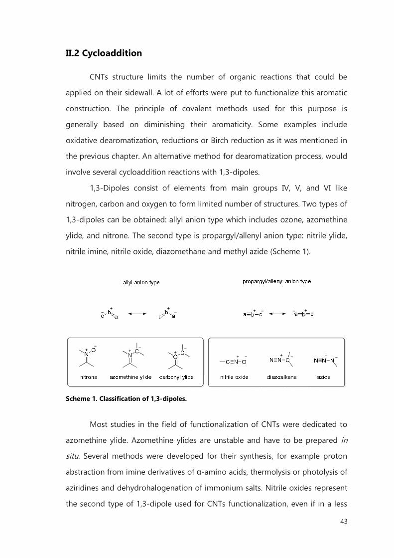
43
II.2 Cycloaddition
CNTs structure limits the number of organic reactions that could be
applied on their sidewall. A lot of efforts were put to functionalize this aromatic
construction. The principle of covalent methods used for this purpose is
generally based on diminishing their aromaticity. Some examples include
oxidative dearomatization, reductions or Birch reduction as it was mentioned in
the previous chapter. An alternative method for dearomatization process, would
involve several cycloaddition reactions with 1,3-dipoles.
1,3-Dipoles consist of elements from main groups IV, V, and VI like
nitrogen, carbon and oxygen to form limited number of structures. Two types of
1,3-dipoles can be obtained: allyl anion type which includes ozone, azomethine
ylide, and nitrone. The second type is propargyl/allenyl anion type: nitrile ylide,
nitrile imine, nitrile oxide, diazomethane and methyl azide (Scheme 1).
Scheme 1. Classification of 1,3-dipoles.
Most studies in the field of functionalization of CNTs were dedicated to
azomethine ylide. Azomethine ylides are unstable and have to be prepared in
situ. Several methods were developed for their synthesis, for example proton
abstraction from imine derivatives of α-amino acids, thermolysis or photolysis of
aziridines and dehydrohalogenation of immonium salts. Nitrile oxides represent
the second type of 1,3-dipole used for CNTs functionalization, even if in a less
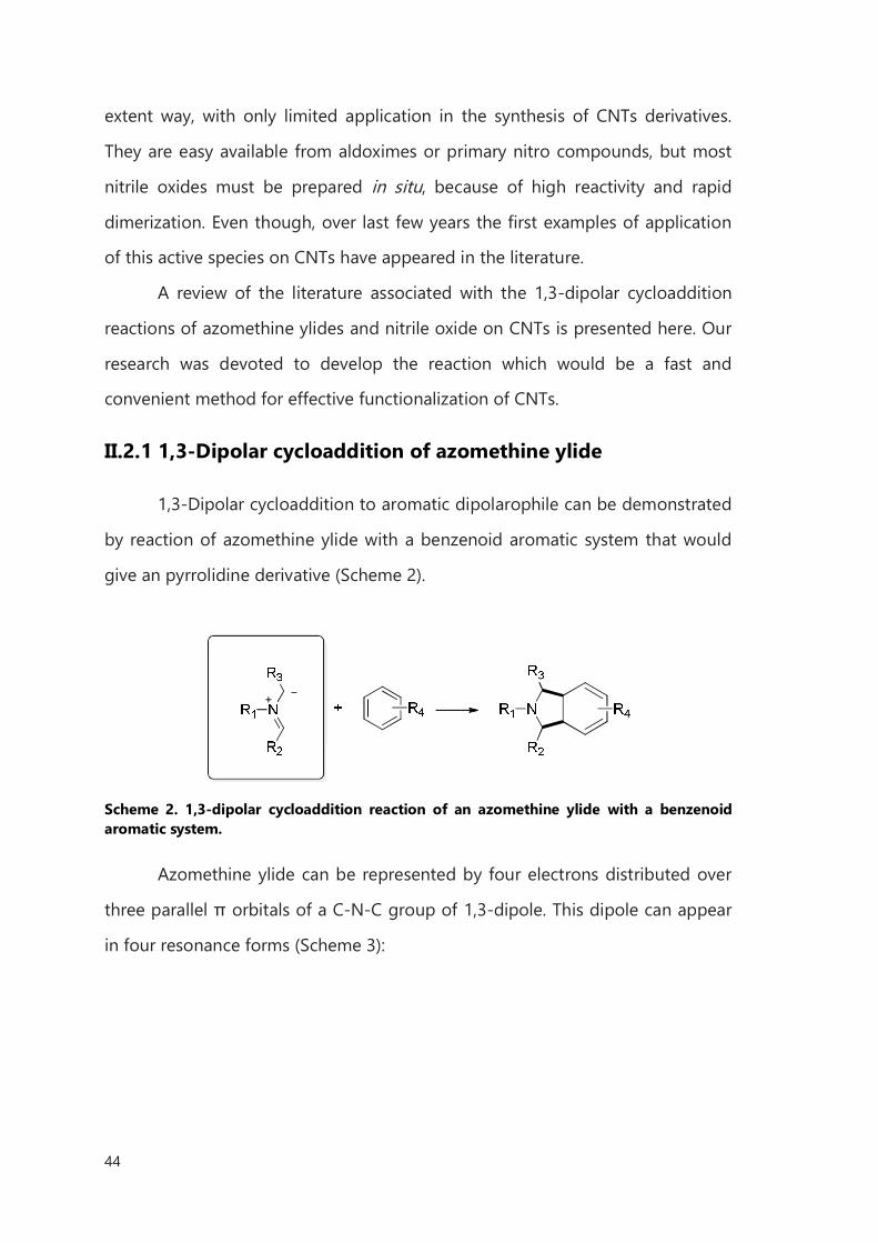
44
extent way, with only limited application in the synthesis of CNTs derivatives.
They are easy available from aldoximes or primary nitro compounds, but most
nitrile oxides must be prepared in situ, because of high reactivity and rapid
dimerization. Even though, over last few years the first examples of application
of this active species on CNTs have appeared in the literature.
A review of the literature associated with the 1,3-dipolar cycloaddition
reactions of azomethine ylides and nitrile oxide on CNTs is presented here. Our
research was devoted to develop the reaction which would be a fast and
convenient method for effective functionalization of CNTs.
II.2.1 1,3-Dipolar cycloaddition of azomethine ylide
1,3-Dipolar cycloaddition to aromatic dipolarophile can be demonstrated
by reaction of azomethine ylide with a benzenoid aromatic system that would
give an pyrrolidine derivative (Scheme 2).
Scheme 2. 1,3-dipolar cycloaddition reaction of an azomethine ylide with a benzenoid aromatic system.
Azomethine ylide can be represented by four electrons distributed over
three parallel π orbitals of a C-N-C group of 1,3-dipole. This dipole can appear
in four resonance forms (Scheme 3):
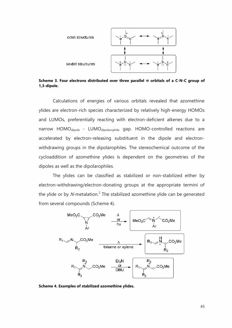
45
Scheme 3. Four electrons distributed over three parallel π orbitals of a C-N-C group of 1,3-dipole.
Calculations of energies of various orbitals revealed that azomethine
ylides are electron-rich species characterized by relatively high-energy HOMOs
and LUMOs, preferentially reacting with electron-deficient alkenes due to a
narrow HOMOdipole - LUMOdipolarophile gap. HOMO-controlled reactions are
accelerated by electron-releasing substituent in the dipole and electron-
withdrawing groups in the dipolarophiles. The stereochemical outcome of the
cycloaddition of azomethine ylides is dependent on the geometries of the
dipoles as well as the dipolarophiles.
The ylides can be classified as stabilized or non-stabilized either by
electron-withdrawing/electron-donating groups at the appropriate termini of
the ylide or by N-metalation.2 The stabilized azomethine ylide can be generated
from several compounds (Scheme 4).
Scheme 4. Examples of stabilized azomethine ylides.
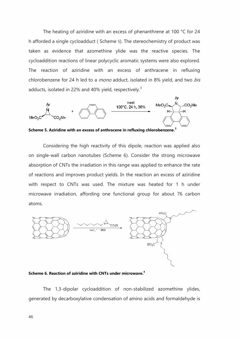
46
The heating of aziridine with an excess of phenanthrene at 100 °C for 24
h afforded a single cycloadduct ( Scheme 5). The stereochemistry of product was
taken as evidence that azomethine ylide was the reactive species. The
cycloaddition reactions of linear polycyclic aromatic systems were also explored.
The reaction of aziridine with an excess of anthracene in refluxing
chlorobenzene for 24 h led to a mono adduct, isolated in 8% yield, and two bis
adducts, isolated in 22% and 40% yield, respectively.3
Scheme 5. Aziridine with an excess of anthracene in refluxing chlorobenzene.3
Considering the high reactivity of this dipole, reaction was applied also
on single-wall carbon nanotubes (Scheme 6). Consider the strong microwave
absorption of CNTs the irradiation in this range was applied to enhance the rate
of reactions and improves product yields. In the reaction an excess of aziridine
with respect to CNTs was used. The mixture was heated for 1 h under
microwave irradiation, affording one functional group for about 76 carbon
atoms.
Scheme 6. Reaction of aziridine with CNTs under microwave.4
The 1,3-dipolar cycloaddition of non-stabilized azomethine ylides,
generated by decarboxylative condensation of amino acids and formaldehyde is
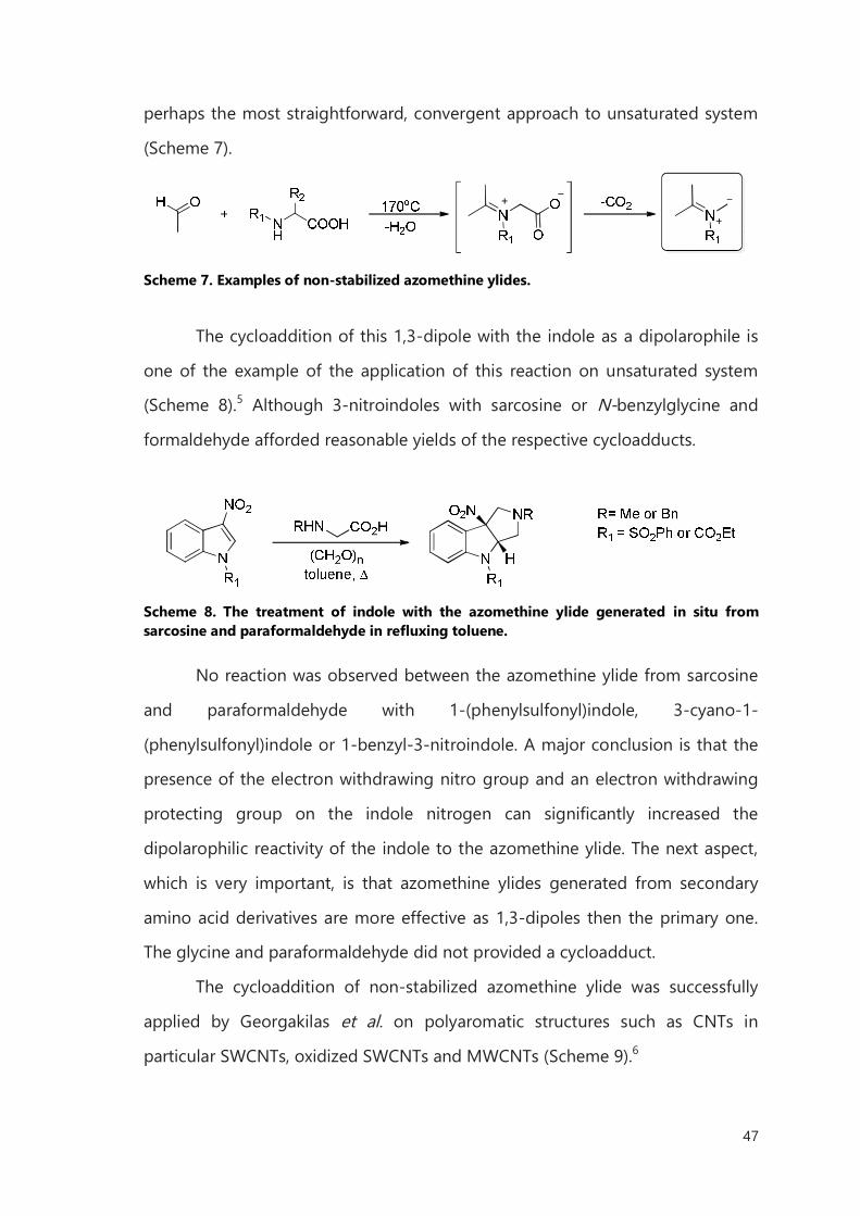
47
perhaps the most straightforward, convergent approach to unsaturated system
(Scheme 7).
Scheme 7. Examples of non-stabilized azomethine ylides.
The cycloaddition of this 1,3-dipole with the indole as a dipolarophile is
one of the example of the application of this reaction on unsaturated system
(Scheme 8).5 Although 3-nitroindoles with sarcosine or N-benzylglycine and
formaldehyde afforded reasonable yields of the respective cycloadducts.
Scheme 8. The treatment of indole with the azomethine ylide generated in situ from sarcosine and paraformaldehyde in refluxing toluene.
No reaction was observed between the azomethine ylide from sarcosine
and paraformaldehyde with 1-(phenylsulfonyl)indole, 3-cyano-1-
(phenylsulfonyl)indole or 1-benzyl-3-nitroindole. A major conclusion is that the
presence of the electron withdrawing nitro group and an electron withdrawing
protecting group on the indole nitrogen can significantly increased the
dipolarophilic reactivity of the indole to the azomethine ylide. The next aspect,
which is very important, is that azomethine ylides generated from secondary
amino acid derivatives are more effective as 1,3-dipoles then the primary one.
The glycine and paraformaldehyde did not provided a cycloadduct.
The cycloaddition of non-stabilized azomethine ylide was successfully
applied by Georgakilas et al. on polyaromatic structures such as CNTs in
particular SWCNTs, oxidized SWCNTs and MWCNTs (Scheme 9).6
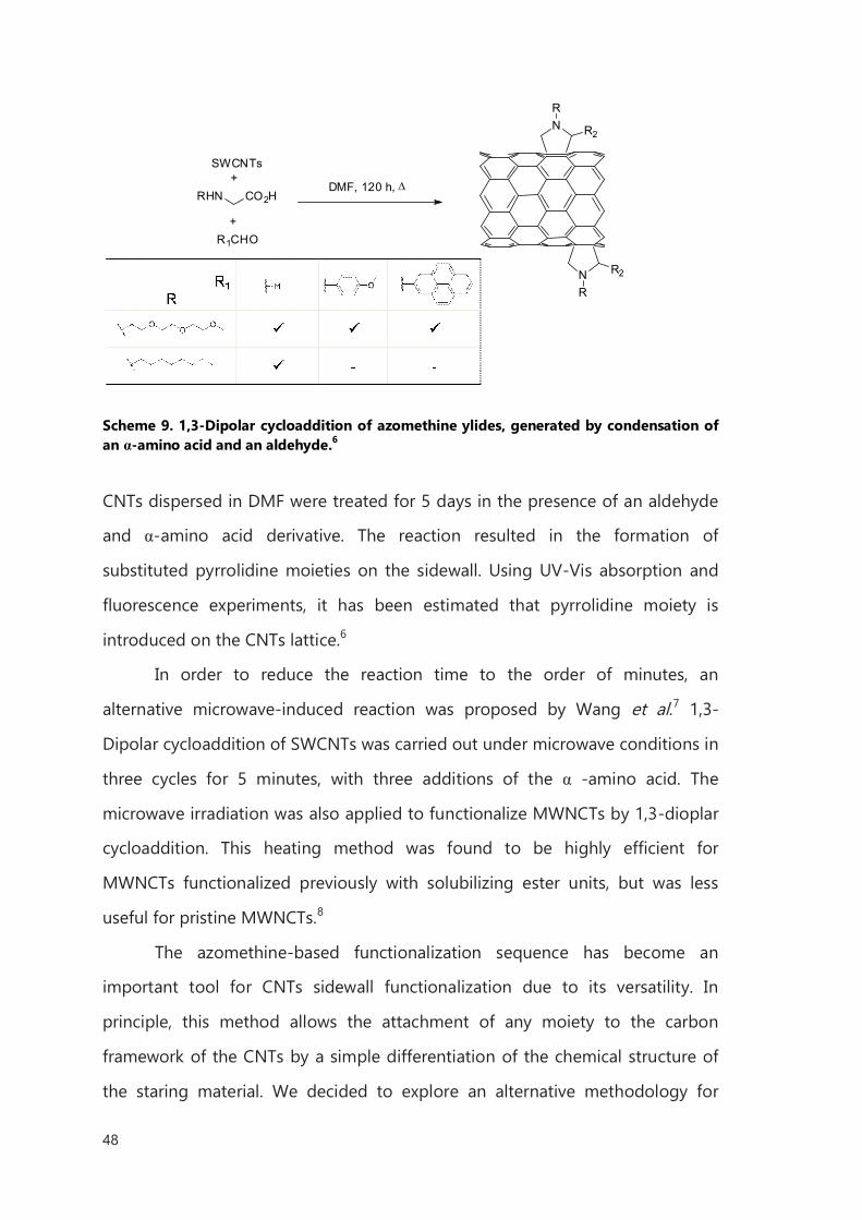
48
RHN CO2HDMF, 120 h,
N
N
R2
R2
R
R
R1CHO
SWCNTs+
+
Scheme 9. 1,3-Dipolar cycloaddition of azomethine ylides, generated by condensation of an α-amino acid and an aldehyde.6
CNTs dispersed in DMF were treated for 5 days in the presence of an aldehyde
and α-amino acid derivative. The reaction resulted in the formation of
substituted pyrrolidine moieties on the sidewall. Using UV-Vis absorption and
fluorescence experiments, it has been estimated that pyrrolidine moiety is
introduced on the CNTs lattice.6
In order to reduce the reaction time to the order of minutes, an
alternative microwave-induced reaction was proposed by Wang et al.7 1,3-
Dipolar cycloaddition of SWCNTs was carried out under microwave conditions in
three cycles for 5 minutes, with three additions of the α -amino acid. The
microwave irradiation was also applied to functionalize MWNCTs by 1,3-dioplar
cycloaddition. This heating method was found to be highly efficient for
MWNCTs functionalized previously with solubilizing ester units, but was less
useful for pristine MWNCTs.8
The azomethine-based functionalization sequence has become an
important tool for CNTs sidewall functionalization due to its versatility. In
principle, this method allows the attachment of any moiety to the carbon
framework of the CNTs by a simple differentiation of the chemical structure of
the staring material. We decided to explore an alternative methodology for
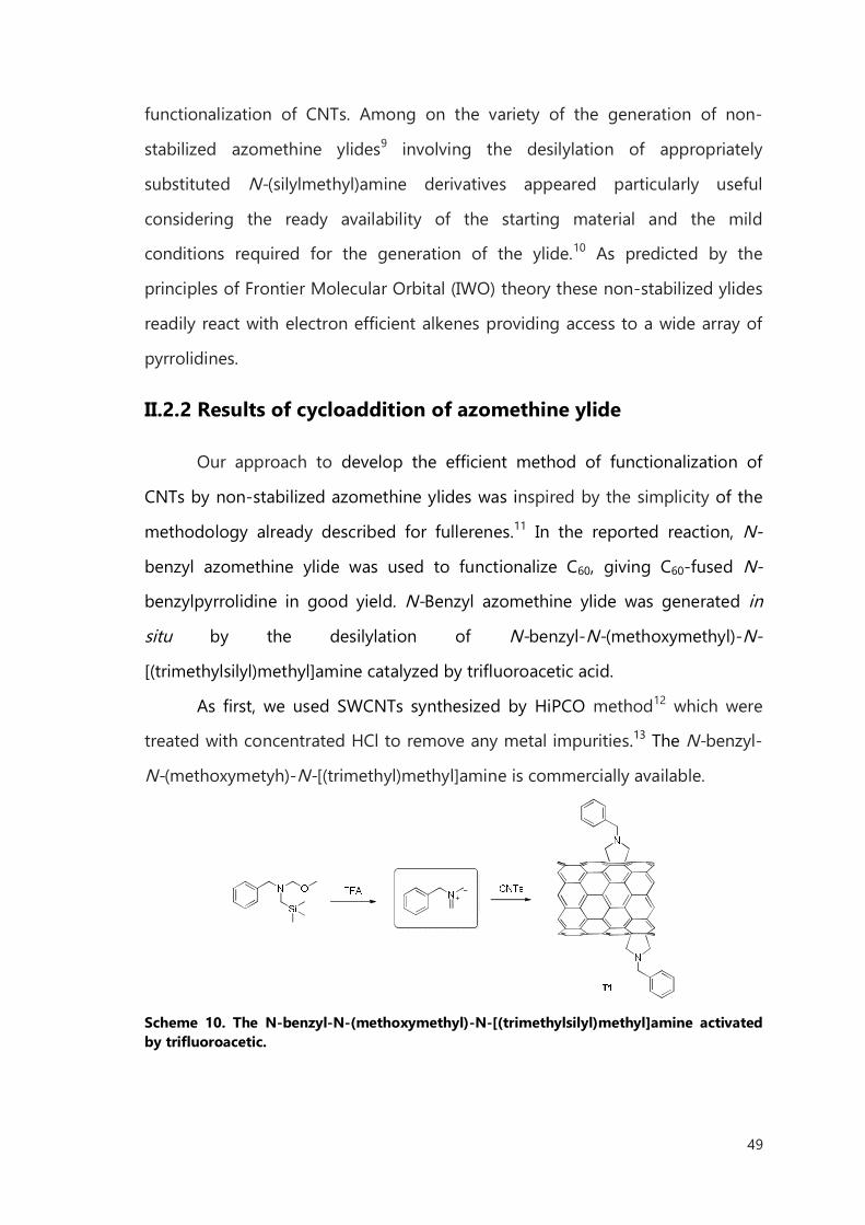
49
functionalization of CNTs. Among on the variety of the generation of non-
stabilized azomethine ylides9 involving the desilylation of appropriately
substituted N-(silylmethyl)amine derivatives appeared particularly useful
considering the ready availability of the starting material and the mild
conditions required for the generation of the ylide.10 As predicted by the
principles of Frontier Molecular Orbital (IWO) theory these non-stabilized ylides
readily react with electron efficient alkenes providing access to a wide array of
pyrrolidines.
II.2.2 Results of cycloaddition of azomethine ylide
Our approach to develop the efficient method of functionalization of
CNTs by non-stabilized azomethine ylides was inspired by the simplicity of the
methodology already described for fullerenes.11 In the reported reaction, N-
benzyl azomethine ylide was used to functionalize C60, giving C60-fused N-
benzylpyrrolidine in good yield. N-Benzyl azomethine ylide was generated in
situ by the desilylation of N-benzyl-N-(methoxymethyl)-N-
[(trimethylsilyl)methyl]amine catalyzed by trifluoroacetic acid.
As first, we used SWCNTs synthesized by HiPCO method12 which were
treated with concentrated HCl to remove any metal impurities.13 The N-benzyl-
N-(methoxymetyh)-N-[(trimethyl)methyl]amine is commercially available.
Scheme 10. The N-benzyl-N-(methoxymethyl)-N-[(trimethylsilyl)methyl]amine activated by trifluoroacetic.
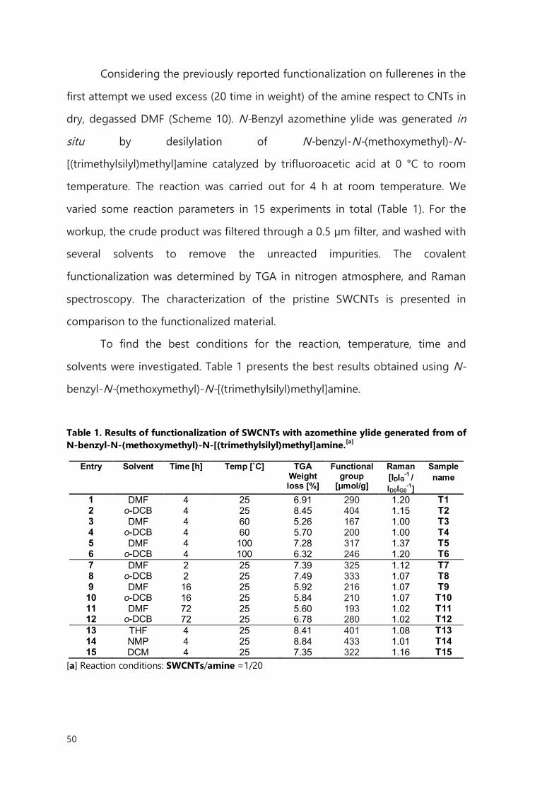
50
Considering the previously reported functionalization on fullerenes in the
first attempt we used excess (20 time in weight) of the amine respect to CNTs in
dry, degassed DMF (Scheme 10). N-Benzyl azomethine ylide was generated in
situ by desilylation of N-benzyl-N-(methoxymethyl)-N-
[(trimethylsilyl)methyl]amine catalyzed by trifluoroacetic acid at 0 °C to room
temperature. The reaction was carried out for 4 h at room temperature. We
varied some reaction parameters in 15 experiments in total (Table 1). For the
workup, the crude product was filtered through a 0.5 µm filter, and washed with
several solvents to remove the unreacted impurities. The covalent
functionalization was determined by TGA in nitrogen atmosphere, and Raman
spectroscopy. The characterization of the pristine SWCNTs is presented in
comparison to the functionalized material.
To find the best conditions for the reaction, temperature, time and
solvents were investigated. Table 1 presents the best results obtained using N-
benzyl-N-(methoxymethyl)-N-[(trimethylsilyl)methyl]amine.
Table 1. Results of functionalization of SWCNTs with azomethine ylide generated from of N-benzyl-N-(methoxymethyl)-N-[(trimethylsilyl)methyl]amine.[a]
Entry Solvent Time [h] Temp [˚C] TGA
Weight loss [%]
Functional group
[μmol/g]
Raman [IDIG-1 / ID0IG0
-1]
Sample name
1 DMF 4 25 6.91 290 1.20 T1 2 o-DCB 4 25 8.45 404 1.15 T2 3 DMF 4 60 5.26 167 1.00 T3 4 o-DCB 4 60 5.70 200 1.00 T4 5 DMF 4 100 7.28 317 1.37 T5 6 o-DCB 4 100 6.32 246 1.20 T6 7 DMF 2 25 7.39 325 1.12 T7 8 o-DCB 2 25 7.49 333 1.07 T8 9 DMF 16 25 5.92 216 1.07 T9 10 o-DCB 16 25 5.84 210 1.07 T10 11 DMF 72 25 5.60 193 1.02 T11 12 o-DCB 72 25 6.78 280 1.02 T12 13 THF 4 25 8.41 401 1.08 T13 14 NMP 4 25 8.84 433 1.01 T14 15 DCM 4 25 7.35 322 1.16 T15
[a] Reaction conditions: SWCNTs/amine =1/20
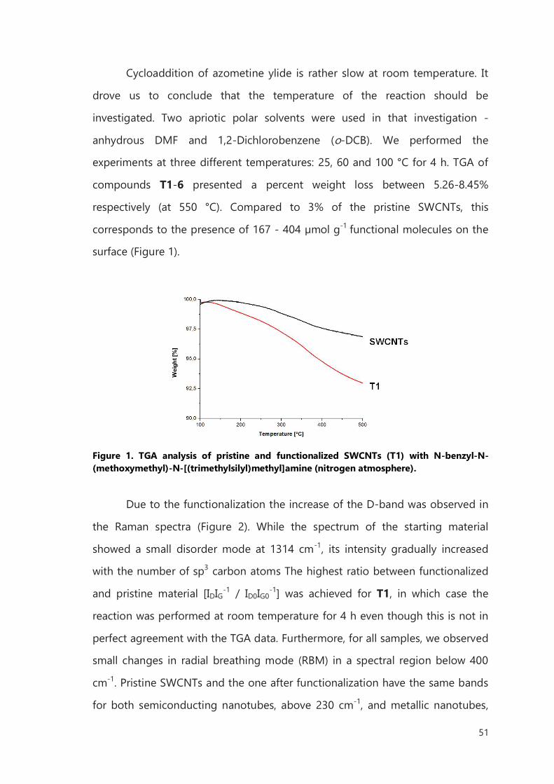
51
Cycloaddition of azometine ylide is rather slow at room temperature. It
drove us to conclude that the temperature of the reaction should be
investigated. Two apriotic polar solvents were used in that investigation -
anhydrous DMF and 1,2-Dichlorobenzene (o-DCB). We performed the
experiments at three different temperatures: 25, 60 and 100 °C for 4 h. TGA of
compounds T1-6 presented a percent weight loss between 5.26-8.45%
respectively (at 550 °C). Compared to 3% of the pristine SWCNTs, this
corresponds to the presence of 167 - 404 µmol g-1 functional molecules on the
surface (Figure 1).
Figure 1. TGA analysis of pristine and functionalized SWCNTs (T1) with N-benzyl-N-(methoxymethyl)-N-[(trimethylsilyl)methyl]amine (nitrogen atmosphere).
Due to the functionalization the increase of the D-band was observed in
the Raman spectra (Figure 2). While the spectrum of the starting material
showed a small disorder mode at 1314 cm-1, its intensity gradually increased
with the number of sp3 carbon atoms The highest ratio between functionalized
and pristine material [IDIG-1 / ID0IG0-1] was achieved for T1, in which case the
reaction was performed at room temperature for 4 h even though this is not in
perfect agreement with the TGA data. Furthermore, for all samples, we observed
small changes in radial breathing mode (RBM) in a spectral region below 400
cm-1. Pristine SWCNTs and the one after functionalization have the same bands
for both semiconducting nanotubes, above 230 cm-1, and metallic nanotubes,
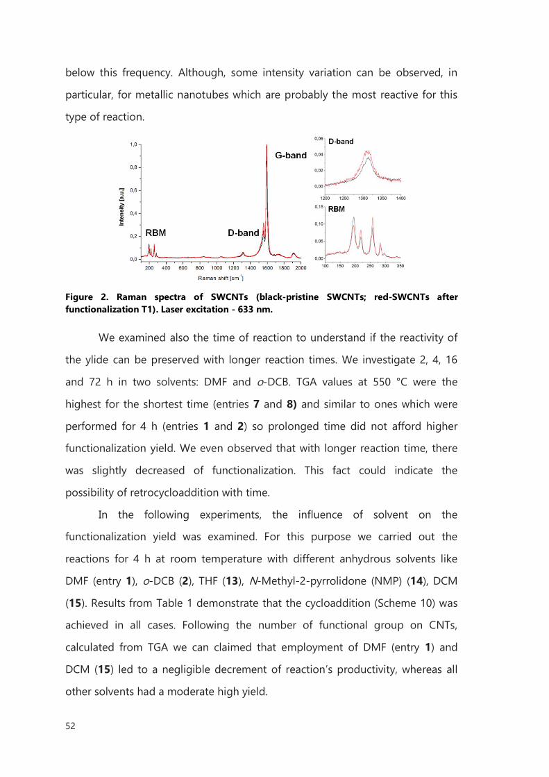
52
below this frequency. Although, some intensity variation can be observed, in
particular, for metallic nanotubes which are probably the most reactive for this
type of reaction.
Figure 2. Raman spectra of SWCNTs (black-pristine SWCNTs; red-SWCNTs after functionalization T1). Laser excitation - 633 nm.
We examined also the time of reaction to understand if the reactivity of
the ylide can be preserved with longer reaction times. We investigate 2, 4, 16
and 72 h in two solvents: DMF and o-DCB. TGA values at 550 °C were the
highest for the shortest time (entries 7 and 8) and similar to ones which were
performed for 4 h (entries 1 and 2) so prolonged time did not afford higher
functionalization yield. We even observed that with longer reaction time, there
was slightly decreased of functionalization. This fact could indicate the
possibility of retrocycloaddition with time.
In the following experiments, the influence of solvent on the
functionalization yield was examined. For this purpose we carried out the
reactions for 4 h at room temperature with different anhydrous solvents like
DMF (entry 1), o-DCB (2), THF (13), N-Methyl-2-pyrrolidone (NMP) (14), DCM
(15). Results from Table 1 demonstrate that the cycloaddition (Scheme 10) was
achieved in all cases. Following the number of functional group on CNTs,
calculated from TGA we can claimed that employment of DMF (entry 1) and
DCM (15) led to a negligible decrement of reaction’s productivity, whereas all
other solvents had a moderate high yield.
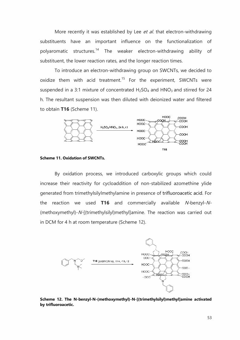
53
More recently it was established by Lee et al. that electron-withdrawing
substituents have an important influence on the functionalization of
polyaromatic structures.14 The weaker electron-withdrawing ability of
substituent, the lower reaction rates, and the longer reaction times.
To introduce an electron-withdrawing group on SWCNTs, we decided to
oxidize them with acid treatment.15 For the experiment, SWCNTs were
suspended in a 3:1 mixture of concentrated H2SO4 and HNO3 and stirred for 24
h. The resultant suspension was then diluted with deionized water and filtered
to obtain T16 (Scheme 11).
Scheme 11. Oxidation of SWCNTs.
By oxidation process, we introduced carboxylic groups which could
increase their reactivity for cycloaddition of non-stabilized azomethine ylide
generated from trimethylsilylmethylamine in presence of trifluoroacetic acid. For
the reaction we used T16 and commercially available N-benzyl-N-
(methoxymethyl)-N-[(trimethylsilyl)methyl]amine. The reaction was carried out
in DCM for 4 h at room temperature (Scheme 12).
Scheme 12. The N-benzyl-N-(methoxymethyl)-N-[(trimethylsilyl)methyl]amine activated by trifluoroacetic.
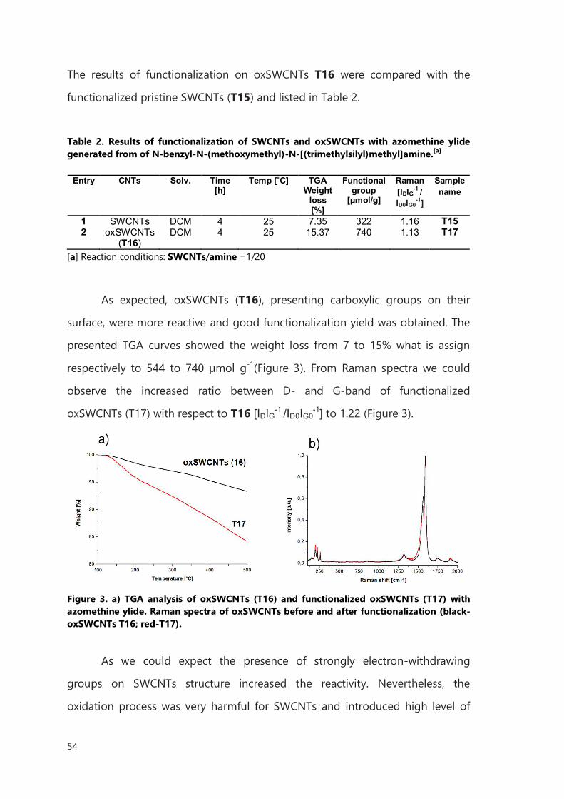
54
The results of functionalization on oxSWCNTs T16 were compared with the
functionalized pristine SWCNTs (T15) and listed in Table 2.
Table 2. Results of functionalization of SWCNTs and oxSWCNTs with azomethine ylide generated from of N-benzyl-N-(methoxymethyl)-N-[(trimethylsilyl)methyl]amine.[a]
Entry CNTs Solv. Time
[h] Temp [˚C] TGA
Weight loss [%]
Functional group
[μmol/g]
Raman [IDIG-1 / ID0IG0
-1]
Sample name
1 SWCNTs DCM 4 25 7.35 322 1.16 T15 2 oxSWCNTs
(T16) DCM 4 25 15.37 740 1.13 T17
[a] Reaction conditions: SWCNTs/amine =1/20
As expected, oxSWCNTs (T16), presenting carboxylic groups on their
surface, were more reactive and good functionalization yield was obtained. The
presented TGA curves showed the weight loss from 7 to 15% what is assign
respectively to 544 to 740 µmol g-1(Figure 3). From Raman spectra we could
observe the increased ratio between D- and G-band of functionalized
oxSWCNTs (T17) with respect to T16 [IDIG-1 /ID0IG0-1] to 1.22 (Figure 3).
Figure 3. a) TGA analysis of oxSWCNTs (T16) and functionalized oxSWCNTs (T17) with azomethine ylide. Raman spectra of oxSWCNTs before and after functionalization (black-oxSWCNTs T16; red-T17).
As we could expect the presence of strongly electron-withdrawing
groups on SWCNTs structure increased the reactivity. Nevertheless, the
oxidation process was very harmful for SWCNTs and introduced high level of
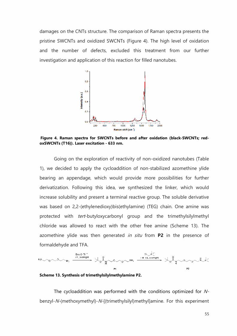
55
damages on the CNTs structure. The comparison of Raman spectra presents the
pristine SWCNTs and oxidized SWCNTs (Figure 4). The high level of oxidation
and the number of defects, excluded this treatment from our further
investigation and application of this reaction for filled nanotubes.
Figure 4. Raman spectra for SWCNTs before and after oxidation (black-SWCNTs; red-oxSWCNTs (T16)). Laser excitation - 633 nm.
Going on the exploration of reactivity of non-oxidized nanotubes (Table
1), we decided to apply the cycloaddition of non-stabilized azomethine ylide
bearing an appendage, which would provide more possibilities for further
derivatization. Following this idea, we synthesized the linker, which would
increase solubility and present a terminal reactive group. The soluble derivative
was based on 2,2-(ethylenedioxy)bis(ethylamine) (TEG) chain. One amine was
protected with tert-butyloxycarbonyl group and the trimethylsilylmethyl
chloride was allowed to react with the other free amine (Scheme 13). The
azomethine ylide was then generated in situ from P2 in the presence of
formaldehyde and TFA.
Scheme 13. Synthesis of trimethylsilylmethylamine P2.
The cycloaddition was performed with the conditions optimized for N-
benzyl-N-(methoxymethyl)-N-[(trimethylsilyl)methyl]amine. For this experiment

56
we have used 10 mg of CNTs (HiPCO SWCNTs purified with HCl treatment, or
MWCNTs), an excess of trimethylsilylmethylamine P2 (26 times in weight) with
respect to carbon nanotubes and catalytic amount of TFA (Scheme 14).
Scheme 14. 1,3-dipolar cycloaddition of azomethine ylide on CNTs, generated from trimethylsilylmethylamine.
Characterization of functionalized CNTs was performed by Raman
spectroscopy, TGA and Kaiser test. The results are listed in Table 3.
Table 3. 1,3-dipolar cycloaddition of azomethine ylide generated from trimethylsilylmethylamine on SWCNTs and MWCNTs.[a]
Entry CNTs Solv. TGA
Weight loss [%]
Functional
group [μmol/g]
Kaiser test
[µmol/g]
Raman [IDIG-1 / ID0IG0
-1]
Sample name
1 SWCNTs DMF 11.00 289 15 1.15 T19 2 MWCNT
s DMF 6.69
190 10 1.04 T20
[a] Reaction conditions: SWCNTs/amine =1/26
To estimate the density of the covalently functionalized molecules on the
sidewall we used TGA. A higher degree of functionalization was obtained for
SWCNTs T19 (Table 5a) when compared to functionalized MWCNTs T20 (Table
5b) under the same reaction conditions. More specifically, the weight loss
measured for the thermal decomposition of the chain on SWCNTs was 11%
compared to 6.69% for functionalized MWCNTs. As expected, the reactivity of
carbon nanostructures was found to depend on their structure and with higher
curvature, SWCNTs proved to be more favorable for covalent chemical reactions
than MWCNTs. However the content in carbon of MWCNTs is much higher than
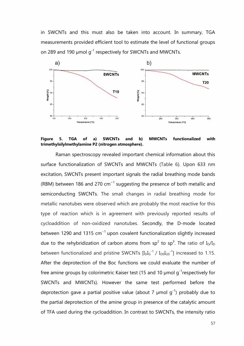
57
in SWCNTs and this must also be taken into account. In summary, TGA
measurements provided efficient tool to estimate the level of functional groups
on 289 and 190 µmol g-1 respectively for SWCNTs and MWCNTs.
Figure 5. TGA of a) SWCNTs and b) MWCNTs functionalized with trimethylsilylmethylamine P2 (nitrogen atmosphere).
Raman spectroscopy revealed important chemical information about this
surface functionalization of SWCNTs and MWCNTs (Table 6). Upon 633 nm
excitation, SWCNTs present important signals the radial breathing mode bands
(RBM) between 186 and 270 cm−1 suggesting the presence of both metallic and
semiconducting SWCNTs. The small changes in radial breathing mode for
metallic nanotubes were observed which are probably the most reactive for this
type of reaction which is in agreement with previously reported results of
cycloaddition of non-oxidized nanotubes. Secondly, the D-mode located
between 1290 and 1315 cm−1 upon covalent functionalization slightly increased
due to the rehybridization of carbon atoms from sp2 to sp3. The ratio of ID/ID
between functionalized and pristine SWCNTs [IDIG-1 / ID0IG0-1] increased to 1.15.
After the deprotection of the Boc functions we could evaluate the number of
free amine groups by colorimetric Kaiser test (15 and 10 µmol g-1respectively for
SWCNTs and MWCNTs). However the same test performed before the
deprotection gave a partial positive value (about 7 µmol g-1) probably due to
the partial deprotection of the amine group in presence of the catalytic amount
of TFA used during the cycloaddition. In contrast to SWCNTs, the intensity ratio
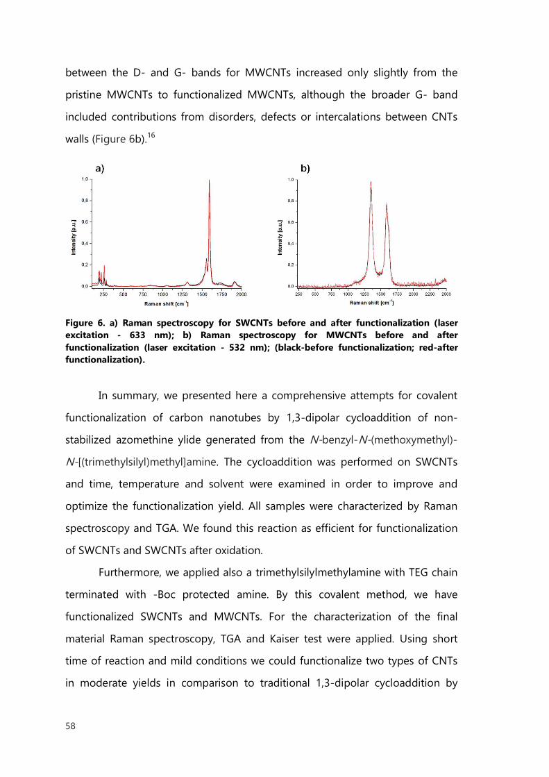
58
between the D- and G- bands for MWCNTs increased only slightly from the
pristine MWCNTs to functionalized MWCNTs, although the broader G- band
included contributions from disorders, defects or intercalations between CNTs
walls (Figure 6b).16
Figure 6. a) Raman spectroscopy for SWCNTs before and after functionalization (laser excitation - 633 nm); b) Raman spectroscopy for MWCNTs before and after functionalization (laser excitation - 532 nm); (black-before functionalization; red-after functionalization).
In summary, we presented here a comprehensive attempts for covalent
functionalization of carbon nanotubes by 1,3-dipolar cycloaddition of non-
stabilized azomethine ylide generated from the N-benzyl-N-(methoxymethyl)-
N-[(trimethylsilyl)methyl]amine. The cycloaddition was performed on SWCNTs
and time, temperature and solvent were examined in order to improve and
optimize the functionalization yield. All samples were characterized by Raman
spectroscopy and TGA. We found this reaction as efficient for functionalization
of SWCNTs and SWCNTs after oxidation.
Furthermore, we applied also a trimethylsilylmethylamine with TEG chain
terminated with -Boc protected amine. By this covalent method, we have
functionalized SWCNTs and MWCNTs. For the characterization of the final
material Raman spectroscopy, TGA and Kaiser test were applied. Using short
time of reaction and mild conditions we could functionalize two types of CNTs
in moderate yields in comparison to traditional 1,3-dipolar cycloaddition by

59
azomethine ylide generated by decarboxylative condensation of amino acids
and formaldehyde.
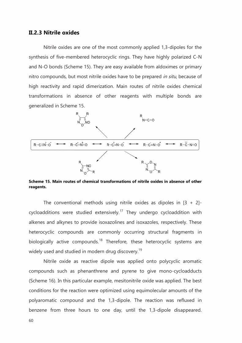
60
II.2.3 Nitrile oxides
Nitrile oxides are one of the most commonly applied 1,3-dipoles for the
synthesis of five-membered heterocyclic rings. They have highly polarized C-N
and N-O bonds (Scheme 15). They are easy available from aldoximes or primary
nitro compounds, but most nitrile oxides have to be prepared in situ, because of
high reactivity and rapid dimerization. Main routes of nitrile oxides chemical
transformations in absence of other reagents with multiple bonds are
generalized in Scheme 15.
Scheme 15. Main routes of chemical transformations of nitrile oxides in absence of other reagents.
The conventional methods using nitrile oxides as dipoles in [3 + 2]-
cycloadditions were studied extensively.17 They undergo cycloaddition with
alkenes and alkynes to provide isoxazolines and isoxazoles, respectively. These
heterocyclic compounds are commonly occurring structural fragments in
biologically active compounds.18 Therefore, these heterocyclic systems are
widely used and studied in modern drug discovery.19
Nitrile oxide as reactive dipole was applied onto polycyclic aromatic
compounds such as phenanthrene and pyrene to give mono-cycloadducts
(Scheme 16). In this particular example, mesitonitrile oxide was applied. The best
conditions for the reaction were optimized using equimolecular amounts of the
polyaromatic compound and the 1,3-dipole. The reaction was refluxed in
benzene from three hours to one day, until the 1,3-dipole disappeared.
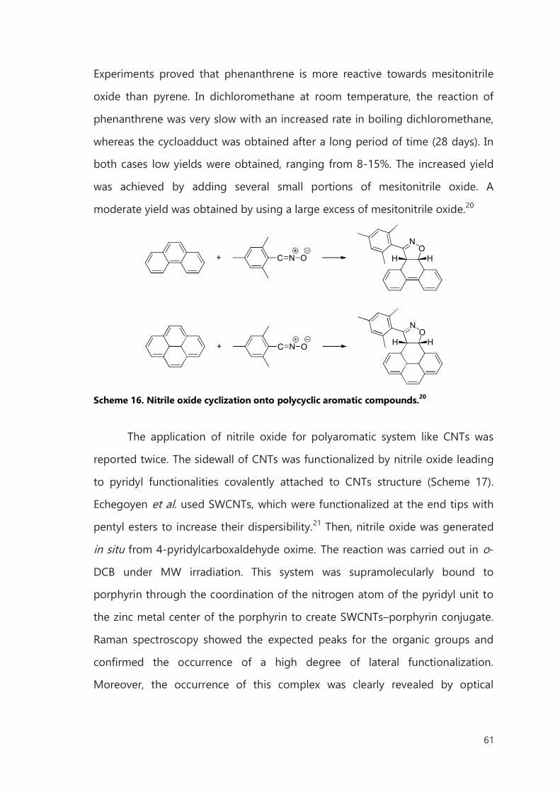
61
Experiments proved that phenanthrene is more reactive towards mesitonitrile
oxide than pyrene. In dichloromethane at room temperature, the reaction of
phenanthrene was very slow with an increased rate in boiling dichloromethane,
whereas the cycloadduct was obtained after a long period of time (28 days). In
both cases low yields were obtained, ranging from 8-15%. The increased yield
was achieved by adding several small portions of mesitonitrile oxide. A
moderate yield was obtained by using a large excess of mesitonitrile oxide.20
C N O+O
N
HH
C N O+O
N
HH
Scheme 16. Nitrile oxide cyclization onto polycyclic aromatic compounds.20
The application of nitrile oxide for polyaromatic system like CNTs was
reported twice. The sidewall of CNTs was functionalized by nitrile oxide leading
to pyridyl functionalities covalently attached to CNTs structure (Scheme 17).
Echegoyen et al. used SWCNTs, which were functionalized at the end tips with
pentyl esters to increase their dispersibility.21 Then, nitrile oxide was generated
in situ from 4-pyridylcarboxaldehyde oxime. The reaction was carried out in o-
DCB under MW irradiation. This system was supramolecularly bound to
porphyrin through the coordination of the nitrogen atom of the pyridyl unit to
the zinc metal center of the porphyrin to create SWCNTs–porphyrin conjugate.
Raman spectroscopy showed the expected peaks for the organic groups and
confirmed the occurrence of a high degree of lateral functionalization.
Moreover, the occurrence of this complex was clearly revealed by optical
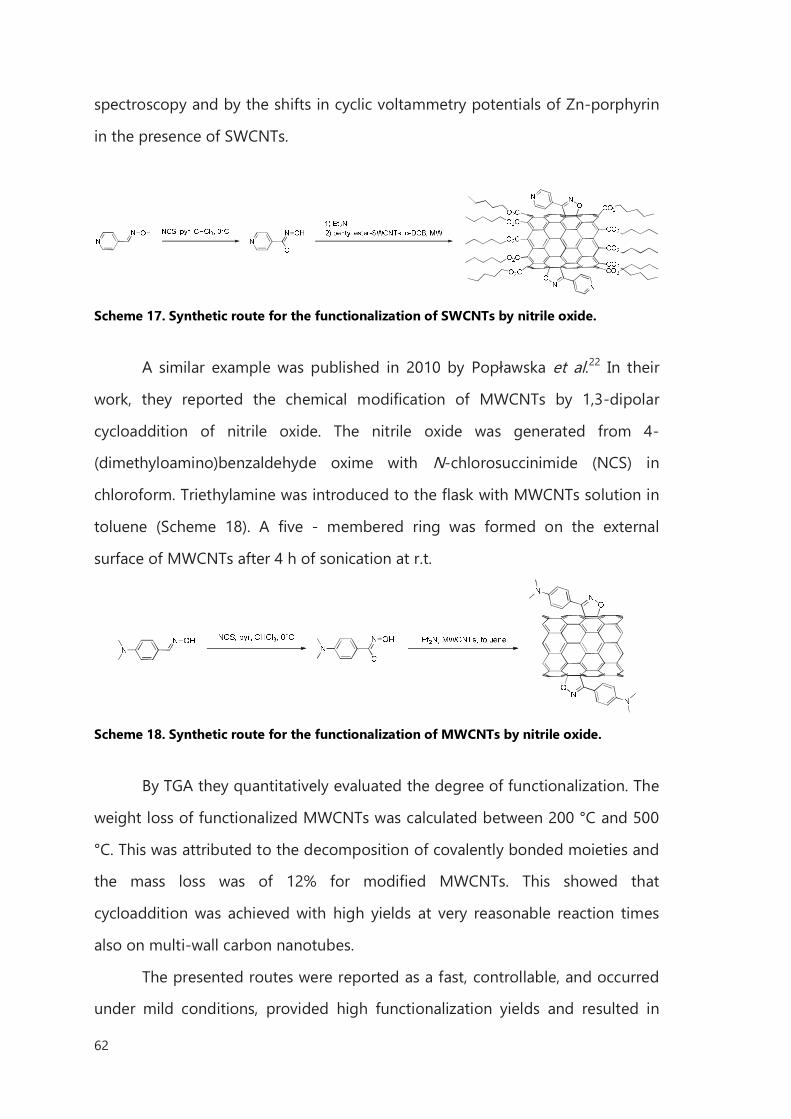
62
spectroscopy and by the shifts in cyclic voltammetry potentials of Zn-porphyrin
in the presence of SWCNTs.
Scheme 17. Synthetic route for the functionalization of SWCNTs by nitrile oxide.
A similar example was published in 2010 by Popławska et al.22 In their
work, they reported the chemical modification of MWCNTs by 1,3-dipolar
cycloaddition of nitrile oxide. The nitrile oxide was generated from 4-
(dimethyloamino)benzaldehyde oxime with N-chlorosuccinimide (NCS) in
chloroform. Triethylamine was introduced to the flask with MWCNTs solution in
toluene (Scheme 18). A five - membered ring was formed on the external
surface of MWCNTs after 4 h of sonication at r.t.
Scheme 18. Synthetic route for the functionalization of MWCNTs by nitrile oxide.
By TGA they quantitatively evaluated the degree of functionalization. The
weight loss of functionalized MWCNTs was calculated between 200 °C and 500
°C. This was attributed to the decomposition of covalently bonded moieties and
the mass loss was of 12% for modified MWCNTs. This showed that
cycloaddition was achieved with high yields at very reasonable reaction times
also on multi-wall carbon nanotubes.
The presented routes were reported as a fast, controllable, and occurred
under mild conditions, provided high functionalization yields and resulted in
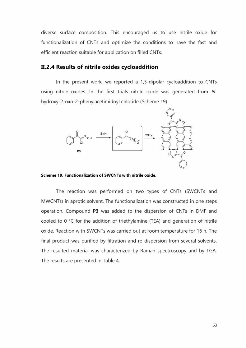
63
diverse surface composition. This encouraged us to use nitrile oxide for
functionalization of CNTs and optimize the conditions to have the fast and
efficient reaction suitable for application on filled CNTs.
II.2.4 Results of nitrile oxides cycloaddition
In the present work, we reported a 1,3-dipolar cycloaddition to CNTs
using nitrile oxides. In the first trials nitrile oxide was generated from N-
hydroxy-2-oxo-2-phenylacetimidoyl chloride (Scheme 19).
NO
ON
CNTsNOH
O
Cl
O
O
Et2N
NO
O
P3
Scheme 19. Functionalization of SWCNTs with nitrile oxide.
The reaction was performed on two types of CNTs (SWCNTs and
MWCNTs) in aprotic solvent. The functionalization was constructed in one steps
operation. Compound P3 was added to the dispersion of CNTs in DMF and
cooled to 0 °C for the addition of triethylamine (TEA) and generation of nitrile
oxide. Reaction with SWCNTs was carried out at room temperature for 16 h. The
final product was purified by filtration and re-dispersion from several solvents.
The resulted material was characterized by Raman spectroscopy and by TGA.
The results are presented in Table 4.
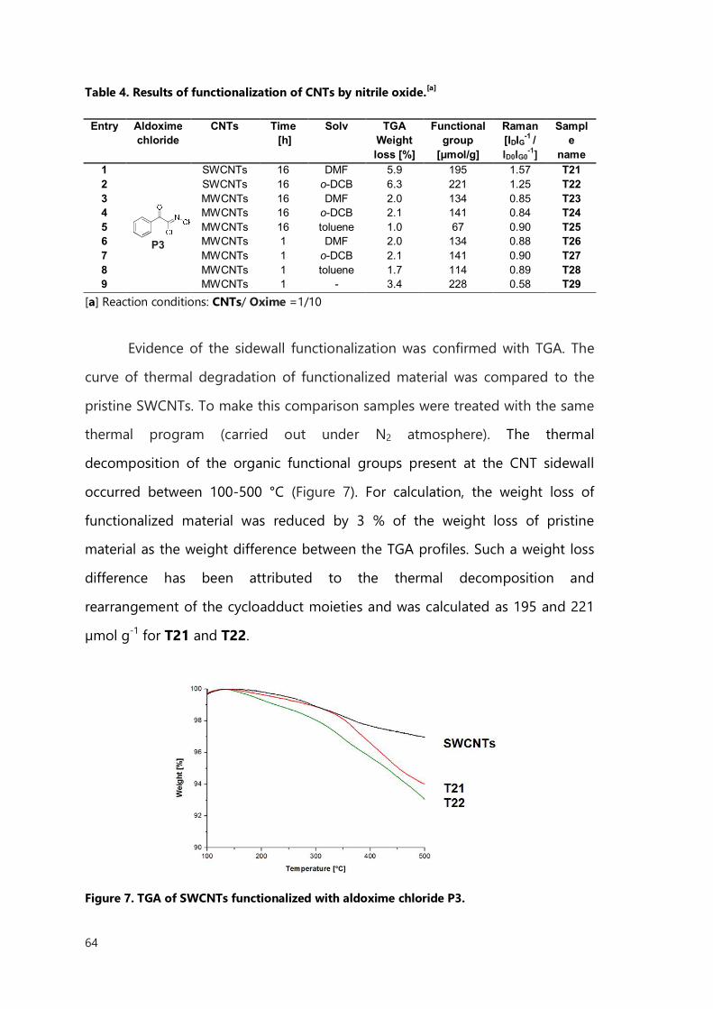
64
Table 4. Results of functionalization of CNTs by nitrile oxide.[a]
Entry Aldoxime
chloride CNTs Time
[h] Solv TGA
Weight loss [%]
Functional group
[μmol/g]
Raman [IDIG-1 / ID0IG0
-1]
Sample
name 1
P3
SWCNTs 16 DMF 5.9 195 1.57 T21 2 SWCNTs 16 o-DCB 6.3 221 1.25 T22 3 MWCNTs 16 DMF 2.0 134 0.85 T23 4 MWCNTs 16 o-DCB 2.1 141 0.84 T24 5 MWCNTs 16 toluene 1.0 67 0.90 T25 6 MWCNTs 1 DMF 2.0 134 0.88 T26 7 MWCNTs 1 o-DCB 2.1 141 0.90 T27 8 MWCNTs 1 toluene 1.7 114 0.89 T28 9 MWCNTs 1 - 3.4 228 0.58 T29
[a] Reaction conditions: CNTs/ Oxime =1/10
Evidence of the sidewall functionalization was confirmed with TGA. The
curve of thermal degradation of functionalized material was compared to the
pristine SWCNTs. To make this comparison samples were treated with the same
thermal program (carried out under N2 atmosphere). The thermal
decomposition of the organic functional groups present at the CNT sidewall
occurred between 100-500 °C (Figure 7). For calculation, the weight loss of
functionalized material was reduced by 3 % of the weight loss of pristine
material as the weight difference between the TGA profiles. Such a weight loss
difference has been attributed to the thermal decomposition and
rearrangement of the cycloadduct moieties and was calculated as 195 and 221
µmol g-1 for T21 and T22.
Figure 7. TGA of SWCNTs functionalized with aldoxime chloride P3.
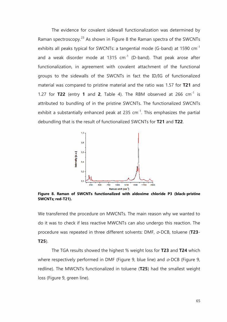
65
The evidence for covalent sidewall functionalization was determined by
Raman spectroscopy.23 As shown in Figure 8 the Raman spectra of the SWCNTs
exhibits all peaks typical for SWCNTs: a tangential mode (G-band) at 1590 cm-1
and a weak disorder mode at 1315 cm-1 (D-band). That peak arose after
functionalization, in agreement with covalent attachment of the functional
groups to the sidewalls of the SWCNTs in fact the ID/IG of functionalized
material was compared to pristine material and the ratio was 1.57 for T21 and
1.27 for T22 (entry 1 and 2, Table 4). The RBM observed at 266 cm-1 is
attributed to bundling of in the pristine SWCNTs. The functionalized SWCNTs
exhibit a substantially enhanced peak at 235 cm-1. This emphasizes the partial
debundling that is the result of functionalized SWCNTs for T21 and T22.
Figure 8. Raman of SWCNTs functionalized with aldoxime chloride P3 (black-pristine SWCNTs; red-T21).
We transferred the procedure on MWCNTs. The main reason why we wanted to
do it was to check if less reactive MWCNTs can also undergo this reaction. The
procedure was repeated in three different solvents: DMF, o-DCB, toluene (T23-
T25).
The TGA results showed the highest % weight loss for T23 and T24 which
where respectively performed in DMF (Figure 9, blue line) and o-DCB (Figure 9,
redline). The MWCNTs functionalized in toluene (T25) had the smallest weight
loss (Figure 9, green line).
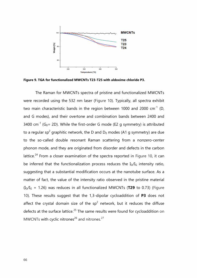
66
Figure 9. TGA for functionalized MWCNTs T23-T25 with aldoxime chloride P3.
The Raman for MWCNTs spectra of pristine and functionalized MWCNTs
were recorded using the 532 nm laser (Figure 10). Typically, all spectra exhibit
two main characteristic bands in the region between 1000 and 2000 cm-1 (D,
and G modes), and their overtone and combination bands between 2400 and
3400 cm-1 (G0= 2D). While the first-order G mode (E2 g symmetry) is attributed
to a regular sp2 graphitic network, the D and D0 modes (A1 g symmetry) are due
to the so-called double resonant Raman scattering from a nonzero-center
phonon mode, and they are originated from disorder and defects in the carbon
lattice.24 From a closer examination of the spectra reported in Figure 10, it can
be inferred that the functionalization process reduces the ID/IG intensity ratio,
suggesting that a substantial modification occurs at the nanotube surface. As a
matter of fact, the value of the intensity ratio observed in the pristine material
(ID/IG = 1.26) was reduces in all functionalized MWCNTs (T29 to 0.73) (Figure
10). These results suggest that the 1,3-dipolar cycloaddition of P3 does not
affect the crystal domain size of the sp2 network, but it reduces the diffuse
defects at the surface lattice.25 The same results were found for cycloaddition on
MWCNTs with cyclic nitrones26 and nitrones.27
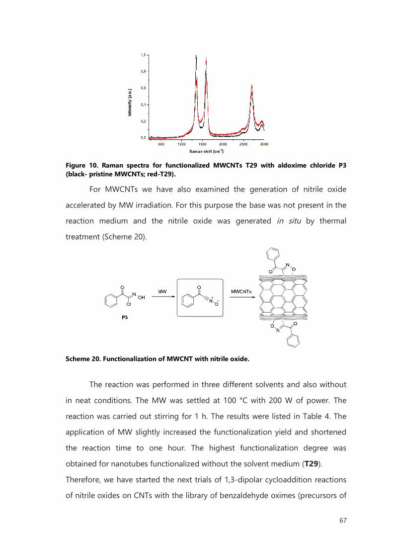
67
Figure 10. Raman spectra for functionalized MWCNTs T29 with aldoxime chloride P3 (black- pristine MWCNTs; red-T29).
For MWCNTs we have also examined the generation of nitrile oxide
accelerated by MW irradiation. For this purpose the base was not present in the
reaction medium and the nitrile oxide was generated in situ by thermal
treatment (Scheme 20).
Scheme 20. Functionalization of MWCNT with nitrile oxide.
The reaction was performed in three different solvents and also without
in neat conditions. The MW was settled at 100 °C with 200 W of power. The
reaction was carried out stirring for 1 h. The results were listed in Table 4. The
application of MW slightly increased the functionalization yield and shortened
the reaction time to one hour. The highest functionalization degree was
obtained for nanotubes functionalized without the solvent medium (T29).
Therefore, we have started the next trials of 1,3-dipolar cycloaddition reactions
of nitrile oxides on CNTs with the library of benzaldehyde oximes (precursors of
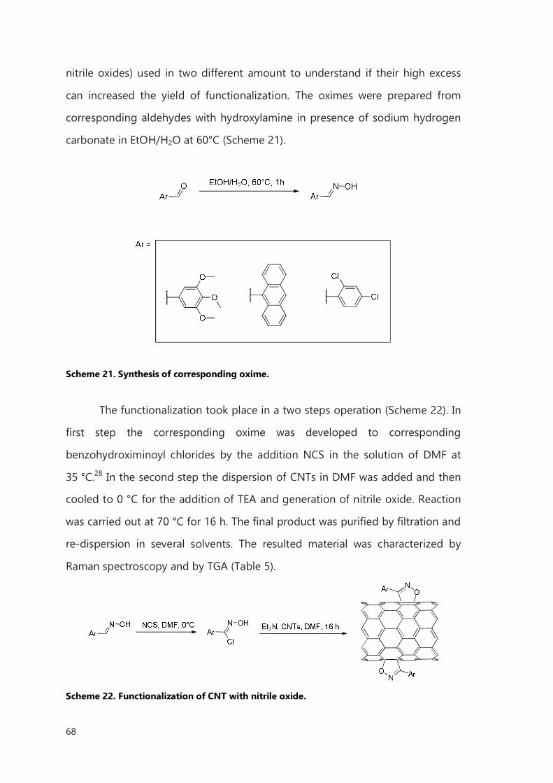
68
nitrile oxides) used in two different amount to understand if their high excess
can increased the yield of functionalization. The oximes were prepared from
corresponding aldehydes with hydroxylamine in presence of sodium hydrogen
carbonate in EtOH/H2O at 60°C (Scheme 21).
Scheme 21. Synthesis of corresponding oxime.
The functionalization took place in a two steps operation (Scheme 22). In
first step the corresponding oxime was developed to corresponding
benzohydroximinoyl chlorides by the addition NCS in the solution of DMF at
35 °C.28 In the second step the dispersion of CNTs in DMF was added and then
cooled to 0 °C for the addition of TEA and generation of nitrile oxide. Reaction
was carried out at 70 °C for 16 h. The final product was purified by filtration and
re-dispersion in several solvents. The resulted material was characterized by
Raman spectroscopy and by TGA (Table 5).
Scheme 22. Functionalization of CNT with nitrile oxide.
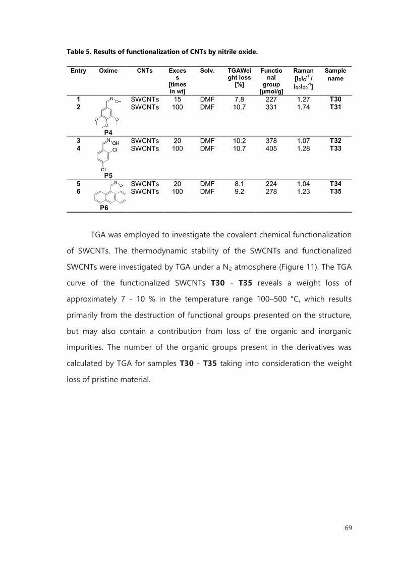
69
Table 5. Results of functionalization of CNTs by nitrile oxide.
Entry Oxime CNTs Exces
s [times in wt]
Solv. TGAWeight loss
[%]
Functional
group [μmol/g]
Raman [IDIG-1 / ID0IG0
-1]
Sample name
1
P4
SWCNTs 15 DMF 7.8 227 1.27 T30 2 SWCNTs 100 DMF 10.7 331 1.74 T31
3
P5
SWCNTs 20 DMF 10.2 378 1.07 T32 4 SWCNTs 100 DMF 10.7 405 1.28 T33
5
P6
SWCNTs 20 DMF 8.1 224 1.04 T34 6 SWCNTs 100 DMF 9.2 278 1.23 T35
TGA was employed to investigate the covalent chemical functionalization
of SWCNTs. The thermodynamic stability of the SWCNTs and functionalized
SWCNTs were investigated by TGA under a N2 atmosphere (Figure 11). The TGA
curve of the functionalized SWCNTs T30 - T35 reveals a weight loss of
approximately 7 - 10 % in the temperature range 100–500 °C, which results
primarily from the destruction of functional groups presented on the structure,
but may also contain a contribution from loss of the organic and inorganic
impurities. The number of the organic groups present in the derivatives was
calculated by TGA for samples T30 - T35 taking into consideration the weight
loss of pristine material.
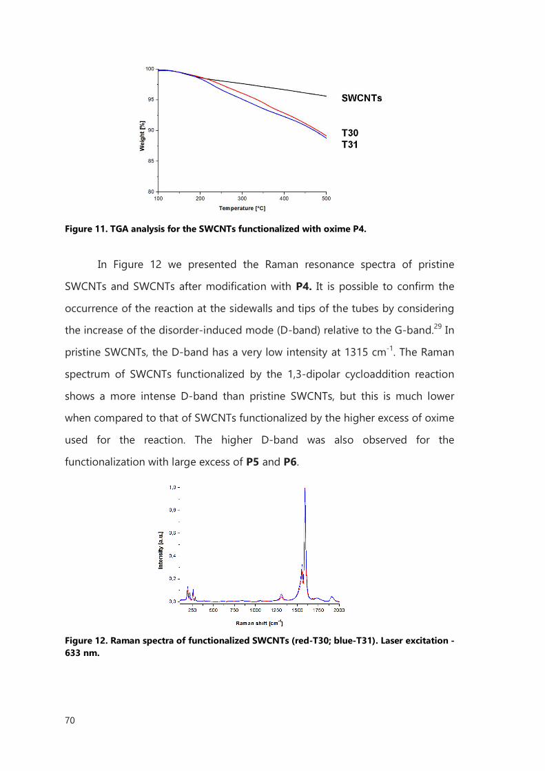
70
Figure 11. TGA analysis for the SWCNTs functionalized with oxime P4.
In Figure 12 we presented the Raman resonance spectra of pristine
SWCNTs and SWCNTs after modification with P4. It is possible to confirm the
occurrence of the reaction at the sidewalls and tips of the tubes by considering
the increase of the disorder-induced mode (D-band) relative to the G-band.29 In
pristine SWCNTs, the D-band has a very low intensity at 1315 cm-1. The Raman
spectrum of SWCNTs functionalized by the 1,3-dipolar cycloaddition reaction
shows a more intense D-band than pristine SWCNTs, but this is much lower
when compared to that of SWCNTs functionalized by the higher excess of oxime
used for the reaction. The higher D-band was also observed for the
functionalization with large excess of P5 and P6.
Figure 12. Raman spectra of functionalized SWCNTs (red-T30; blue-T31). Laser excitation - 633 nm.
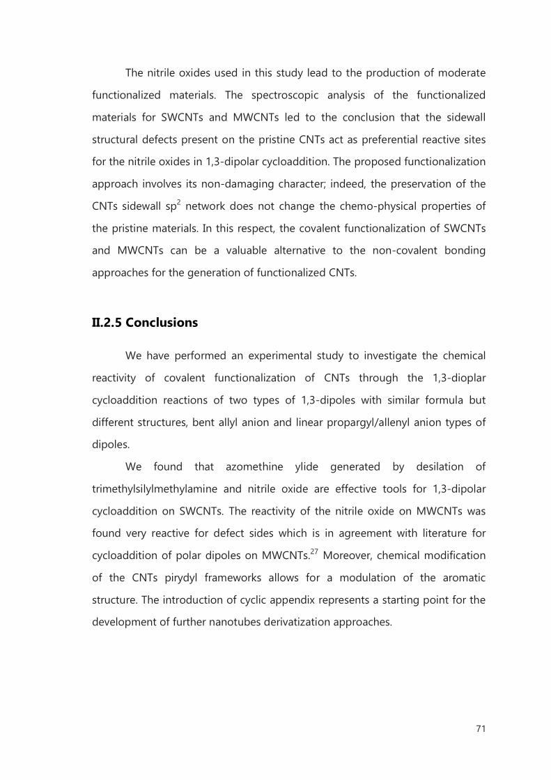
71
The nitrile oxides used in this study lead to the production of moderate
functionalized materials. The spectroscopic analysis of the functionalized
materials for SWCNTs and MWCNTs led to the conclusion that the sidewall
structural defects present on the pristine CNTs act as preferential reactive sites
for the nitrile oxides in 1,3-dipolar cycloaddition. The proposed functionalization
approach involves its non-damaging character; indeed, the preservation of the
CNTs sidewall sp2 network does not change the chemo-physical properties of
the pristine materials. In this respect, the covalent functionalization of SWCNTs
and MWCNTs can be a valuable alternative to the non-covalent bonding
approaches for the generation of functionalized CNTs.
II.2.5 Conclusions
We have performed an experimental study to investigate the chemical
reactivity of covalent functionalization of CNTs through the 1,3-dioplar
cycloaddition reactions of two types of 1,3-dipoles with similar formula but
different structures, bent allyl anion and linear propargyl/allenyl anion types of
dipoles.
We found that azomethine ylide generated by desilation of
trimethylsilylmethylamine and nitrile oxide are effective tools for 1,3-dipolar
cycloaddition on SWCNTs. The reactivity of the nitrile oxide on MWCNTs was
found very reactive for defect sides which is in agreement with literature for
cycloaddition of polar dipoles on MWCNTs.27 Moreover, chemical modification
of the CNTs pirydyl frameworks allows for a modulation of the aromatic
structure. The introduction of cyclic appendix represents a starting point for the
development of further nanotubes derivatization approaches.
72
II.3 Diazonium-Based Functionalization
The diazonium-based functionalization of CNTs is a very useful method
(described in Introduction). Since 2001, when Tour and coworkers discovered a
convenient route to sidewall-functionalized carbon nanotubes by reacting aryl
diazonium salts with SWCNTs in an electrochemical reaction,30 a variety of the
aryl diazonium salts was applied for covalent modification of CNT. The
corresponding reactive aryl radicals can be generated from the diazonium salt
by electrochemical one-electron reduction or in situ from the diazonium
compound by reacting of aniline derivatives with isopentyl nitrite.31 Another
route was developed after the work of Tour and coworkers. The SWCNTs were
functionalized for minutes at room temperature with aryl diazonium salts in the
presence of ionic liquids and potassium carbonate.32 The functionalized material
was comparable to those derived from other processes using harsh reaction
conditions. This functionalization was also performed in water as a solvent to
reduce harmful wastes.33 The range of solvents for this type of CNTs
functionalization reaction was extended to Caro’s acid, a mixture of 96% sulfuric
acid and ammonium persulfate.34 Lately, in situ generation of aryl diazonium
salts in the presence of ammonium persulfate in a solvent-free medium was
reported for the derivatization of MWCNTs, with high degree of
functionalization.35 Another comprehensive example on MWCNTs was published
recently.36 The covalent functionalization of MWCNTs was performed with the
well-established procedure in water reported by Tour et al.33
As an alternative for aniline derivative, triazene compounds were used.
This stable diazonium salt precursor was proposed for the functionalization of
SWCNTs in aqueous media37 and the authors claimed that is particularly useful
for biological relevant molecules.
In literature there are also examples of application of MWs for the
acceleration of the reaction.38 The authors reported the results in comparison to
73
the functionalization with classical heating. In principle, under MW irradiation,
the reaction leads to shorter reaction time and higher functionalization degrees.
Meanwhile, the diazonium-based functionalization became the most
frequently used reaction to derivatized CNTs, for chemists and material
scientists. For example short linkers like 4-aminobenzylamine, were covalently
bind to CNTs sidewalls, by the diazonium route, and used for further
modifications. Burghard et al. reported the attachment of gold nanoparticles to
carbon nanotubes functionalized with 4-diazo-N,N-diethylaniline
tetrafluoroborate for enhancing the Raman response.39 Other nanoparticles -
CdSe–ZnS quantum dots were conjugated to the sidewall functionalized
SWCNTs templates for enhancing photophysical properties for light harvesting
systems. 40Diazonium-based functionalization was also used to bind nickel to
the surface of the nanotube to direct the self-assembly of histidine-tagged
proteins at controlled orientation.41 This approach has application for metal
interacting protein systems where the redox center of the protein is inaccessible
through conventional means of directed assembly onto carbon nanotubes.
The presented examples, of the covalent functionalization of CNTs
through aryl diazonium salts, were demonstrated as an easy and very efficient
way to decorate carbon nanotubes. The fast and efficient way of
functionalization with aniline derivatives gives a possibility for modification of
sidewall of CNTs with versatile molecules. These encourage us to apply this
reaction to develop the most suitable “bridge” between CNTs and biomedical
application.
II.3.1 Results of Diazonium-Based Functionalization
Basing on the aim of the project, we decided to thoroughly investigate
the functionalization of SWCNTs and MWCNTs with diazonium based aniline
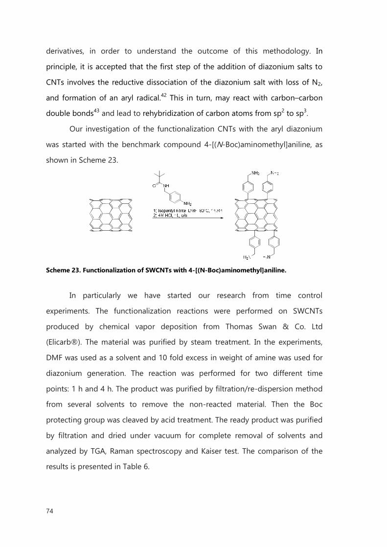
74
derivatives, in order to understand the outcome of this methodology. In
principle, it is accepted that the first step of the addition of diazonium salts to
CNTs involves the reductive dissociation of the diazonium salt with loss of N2,
and formation of an aryl radical.42 This in turn, may react with carbon–carbon
double bonds43 and lead to rehybridization of carbon atoms from sp2 to sp3.
Our investigation of the functionalization CNTs with the aryl diazonium
was started with the benchmark compound 4-[(N-Boc)aminomethyl]aniline, as
shown in Scheme 23.
Scheme 23. Functionalization of SWCNTs with 4-[(N-Boc)aminomethyl]aniline.
In particularly we have started our research from time control
experiments. The functionalization reactions were performed on SWCNTs
produced by chemical vapor deposition from Thomas Swan & Co. Ltd
(Elicarb®). The material was purified by steam treatment. In the experiments,
DMF was used as a solvent and 10 fold excess in weight of amine was used for
diazonium generation. The reaction was performed for two different time
points: 1 h and 4 h. The product was purified by filtration/re-dispersion method
from several solvents to remove the non-reacted material. Then the Boc
protecting group was cleaved by acid treatment. The ready product was purified
by filtration and dried under vacuum for complete removal of solvents and
analyzed by TGA, Raman spectroscopy and Kaiser test. The comparison of the
results is presented in Table 6.
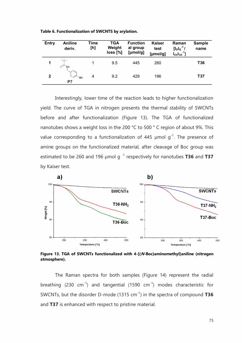
75
Table 6. Functionalization of SWCNTS by arylation.
Entry Aniline
deriv. Time [h]
TGA Weight loss [%]
Functional group [μmol/g]
Kaiser test
[µmol/g]
Raman [IDIG-1 / ID0IG0
-1]
Sample name
1
P7
1 9.5 445 260 T36
2 4 9.2 429 196 T37
Interestingly, lower time of the reaction leads to higher functionalization
yield. The curve of TGA in nitrogen presents the thermal stability of SWCNTs
before and after functionalization (Figure 13). The TGA of functionalized
nanotubes shows a weight loss in the 200 °C to 500 ° C region of about 9%. This
value corresponding to a functionalization of 445 µmol g-1. The presence of
amine groups on the functionalized material, after cleavage of Boc group was
estimated to be 260 and 196 µmol g -1 respectively for nanotubes T36 and T37
by Kaiser test.
Figure 13. TGA of SWCNTs functionalized with 4-[(N-Boc)aminomethyl]aniline (nitrogen atmosphere).
The Raman spectra for both samples (Figure 14) represent the radial
breathing (230 cm-1) and tangential (1590 cm-1) modes characteristic for
SWCNTs, but the disorder D-mode (1315 cm-1) in the spectra of compound T36
and T37 is enhanced with respect to pristine material.
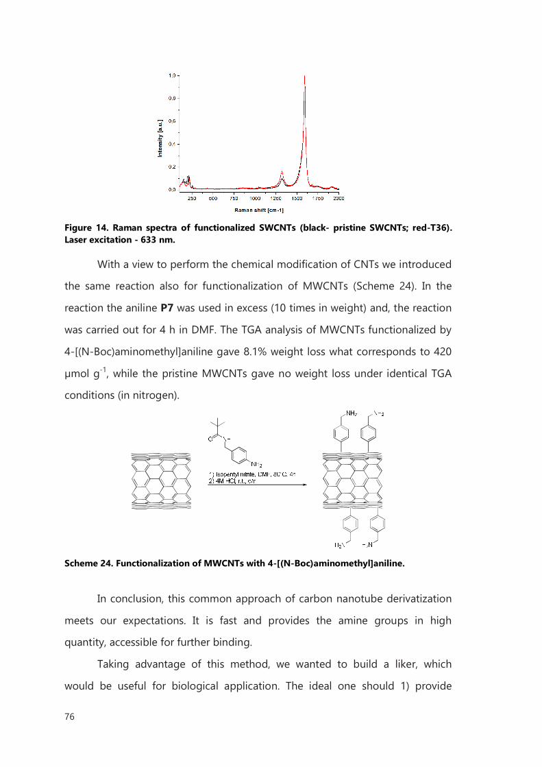
76
Figure 14. Raman spectra of functionalized SWCNTs (black- pristine SWCNTs; red-T36). Laser excitation - 633 nm.
With a view to perform the chemical modification of CNTs we introduced
the same reaction also for functionalization of MWCNTs (Scheme 24). In the
reaction the aniline P7 was used in excess (10 times in weight) and, the reaction
was carried out for 4 h in DMF. The TGA analysis of MWCNTs functionalized by
4-[(N-Boc)aminomethyl]aniline gave 8.1% weight loss what corresponds to 420
µmol g-1, while the pristine MWCNTs gave no weight loss under identical TGA
conditions (in nitrogen).
Scheme 24. Functionalization of MWCNTs with 4-[(N-Boc)aminomethyl]aniline.
In conclusion, this common approach of carbon nanotube derivatization
meets our expectations. It is fast and provides the amine groups in high
quantity, accessible for further binding.
Taking advantage of this method, we wanted to build a liker, which
would be useful for biological application. The ideal one should 1) provide
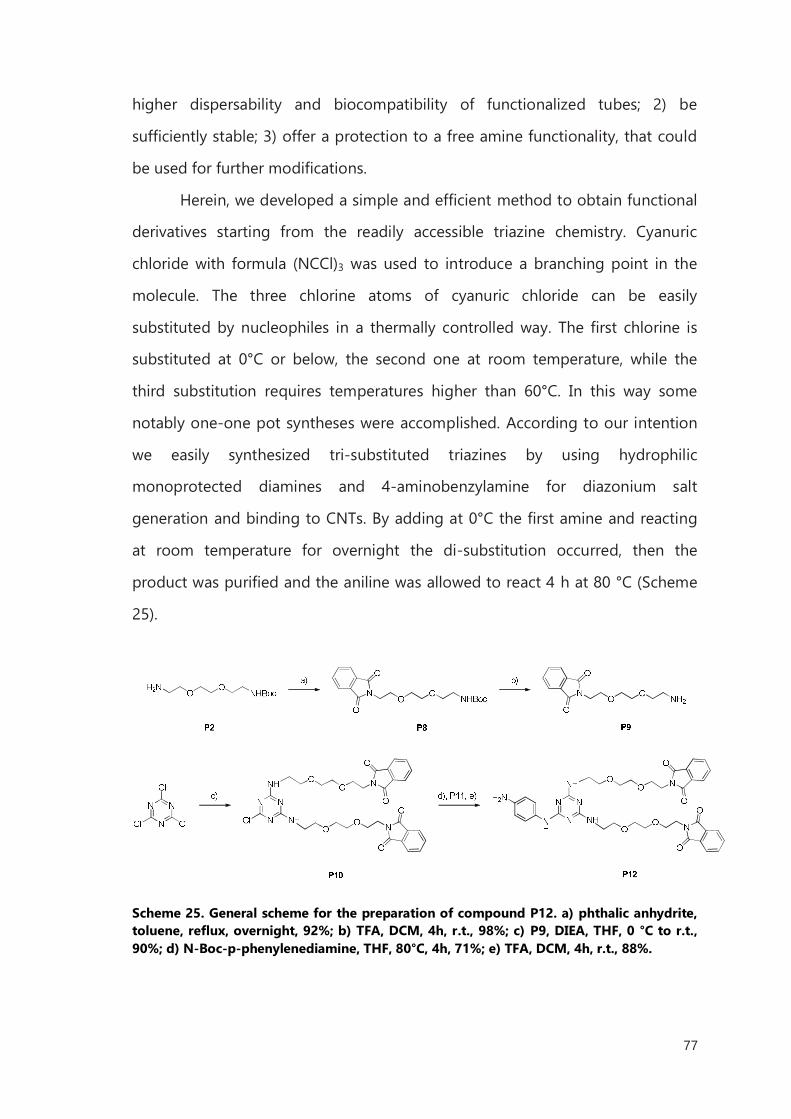
77
higher dispersability and biocompatibility of functionalized tubes; 2) be
sufficiently stable; 3) offer a protection to a free amine functionality, that could
be used for further modifications.
Herein, we developed a simple and efficient method to obtain functional
derivatives starting from the readily accessible triazine chemistry. Cyanuric
chloride with formula (NCCl)3 was used to introduce a branching point in the
molecule. The three chlorine atoms of cyanuric chloride can be easily
substituted by nucleophiles in a thermally controlled way. The first chlorine is
substituted at 0°C or below, the second one at room temperature, while the
third substitution requires temperatures higher than 60°C. In this way some
notably one-one pot syntheses were accomplished. According to our intention
we easily synthesized tri-substituted triazines by using hydrophilic
monoprotected diamines and 4-aminobenzylamine for diazonium salt
generation and binding to CNTs. By adding at 0°C the first amine and reacting
at room temperature for overnight the di-substitution occurred, then the
product was purified and the aniline was allowed to react 4 h at 80 °C (Scheme
25).
Scheme 25. General scheme for the preparation of compound P12. a) phthalic anhydrite, toluene, reflux, overnight, 92%; b) TFA, DCM, 4h, r.t., 98%; c) P9, DIEA, THF, 0 °C to r.t., 90%; d) N-Boc-p-phenylenediamine, THF, 80°C, 4h, 71%; e) TFA, DCM, 4h, r.t., 88%.
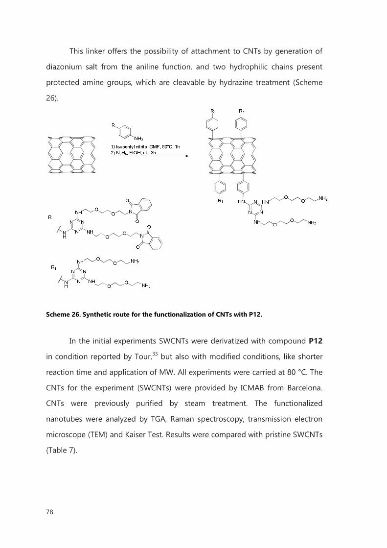
78
This linker offers the possibility of attachment to CNTs by generation of
diazonium salt from the aniline function, and two hydrophilic chains present
protected amine groups, which are cleavable by hydrazine treatment (Scheme
26).
Scheme 26. Synthetic route for the functionalization of CNTs with P12.
In the initial experiments SWCNTs were derivatized with compound P12
in condition reported by Tour,33 but also with modified conditions, like shorter
reaction time and application of MW. All experiments were carried at 80 °C. The
CNTs for the experiment (SWCNTs) were provided by ICMAB from Barcelona.
CNTs were previously purified by steam treatment. The functionalized
nanotubes were analyzed by TGA, Raman spectroscopy, transmission electron
microscope (TEM) and Kaiser Test. Results were compared with pristine SWCNTs
(Table 7).
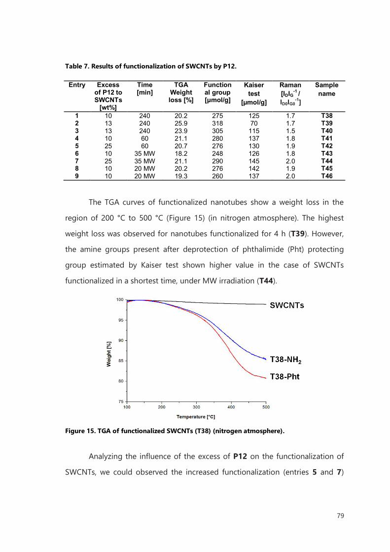
79
Table 7. Results of functionalization of SWCNTs by P12.
Entry Excess
of P12 to SWCNTs
[wt%]
Time [min]
TGA Weight loss [%]
Functional group [μmol/g]
Kaiser test
[µmol/g]
Raman [IDIG-1 / ID0IG0
-1]
Sample name
1 10 240 20.2 275 125 1.7 T38 2 13 240 25.9 318 70 1.7 T39 3 13 240 23.9 305 115 1.5 T40 4 10 60 21.1 280 137 1.8 T41 5 25 60 20.7 276 130 1.9 T42 6 10 35 MW 18.2 248 126 1.8 T43 7 25 35 MW 21.1 290 145 2.0 T44 8 10 20 MW 20.2 276 142 1.9 T45 9 10 20 MW 19.3 260 137 2.0 T46
The TGA curves of functionalized nanotubes show a weight loss in the
region of 200 °C to 500 °C (Figure 15) (in nitrogen atmosphere). The highest
weight loss was observed for nanotubes functionalized for 4 h (T39). However,
the amine groups present after deprotection of phthalimide (Pht) protecting
group estimated by Kaiser test shown higher value in the case of SWCNTs
functionalized in a shortest time, under MW irradiation (T44).
Figure 15. TGA of functionalized SWCNTs (T38) (nitrogen atmosphere).
Analyzing the influence of the excess of P12 on the functionalization of
SWCNTs, we could observed the increased functionalization (entries 5 and 7)
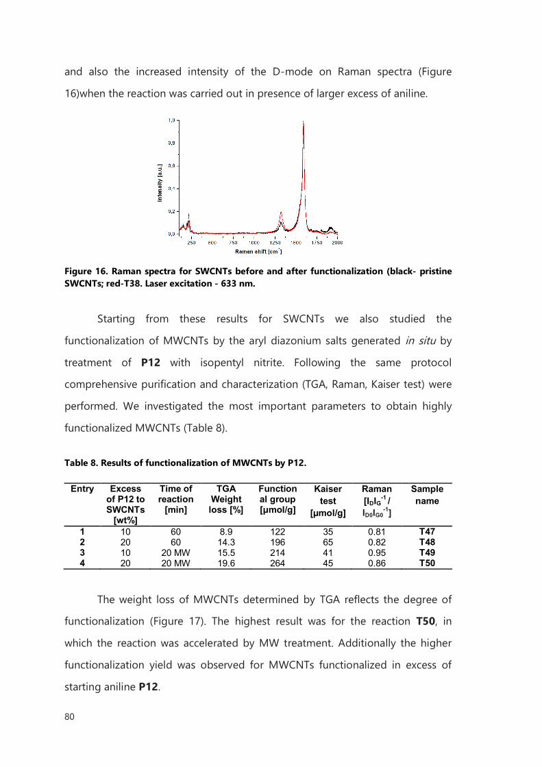
80
and also the increased intensity of the D-mode on Raman spectra (Figure
16)when the reaction was carried out in presence of larger excess of aniline.
Figure 16. Raman spectra for SWCNTs before and after functionalization (black- pristine SWCNTs; red-T38. Laser excitation - 633 nm.
Starting from these results for SWCNTs we also studied the
functionalization of MWCNTs by the aryl diazonium salts generated in situ by
treatment of P12 with isopentyl nitrite. Following the same protocol
comprehensive purification and characterization (TGA, Raman, Kaiser test) were
performed. We investigated the most important parameters to obtain highly
functionalized MWCNTs (Table 8).
Table 8. Results of functionalization of MWCNTs by P12.
Entry Excess
of P12 to SWCNTs
[wt%]
Time of reaction
[min]
TGA Weight loss [%]
Functional group [μmol/g]
Kaiser test
[µmol/g]
Raman [IDIG-1 / ID0IG0
-1]
Sample name
1 10 60 8.9 122 35 0.81 T47 2 20 60 14.3 196 65 0.82 T48 3 10 20 MW 15.5 214 41 0.95 T49 4 20 20 MW 19.6 264 45 0.86 T50
The weight loss of MWCNTs determined by TGA reflects the degree of
functionalization (Figure 17). The highest result was for the reaction T50, in
which the reaction was accelerated by MW treatment. Additionally the higher
functionalization yield was observed for MWCNTs functionalized in excess of
starting aniline P12.
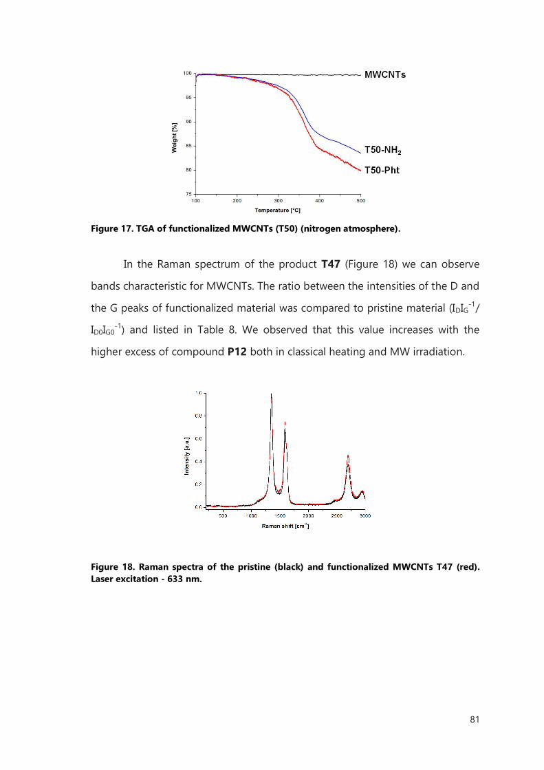
81
Figure 17. TGA of functionalized MWCNTs (T50) (nitrogen atmosphere).
In the Raman spectrum of the product T47 (Figure 18) we can observe
bands characteristic for MWCNTs. The ratio between the intensities of the D and
the G peaks of functionalized material was compared to pristine material (IDIG-1/
ID0IG0-1) and listed in Table 8. We observed that this value increases with the
higher excess of compound P12 both in classical heating and MW irradiation.
Figure 18. Raman spectra of the pristine (black) and functionalized MWCNTs T47 (red). Laser excitation - 633 nm.

82
II.3.2 Conclusions
These positive outcomes from the arylation reaction on both types of CNTs are
the starting point for the design and controlled processing of novel decorated
carbon nanotubes. It would provide an easily and convenient methodology for
functionalization of filled nanotubes.
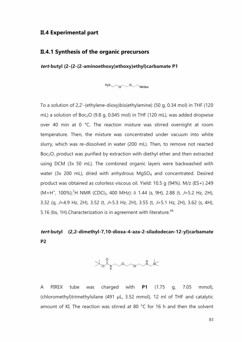
83
II.4 Experimental part
II.4.1 Synthesis of the organic precursors
tert-butyl (2-(2-(2-aminoethoxy)ethoxy)ethyl)carbamate P1
To a solution of 2,2’-(ethylene-dioxy)bis(ethylamine) (50 g, 0.34 mol) in THF (120
mL) a solution of Boc2O (9.8 g, 0.045 mol) in THF (120 mL), was added dropwise
over 40 min at 0 °C. The reaction mixture was stirred overnight at room
temperature. Then, the mixture was concentrated under vacuum into white
slurry, which was re-dissolved in water (200 mL). Then, to remove not reacted
Boc2O, product was purified by extraction with diethyl ether and then extracted
using DCM (3x 50 mL). The combined organic layers were backwashed with
water (3x 200 mL), dried with anhydrous MgSO4 and concentrated. Desired
product was obtained as colorless viscous oil. Yield: 10.5 g (94%). M/z (ES+) 249
(M+H+, 100%);1H NMR (CDCl3, 400 MHz): 1.44 (s, 9H), 2.88 (t, J=5.2 Hz, 2H),
3.32 (q, J=4.9 Hz, 2H), 3.52 (t, J=5.3 Hz, 2H), 3.55 (t, J=5.1 Hz, 2H), 3.62 (s, 4H),
5.16 (bs, 1H).Characterization is in agreement with literature.44
tert-butyl (2,2-dimethyl-7,10-dioxa-4-aza-2-siladodecan-12-yl)carbamate
P2
A PIREX tube was charged with P1 (1.75 g, 7.05 mmol),
(chloromethyl)trimethylsilane (491 µL, 3.52 mmol), 12 ml of THF and catalytic
amount of KI. The reaction was stirred at 80 °C for 16 h and then the solvent
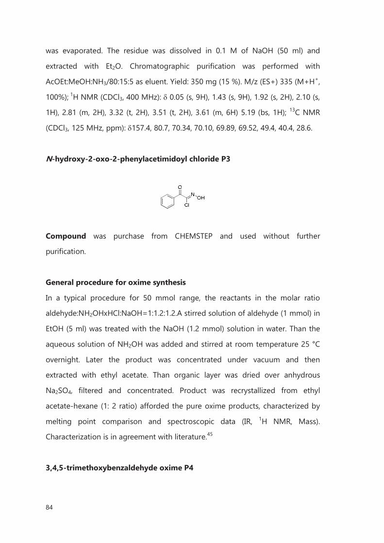
84
was evaporated. The residue was dissolved in 0.1 M of NaOH (50 ml) and
extracted with Et2O. Chromatographic purification was performed with
AcOEt:MeOH:NH3/80:15:5 as eluent. Yield: 350 mg (15 %). M/z (ES+) 335 (M+H+,
100%); 1H NMR (CDCl3, 400 MHz): 0.05 (s, 9H), 1.43 (s, 9H), 1.92 (s, 2H), 2.10 (s,
1H), 2.81 (m, 2H), 3.32 (t, 2H), 3.51 (t, 2H), 3.61 (m, 6H) 5.19 (bs, 1H); 13C NMR
(CDCl3, 125 MHz, ppm): 157.4, 80.7, 70.34, 70.10, 69.89, 69.52, 49.4, 40.4, 28.6.
N-hydroxy-2-oxo-2-phenylacetimidoyl chloride P3
Compound was purchase from CHEMSTEP and used without further
purification.
General procedure for oxime synthesis
In a typical procedure for 50 mmol range, the reactants in the molar ratio
aldehyde:NH2OHxHCl:NaOH=1:1.2:1.2.A stirred solution of aldehyde (1 mmol) in
EtOH (5 ml) was treated with the NaOH (1.2 mmol) solution in water. Than the
aqueous solution of NH2OH was added and stirred at room temperature 25 °C
overnight. Later the product was concentrated under vacuum and then
extracted with ethyl acetate. Than organic layer was dried over anhydrous
Na2SO4, filtered and concentrated. Product was recrystallized from ethyl
acetate-hexane (1: 2 ratio) afforded the pure oxime products, characterized by
melting point comparison and spectroscopic data (IR, 1H NMR, Mass).
Characterization is in agreement with literature.45
3,4,5-trimethoxybenzaldehyde oxime P4
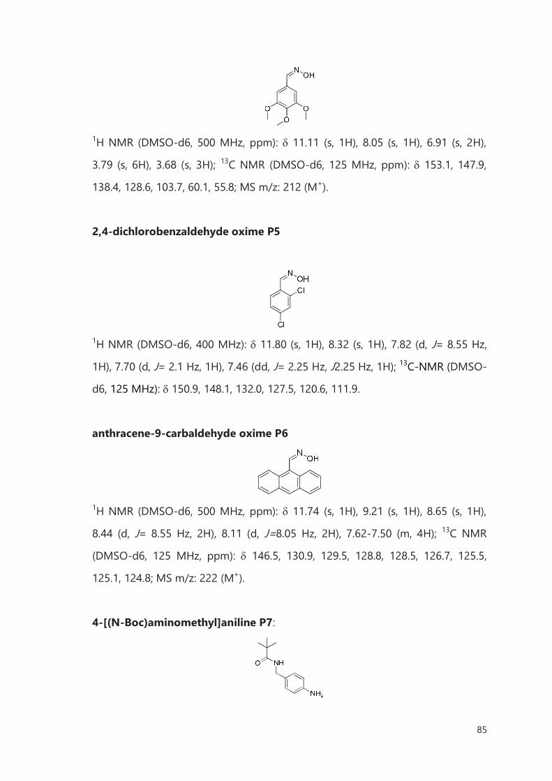
85
1H NMR (DMSO-d6, 500 MHz, ppm): 11.11 (s, 1H), 8.05 (s, 1H), 6.91 (s, 2H),
3.79 (s, 6H), 3.68 (s, 3H); 13C NMR (DMSO-d6, 125 MHz, ppm): 153.1, 147.9,
138.4, 128.6, 103.7, 60.1, 55.8; MS m/z: 212 (M+).
2,4-dichlorobenzaldehyde oxime P5
1H NMR (DMSO-d6, 400 MHz): 11.80 (s, 1H), 8.32 (s, 1H), 7.82 (d, J= 8.55 Hz,
1H), 7.70 (d, J= 2.1 Hz, 1H), 7.46 (dd, J= 2.25 Hz, J2.25 Hz, 1H); 13C-NMR (DMSO-
d6, 125 MHz): 150.9, 148.1, 132.0, 127.5, 120.6, 111.9.
anthracene-9-carbaldehyde oxime P6
1H NMR (DMSO-d6, 500 MHz, ppm): 11.74 (s, 1H), 9.21 (s, 1H), 8.65 (s, 1H),
8.44 (d, J= 8.55 Hz, 2H), 8.11 (d, J=8.05 Hz, 2H), 7.62-7.50 (m, 4H); 13C NMR
(DMSO-d6, 125 MHz, ppm): 146.5, 130.9, 129.5, 128.8, 128.5, 126.7, 125.5,
125.1, 124.8; MS m/z: 222 (M+).
4-[(N-Boc)aminomethyl]aniline P7:
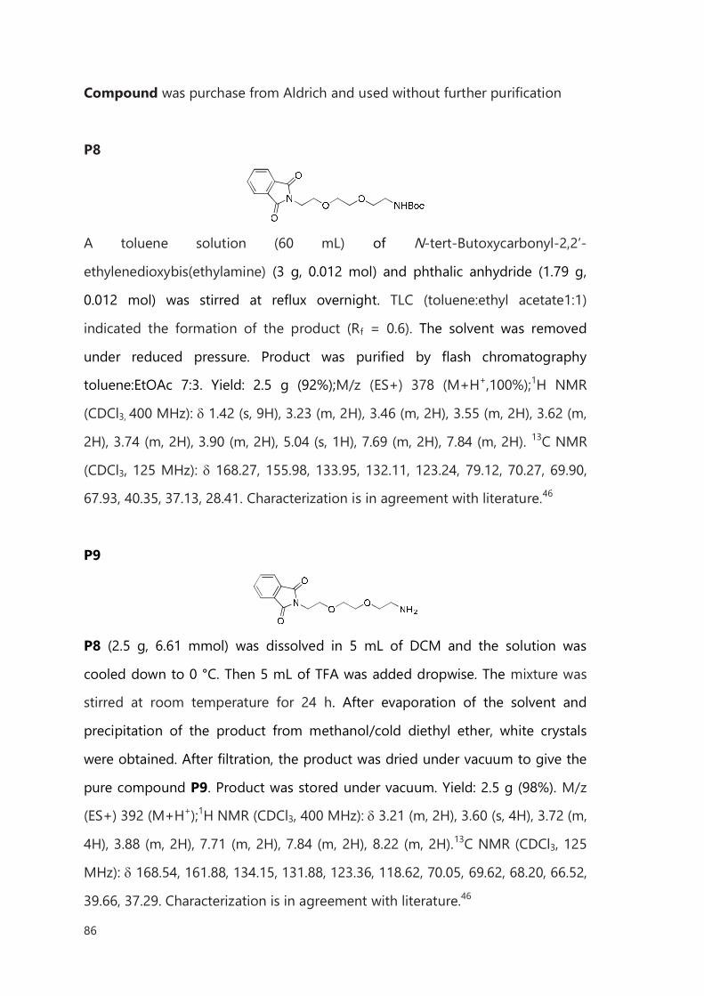
86
Compound was purchase from Aldrich and used without further purification
P8
A toluene solution (60 mL) of N-tert-Butoxycarbonyl-2,2’-
ethylenedioxybis(ethylamine) (3 g, 0.012 mol) and phthalic anhydride (1.79 g,
0.012 mol) was stirred at reflux overnight. TLC (toluene:ethyl acetate1:1)
indicated the formation of the product (Rf = 0.6). The solvent was removed
under reduced pressure. Product was purified by flash chromatography
toluene:EtOAc 7:3. Yield: 2.5 g (92%);M/z (ES+) 378 (M+H+,100%);1H NMR
(CDCl3, 400 MHz): 1.42 (s, 9H), 3.23 (m, 2H), 3.46 (m, 2H), 3.55 (m, 2H), 3.62 (m,
2H), 3.74 (m, 2H), 3.90 (m, 2H), 5.04 (s, 1H), 7.69 (m, 2H), 7.84 (m, 2H). 13C NMR
(CDCl3, 125 MHz): 168.27, 155.98, 133.95, 132.11, 123.24, 79.12, 70.27, 69.90,
67.93, 40.35, 37.13, 28.41. Characterization is in agreement with literature.46
P9
P8 (2.5 g, 6.61 mmol) was dissolved in 5 mL of DCM and the solution was
cooled down to 0 °C. Then 5 mL of TFA was added dropwise. The mixture was
stirred at room temperature for 24 h. After evaporation of the solvent and
precipitation of the product from methanol/cold diethyl ether, white crystals
were obtained. After filtration, the product was dried under vacuum to give the
pure compound P9. Product was stored under vacuum. Yield: 2.5 g (98%). M/z
(ES+) 392 (M+H+);1H NMR (CDCl3, 400 MHz): 3.21 (m, 2H), 3.60 (s, 4H), 3.72 (m,
4H), 3.88 (m, 2H), 7.71 (m, 2H), 7.84 (m, 2H), 8.22 (m, 2H).13C NMR (CDCl3, 125
MHz): 168.54, 161.88, 134.15, 131.88, 123.36, 118.62, 70.05, 69.62, 68.20, 66.52,
39.66, 37.29. Characterization is in agreement with literature.46
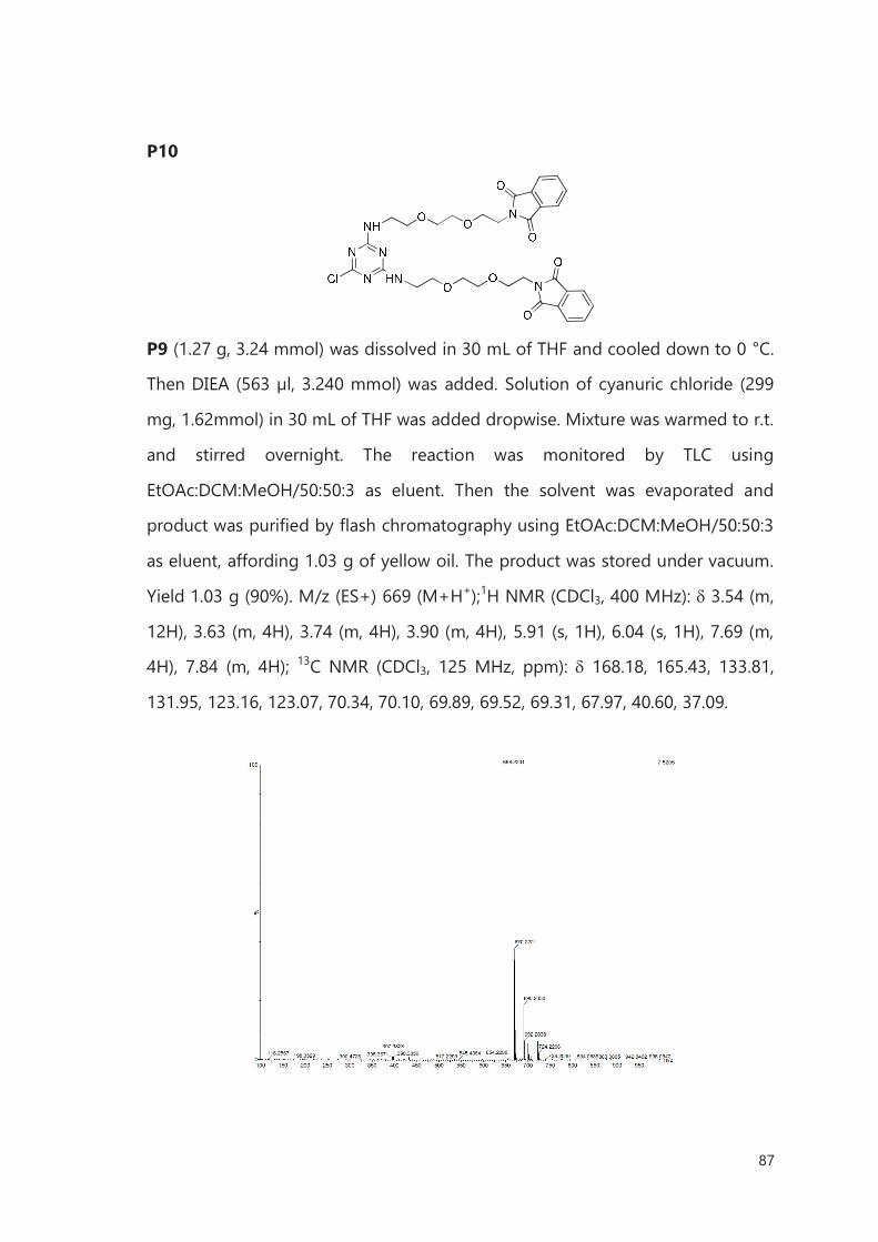
87
P10
P9 (1.27 g, 3.24 mmol) was dissolved in 30 mL of THF and cooled down to 0 °C.
Then DIEA (563 µl, 3.240 mmol) was added. Solution of cyanuric chloride (299
mg, 1.62mmol) in 30 mL of THF was added dropwise. Mixture was warmed to r.t.
and stirred overnight. The reaction was monitored by TLC using
EtOAc:DCM:MeOH/50:50:3 as eluent. Then the solvent was evaporated and
product was purified by flash chromatography using EtOAc:DCM:MeOH/50:50:3
as eluent, affording 1.03 g of yellow oil. The product was stored under vacuum.
Yield 1.03 g (90%). M/z (ES+) 669 (M+H+);1H NMR (CDCl3, 400 MHz): 3.54 (m,
12H), 3.63 (m, 4H), 3.74 (m, 4H), 3.90 (m, 4H), 5.91 (s, 1H), 6.04 (s, 1H), 7.69 (m,
4H), 7.84 (m, 4H); 13C NMR (CDCl3, 125 MHz, ppm): 168.18, 165.43, 133.81,
131.95, 123.16, 123.07, 70.34, 70.10, 69.89, 69.52, 69.31, 67.97, 40.60, 37.09.
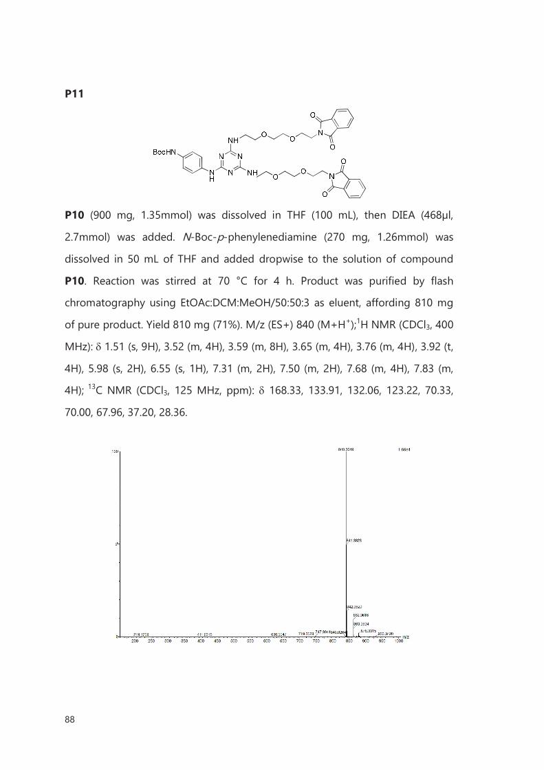
88
P11
P10 (900 mg, 1.35mmol) was dissolved in THF (100 mL), then DIEA (468µl,
2.7mmol) was added. N-Boc-p-phenylenediamine (270 mg, 1.26mmol) was
dissolved in 50 mL of THF and added dropwise to the solution of compound
P10. Reaction was stirred at 70 °C for 4 h. Product was purified by flash
chromatography using EtOAc:DCM:MeOH/50:50:3 as eluent, affording 810 mg
of pure product. Yield 810 mg (71%). M/z (ES+) 840 (M+H+);1H NMR (CDCl3, 400
MHz): 1.51 (s, 9H), 3.52 (m, 4H), 3.59 (m, 8H), 3.65 (m, 4H), 3.76 (m, 4H), 3.92 (t,
4H), 5.98 (s, 2H), 6.55 (s, 1H), 7.31 (m, 2H), 7.50 (m, 2H), 7.68 (m, 4H), 7.83 (m,
4H); 13C NMR (CDCl3, 125 MHz, ppm): 168.33, 133.91, 132.06, 123.22, 70.33,
70.00, 67.96, 37.20, 28.36.
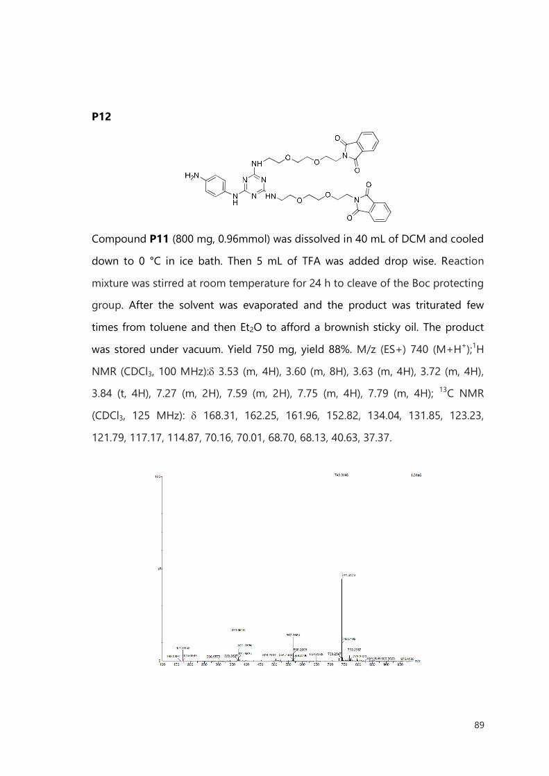
89
P12
Compound P11 (800 mg, 0.96mmol) was dissolved in 40 mL of DCM and cooled
down to 0 °C in ice bath. Then 5 mL of TFA was added drop wise. Reaction
mixture was stirred at room temperature for 24 h to cleave of the Boc protecting
group. After the solvent was evaporated and the product was triturated few
times from toluene and then Et2O to afford a brownish sticky oil. The product
was stored under vacuum. Yield 750 mg, yield 88%. M/z (ES+) 740 (M+H+);1H
NMR (CDCl3, 100 MHz): 3.53 (m, 4H), 3.60 (m, 8H), 3.63 (m, 4H), 3.72 (m, 4H),
3.84 (t, 4H), 7.27 (m, 2H), 7.59 (m, 2H), 7.75 (m, 4H), 7.79 (m, 4H); 13C NMR
(CDCl3, 125 MHz): 168.31, 162.25, 161.96, 152.82, 134.04, 131.85, 123.23,
121.79, 117.17, 114.87, 70.16, 70.01, 68.70, 68.13, 40.63, 37.37.
90
II.4.2 Functionalization
Purification of HiPCO SWCNTs for cycloaddition reactions
The SWCNTs used for cycloaddition study were produced high pressure carbon
monoxide method called (HiPCO) purchased from Carbon Nanotechnologies
(lot #R0510C). The material was purified by concentrated HCl treatment
following a previously reported protocol.47 In this step the as-received SWCNTs
were treated with hydrochloric acid, which is an established method for the
removal of impurities. One gram of SWNCTs was placed in a 500 ml round
bottom flask and 200 mL of HCl was added. The mixture was stirred using
magnetic stirrer for 2 h, then diluted in water, collected by filtration through a
0.2 µm polycarbonate membrane and thoroughly washed with water until
neutral pH. The sample was dried under vacuum.
Purification of SWCNTs for diazonium-based functionalization
The SWCNTs used in this investigation were produced by chemical vapor
deposition from Thomas Swan & Co. Ltd (Elicarb®). The material was purified by
steam treatment following a previously reported protocol.48 Briefly, SWCNTs
were spread inside a silica tube, which was then placed into a furnace alumina
tube. The whole system was initially purged with argon for 2 h, to remove
atmospheric air. Later on, steam was introduced by bubbling argon through a
flask containing hot distilled water and furnace was heated at 900 °C for4 h to
allow the purification of the material. The solid powder was collected and
treated with 6 M HCl (Panreac) at 110 °C overnight to dissolve the exposed iron
nanoparticles (from catalyst). The purified SWCNTs were collected by filtration
through a 0.2 µm polycarbonate membrane and thoroughly washed with water
until neutral pH. The sample was oven-dried at 80 °C overnight.
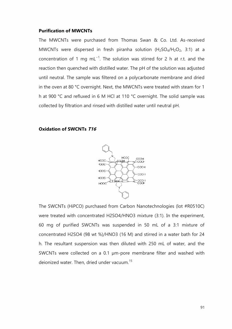
91
Purification of MWCNTs
The MWCNTs were purchased from Thomas Swan & Co. Ltd. As-received
MWCNTs were dispersed in fresh piranha solution (H2SO4/H2O2, 3:1) at a
concentration of 1 mg mL−1. The solution was stirred for 2 h at r.t. and the
reaction then quenched with distilled water. The pH of the solution was adjusted
until neutral. The sample was filtered on a polycarbonate membrane and dried
in the oven at 80 °C overnight. Next, the MWCNTs were treated with steam for 1
h at 900 °C and refluxed in 6 M HCl at 110 °C overnight. The solid sample was
collected by filtration and rinsed with distilled water until neutral pH.
Oxidation of SWCNTs T16
The SWCNTs (HiPCO) purchased from Carbon Nanotechnologies (lot #R0510C)
were treated with concentrated H2SO4/HNO3 mixture (3:1). In the experiment,
60 mg of purified SWCNTs was suspended in 50 mL of a 3:1 mixture of
concentrated H2SO4 (98 wt %)/HNO3 (16 M) and stirred in a water bath for 24
h. The resultant suspension was then diluted with 250 mL of water, and the
SWCNTs were collected on a 0.1 µm-pore membrane filter and washed with
deionized water. Then, dried under vacuum.15
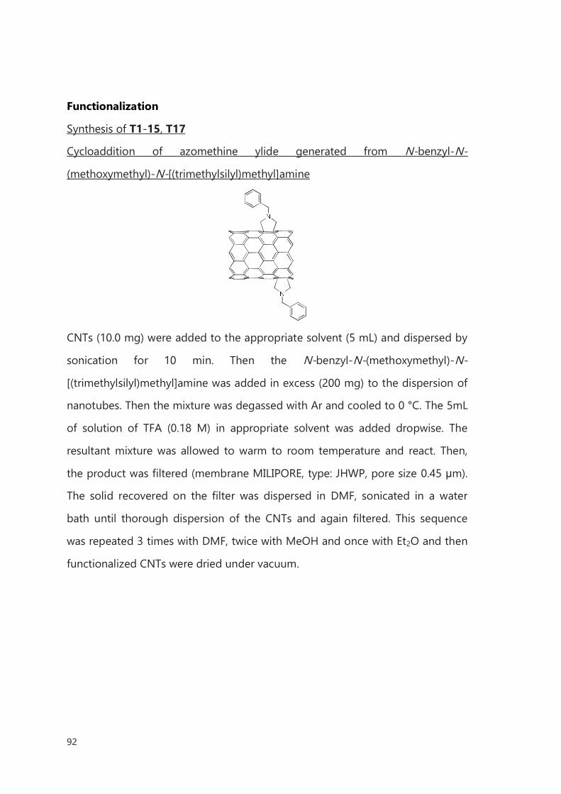
92
Functionalization
Synthesis of T1-15, T17
Cycloaddition of azomethine ylide generated from N-benzyl-N-
(methoxymethyl)-N-[(trimethylsilyl)methyl]amine
CNTs (10.0 mg) were added to the appropriate solvent (5 mL) and dispersed by
sonication for 10 min. Then the N-benzyl-N-(methoxymethyl)-N-
[(trimethylsilyl)methyl]amine was added in excess (200 mg) to the dispersion of
nanotubes. Then the mixture was degassed with Ar and cooled to 0 °C. The 5mL
of solution of TFA (0.18 M) in appropriate solvent was added dropwise. The
resultant mixture was allowed to warm to room temperature and react. Then,
the product was filtered (membrane MILIPORE, type: JHWP, pore size 0.45 μm).
The solid recovered on the filter was dispersed in DMF, sonicated in a water
bath until thorough dispersion of the CNTs and again filtered. This sequence
was repeated 3 times with DMF, twice with MeOH and once with Et2O and then
functionalized CNTs were dried under vacuum.
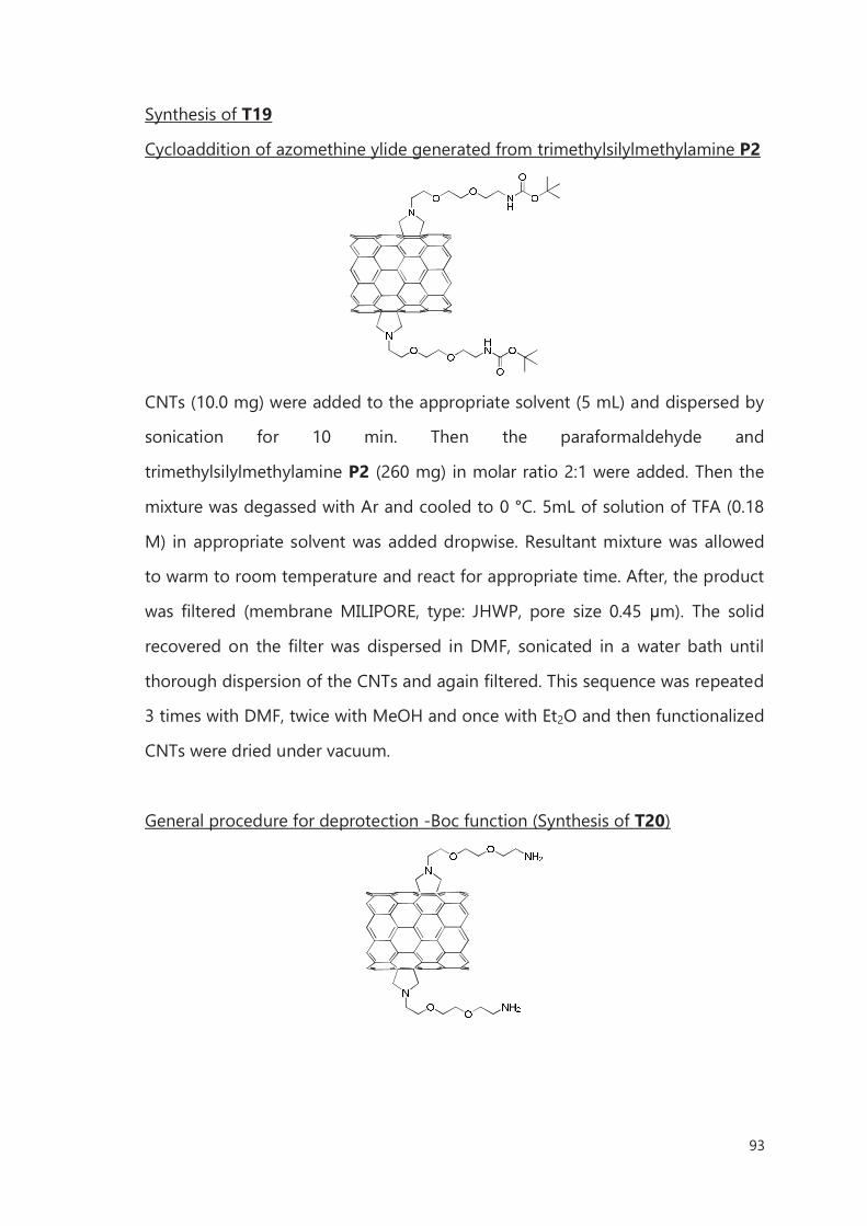
93
Synthesis of T19
Cycloaddition of azomethine ylide generated from trimethylsilylmethylamine P2
CNTs (10.0 mg) were added to the appropriate solvent (5 mL) and dispersed by
sonication for 10 min. Then the paraformaldehyde and
trimethylsilylmethylamine P2 (260 mg) in molar ratio 2:1 were added. Then the
mixture was degassed with Ar and cooled to 0 °C. 5mL of solution of TFA (0.18
M) in appropriate solvent was added dropwise. Resultant mixture was allowed
to warm to room temperature and react for appropriate time. After, the product
was filtered (membrane MILIPORE, type: JHWP, pore size 0.45 μm). The solid
recovered on the filter was dispersed in DMF, sonicated in a water bath until
thorough dispersion of the CNTs and again filtered. This sequence was repeated
3 times with DMF, twice with MeOH and once with Et2O and then functionalized
CNTs were dried under vacuum.
General procedure for deprotection -Boc function (Synthesis of T20)
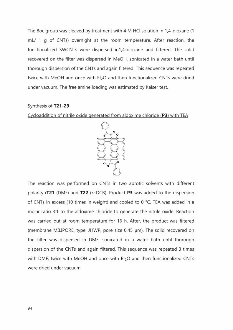
94
The Boc group was cleaved by treatment with 4 M HCl solution in 1,4-dioxane (1
mL/ 1 g of CNTs) overnight at the room temperature. After reaction, the
functionalized SWCNTs were dispersed in1,4-dioxane and filtered. The solid
recovered on the filter was dispersed in MeOH, sonicated in a water bath until
thorough dispersion of the CNTs and again filtered. This sequence was repeated
twice with MeOH and once with Et2O and then functionalized CNTs were dried
under vacuum. The free amine loading was estimated by Kaiser test.
Synthesis of T21-29
Cycloaddition of nitrile oxide generated from aldoxime chloride (P3) with TEA
The reaction was performed on CNTs in two aprotic solvents with different
polarity (T21 (DMF) and T22 (o-DCB). Product P3 was added to the dispersion
of CNTs in excess (10 times in weight) and cooled to 0 °C. TEA was added in a
molar ratio 3:1 to the aldoxime chloride to generate the nitrile oxide. Reaction
was carried out at room temperature for 16 h. After, the product was filtered
(membrane MILIPORE, type: JHWP, pore size 0.45 μm). The solid recovered on
the filter was dispersed in DMF, sonicated in a water bath until thorough
dispersion of the CNTs and again filtered. This sequence was repeated 3 times
with DMF, twice with MeOH and once with Et2O and then functionalized CNTs
were dried under vacuum.
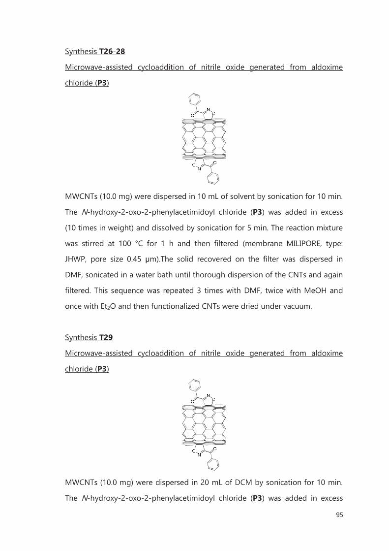
95
Synthesis T26-28
Microwave-assisted cycloaddition of nitrile oxide generated from aldoxime
chloride (P3)
MWCNTs (10.0 mg) were dispersed in 10 mL of solvent by sonication for 10 min.
The N-hydroxy-2-oxo-2-phenylacetimidoyl chloride (P3) was added in excess
(10 times in weight) and dissolved by sonication for 5 min. The reaction mixture
was stirred at 100 °C for 1 h and then filtered (membrane MILIPORE, type:
JHWP, pore size 0.45 μm).The solid recovered on the filter was dispersed in
DMF, sonicated in a water bath until thorough dispersion of the CNTs and again
filtered. This sequence was repeated 3 times with DMF, twice with MeOH and
once with Et2O and then functionalized CNTs were dried under vacuum.
Synthesis T29
Microwave-assisted cycloaddition of nitrile oxide generated from aldoxime
chloride (P3)
MWCNTs (10.0 mg) were dispersed in 20 mL of DCM by sonication for 10 min.
The N-hydroxy-2-oxo-2-phenylacetimidoyl chloride (P3) was added in excess
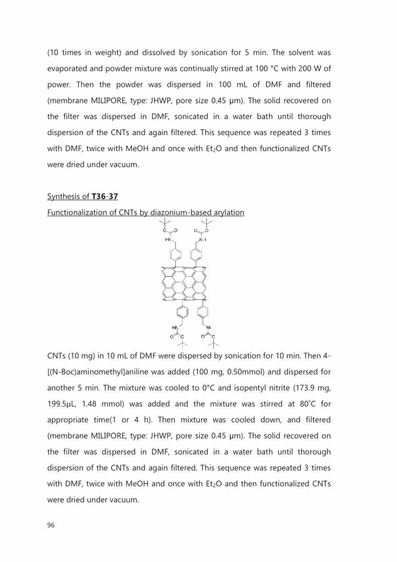
96
(10 times in weight) and dissolved by sonication for 5 min. The solvent was
evaporated and powder mixture was continually stirred at 100 °C with 200 W of
power. Then the powder was dispersed in 100 mL of DMF and filtered
(membrane MILIPORE, type: JHWP, pore size 0.45 μm). The solid recovered on
the filter was dispersed in DMF, sonicated in a water bath until thorough
dispersion of the CNTs and again filtered. This sequence was repeated 3 times
with DMF, twice with MeOH and once with Et2O and then functionalized CNTs
were dried under vacuum.
Synthesis of T36-37
Functionalization of CNTs by diazonium-based arylation
CNTs (10 mg) in 10 mL of DMF were dispersed by sonication for 10 min. Then 4-
[(N-Boc)aminomethyl]aniline was added (100 mg, 0.50mmol) and dispersed for
another 5 min. The mixture was cooled to 0°C and isopentyl nitrite (173.9 mg,
199.5µL, 1.48 mmol) was added and the mixture was stirred at 80˚C for
appropriate time(1 or 4 h). Then mixture was cooled down, and filtered
(membrane MILIPORE, type: JHWP, pore size 0.45 μm). The solid recovered on
the filter was dispersed in DMF, sonicated in a water bath until thorough
dispersion of the CNTs and again filtered. This sequence was repeated 3 times
with DMF, twice with MeOH and once with Et2O and then functionalized CNTs
were dried under vacuum.
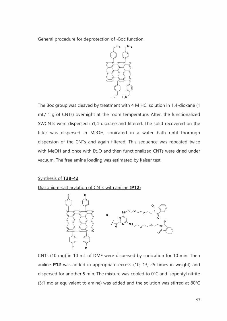
97
General procedure for deprotection of -Boc function
The Boc group was cleaved by treatment with 4 M HCl solution in 1,4-dioxane (1
mL/ 1 g of CNTs) overnight at the room temperature. After, the functionalized
SWCNTs were dispersed in1,4-dioxane and filtered. The solid recovered on the
filter was dispersed in MeOH, sonicated in a water bath until thorough
dispersion of the CNTs and again filtered. This sequence was repeated twice
with MeOH and once with Et2O and then functionalized CNTs were dried under
vacuum. The free amine loading was estimated by Kaiser test.
Synthesis of T38-42
Diazonium-salt arylation of CNTs with aniline (P12)
CNTs (10 mg) in 10 mL of DMF were dispersed by sonication for 10 min. Then
aniline P12 was added in appropriate excess (10, 13, 25 times in weight) and
dispersed for another 5 min. The mixture was cooled to 0°C and isopentyl nitrite
(3:1 molar equivalent to amine) was added and the solution was stirred at 80°C
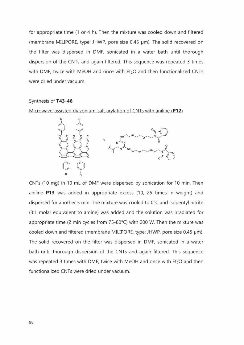
98
for appropriate time (1 or 4 h). Then the mixture was cooled down and filtered
(membrane MILIPORE, type: JHWP, pore size 0.45 μm). The solid recovered on
the filter was dispersed in DMF, sonicated in a water bath until thorough
dispersion of the CNTs and again filtered. This sequence was repeated 3 times
with DMF, twice with MeOH and once with Et2O and then functionalized CNTs
were dried under vacuum.
Synthesis of T43-46
Microwave-assisted diazonium-salt arylation of CNTs with aniline (P12)
N
N
N
NH
NH
OO
OO N
O
O
N
O
O
R
R
NH
R
R
R
CNTs (10 mg) in 10 mL of DMF were dispersed by sonication for 10 min. Then
aniline P13 was added in appropriate excess (10, 25 times in weight) and
dispersed for another 5 min. The mixture was cooled to 0°C and isopentyl nitrite
(3:1 molar equivalent to amine) was added and the solution was irradiated for
appropriate time (2 min cycles from 75-80°C) with 200 W. Then the mixture was
cooled down and filtered (membrane MILIPORE, type: JHWP, pore size 0.45 μm).
The solid recovered on the filter was dispersed in DMF, sonicated in a water
bath until thorough dispersion of the CNTs and again filtered. This sequence
was repeated 3 times with DMF, twice with MeOH and once with Et2O and then
functionalized CNTs were dried under vacuum.
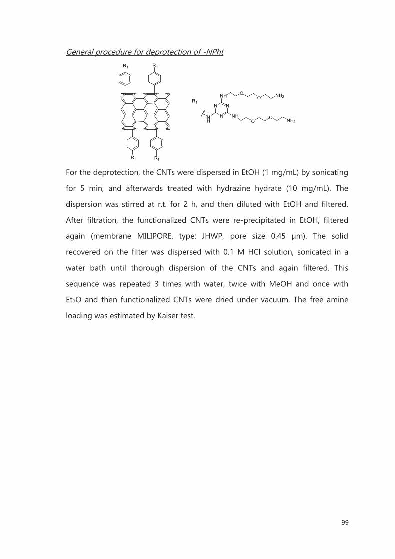
99
General procedure for deprotection of -NPht
N
N
N
NH
NH
OO
OO NH2
NH2
R1
R1
NH
R1
R1
R1
For the deprotection, the CNTs were dispersed in EtOH (1 mg/mL) by sonicating
for 5 min, and afterwards treated with hydrazine hydrate (10 mg/mL). The
dispersion was stirred at r.t. for 2 h, and then diluted with EtOH and filtered.
After filtration, the functionalized CNTs were re-precipitated in EtOH, filtered
again (membrane MILIPORE, type: JHWP, pore size 0.45 µm). The solid
recovered on the filter was dispersed with 0.1 M HCl solution, sonicated in a
water bath until thorough dispersion of the CNTs and again filtered. This
sequence was repeated 3 times with water, twice with MeOH and once with
Et2O and then functionalized CNTs were dried under vacuum. The free amine
loading was estimated by Kaiser test.
100
Bibliography
1 V. Georgakilas, K. Kordatos, M. Prato, D. M. Guldi, M. Holzinger, A. Hirsch; J. Am. Chem. Soc.
2002, 124, 760–761. 2 G. Pandey, P. Banerjee, S. R. Gadre; Chem. Rev. 2006, 106, 4484–4517. 3 R. Huisgen, W. Scheer; Tetrahedron Lett. 1971, 12, 481–484.
4 F. G. Brunetti, M. A. Herrero, J. M. Muñoz, S. Giordani, A. Díaz-Ortiz, S. Filippone, G. Ruaro, M.
Meneghetti, Maurizio Prato, and Ester Vázquez; J. Am. Chem. Soc. 2007, 129, 14580–14581. 5 S. Roy, T. L. S. Kishbaugh, J. P. Jasinski, G. Gribble; Tetrahedron Lett. 2007, 48, 1313–1316. 6 V. Georgakilas, K. Kordatos, M. Prato. D. M. Guldi, M. Holzinger, A. Hirsch; J. Am. Chem. Soc.
2002, 124, 760–761. 7 Y. Wang, Z. Iqbal, S. Mitra; Carbon 2005, 43, 1015–1020. 8 J. Li, H. Grennberg; Chem. Eur. J. 2006, 12, 386–3875. 9 Vedejs, E., In Advances in Cycloaddition, Curran, D. P., Ed.; JAI Press: London, 1988.
10 E. Laborde; Tetrahedron Lett. 1992, 33, 6607–6610. 11 X. Zhang, M. Willems, C. S. Foote; Tetrahedron Lett. 1993, 34, 8187–8188. 12 Z. Li, L. Zheng, W. Yan, Z. Pan, S. Wei; Chem. Phys. Chem. 2009, 10, 2296–2304. 13 V. Datsyuk, M. Kalyva, K. Papagelis, J. Parthenios, D. Tasis, A. Siokou, I. Kallitsis, C. Galiotis;
Carbon 2008, 46, 833–840. 14 S. Lee, S. Diab, P. Queval, M. Sebban, I. Chataigner, S. R. Piettre; Chem. Eur. J. 2013, 19, 7181 –
7192. 15 J. Zhang, H. Zou, Q. Qing, Y. Yang, Q. Li, Z. Liu, X. Guo, Z. Du; J. Phys. Chem. B 2003, 107, 3712–
3718. 16 M L. M.alard, M. A. Pimenta, G. Dresselhaus, M. S. Dresselhaus; Phys. Rep. 2009, 473, 51. 17 K.B.G. Torssell, Nitrile Oxides, Nitrones and Nitronates in Organic Synthesis, VCH Verlag
GmbH, New York 1988 18 J. J. Talley, D. L. Brown, J. S. Carter, M. J. Graneto, C. M. Koboldt, J. L. Masferrer, W. E. Perkins, R.
S. Rogers, A. F. Shaffer, Y. Y. Zhang, B. S. Zweifel, K. Seibert; J. Med. Chem., 2000, 43, 775–777. 19 D. J Burkhart, B. Twamley, N. R Natale; Tetrahedron Lett. 2001, 42, 8415–8418.
20 V. Librando, U. Chiacchio, A. Corsaro, G. Gumina; Polycydic Aromatic Compounds 1996, 11,
313–316. 21 M. Alvaro, P. Atienzar, P. de la Cruz,J. L. Delgado, V.Troiani, H. Garcia, F. Langa, A. Palkar, L.
Echegoyen; J. Am. Chem. Soc. 2006, 128, 6626–6635. 22 M. Popławska, G.Z. Żukowska, S. Cudziło, M. Bystrzejewski; Carbon 2010, 48, 1318–1320.
101
23 R. K. Saini, I. W. Chiang, H. Peng, R. E. Smalley, W. E. Billups, R. H. Hauge, J. L. Margrave; J. Am.
Chem. Soc. 2003, 125, 3617–3621. 24 A. C. Ferrari, J. Robertson; J. Phys. Rev. B 2000, 61, 14095–14107. 25 De-Q. Yang, J.-F. Rochette, E. Sacher; Langmuir 2005, 21, 8539–8545. 26 G. Giambastiani, S. Cicchi, A. Giannasi, L. Luconi, A. Rossin, F. Mercuri, C. Bianchini, A. Brandi,
M. Melucci, G. Ghini, P. Stagnaro, L. Conzatti, E. Passaglia, M. Zoppi, T. Montini, P. Fornasiero;
Chem. Mater. 2011, 23, 1923–1938. 27 G. Ghini,a L. Luconi, A. Rossin, C. Bianchini, G. Giambastiani, S. Cicchi, L. Lascialfari, A. Brandi, A.
Giannasic; Chem. Commun. 2010, 46, 252–254. 28 K.-C. Liu, B. R. Shelton, R. K. Howe; J. Org. Chem. 1980, 45, 3916–3918. 29 R. Graupner; Raman Spectrosc. 2007, 38, 673–683. 30 J. L. Bahr, J. Yang, D. V. Kosynkin, M. J. Bronikowski, R. E. Smalley, J. M. Tour; J. Am. Chem. Soc.
2001, 123, 6536–6542.
31 J. L. Bahr, J. M. Tour; Chem. Mater. 2001, 13, 3823–3824. 32 B. K. Price, J. L. Hudson, J. M. Tour; J. Am. Chem. Soc. 2005, 127, 14867–14870. 33 B. K. Price, J. M. Tour; J. Am. Chem. Soc. 2006, 128, 12899–12904. 34 J. J. Stephenson, J. L. Hudson, S. Azad, J. M. Tour; Chem. Mater. 2006, 18, 374–377. 35 X. Chen, J. Wang, W. Zhong, T. Feng, X. Yang, J. Chen; Macromol. Chem. Phys. 2008, 209, 846–
853. 36 J. Mateos-Gil, L. Rodríguez-Pérez, M. Moreno Oliva, G. Katsukis, C. Romero-Nieto, M. Á.
Herranz, D. M. Guldi, N. Martín; Nanoscale, 2015, 7, 1193–1200.
37 J. L. Hudson, H. Jian, A. D. Leonard, J. J. Stephenson, J. M. Tour; Chem. Mater. 2006, 18, 2766–
2770. 38 J. Liu, M. Rodriguez i Zubiri, B. Vigolo, M. Dossot, B. Humbert, Y. Fort, E. McRae; J. Nanosci.
Nanotechnol. 2007, 7, 3519–3523. 39 M. Burghard, A. Maroto, K. Balasubramanian, T. Assmus, A. Forment-Aliaga, E. J. H. Lee, R. T.
Weitz, M. Scolari, F. Nan, A. Mews, K. Kern; Phys. Stat. Sol. B 2007, 244, 4021–4025. 40 V. Biju, T. Itoh, Y. Makita, M. Ishikawa; J. Photochem. Photobiol. A: Chem. 2006, 183, 315–321. 41 R. A. Graff, T. M. Swanson, M. S. Strano; Chem. Mater. 2008, 20, 1824–1829.
42 M. S. Strano, C. A. Dyke, M. L. Usrey, P. W. Barone, M. J. Allen, H. W. Shan, C. Kittrell, R. H.
Hauge, J. M. Tour, R. E. Smalley; Science 2003, 301, 1519–22. 43 M. R. Heinrich; Chem. Eur. J. 2009, 15, 820–833. 44 K. Kordatos, T. Da Ros, S. Bosi, E. Vázquez, M. Bergamin, C. Cusan, F. Pellarini, V. Tomberli, B.
Baiti, D. Pantarotto, V. Georgakilas, G. Spalluto, and M. Prato; J. Org. Chem. 2001, 66, 4915–4920.

102
45 J. Yua, M. Lu; Adv. Synth. Catal. 2015, 357, 1175–1180. 46 W. Wu, S. Wieckowski, G. Pastorin, M. Benincasa, C. Klumpp, J. P. Briand, R. Gennaro, M. Prato,
A. Bianco; Angew. Chem. Int. Ed. Engl. 2005, 44, 6358–6362 47 V. Datsyuk, M. Kalyva, K. Papagelis, J. Parthenios, D. Tasis, A. Siokou, I. Kallitsis, C. Galiotis;
Carbon 2008, 46, 833–840. 48 B. Ballesteros, G. Tobias, L. Shao, E. Pellicer, J. Nogués, E. Mendoza, M. L.H. Green; Small 2008,
4, 1501–1506.
103
III. Functionalization of filled CNTs for
radioactivity delivery
III.1 Introduction
Among all carbon nanostructures there is a growing interest to combine
carbon nanotubes with foreign materials to explore their properties in potential
biological application. Taking advantage of CNTs’ ability to cross many
biological barriers with a small cytotoxic effect, the internal part of CNTs
provides an excellent host matrix for several therapeutic agents. Filling of both
SWCNTs and MWCNTs were reported in the literature1 and the activity of such
system in a biological media was reported, for the first time, on SWCNTs filled
with NaI. Hong et al. presented single-walled CNTs filled with Na125I (Auger and
γ-emitter), functionalized and glycosylated for in vitro and in vivo study (Figure
1).2
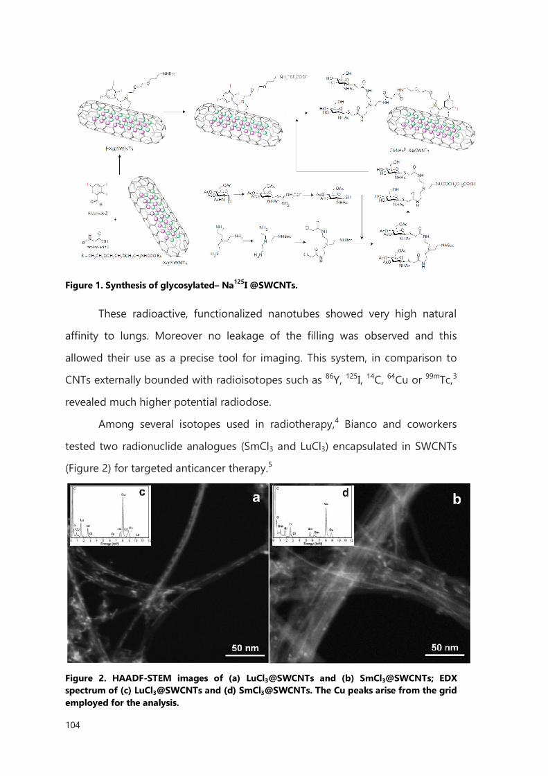
104
Figure 1. Synthesis of glycosylated– Na125I @SWCNTs.
These radioactive, functionalized nanotubes showed very high natural
affinity to lungs. Moreover no leakage of the filling was observed and this
allowed their use as a precise tool for imaging. This system, in comparison to
CNTs externally bounded with radioisotopes such as 86Y, 125I, 14C, 64Cu or 99mTc,3
revealed much higher potential radiodose.
Among several isotopes used in radiotherapy,4 Bianco and coworkers
tested two radionuclide analogues (SmCl3 and LuCl3) encapsulated in SWCNTs
(Figure 2) for targeted anticancer therapy.5
Figure 2. HAADF-STEM images of (a) LuCl3@SWCNTs and (b) SmCl3@SWCNTs; EDX spectrum of (c) LuCl3@SWCNTs and (d) SmCl3@SWCNTs. The Cu peaks arise from the grid employed for the analysis.
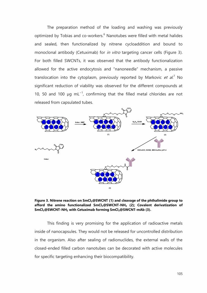
105
The preparation method of the loading and washing was previously
optimized by Tobias and co-workers.6 Nanotubes were filled with metal halides
and sealed, then functionalized by nitrene cycloaddition and bound to
monoclonal antibody (Cetuximab) for in vitro targeting cancer cells (Figure 3).
For both filled SWCNTs, it was observed that the antibody functionalization
allowed for the active endocytosis and “nanoneedle” mechanism, a passive
translocation into the cytoplasm, previously reported by Markovic et al.7 No
significant reduction of viability was observed for the different compounds at
10, 50 and 100 µg mL−1, confirming that the filled metal chlorides are not
released from capsulated tubes.
NO
ON
O
O
N3O
O N
O
O
linker
linker, NMP
NO
ONH2
N2H4, EtOH
N O OHN
O
EDCxHCl, NHSS, MES buffer pH7.4
(1) (2)
(3)
Figure 3. Nitrene reaction on SmCl3@SWCNT (1) and cleavage of the phthalimide group to afford the amino functionalized SmCl3@SWCNT-NH2 (2); Covalent derivatization of SmCl3@SWCNT-NH2 with Cetuximab forming SmCl3@SWCNT-mAb (3).
This finding is very promising for the application of radioactive metals
inside of nanocapsules. They would not be released for uncontrolled distribution
in the organism. Also after sealing of radionuclides, the external walls of the
closed-ended filled carbon nanotubes can be decorated with active molecules
for specific targeting enhancing their biocompatibility.
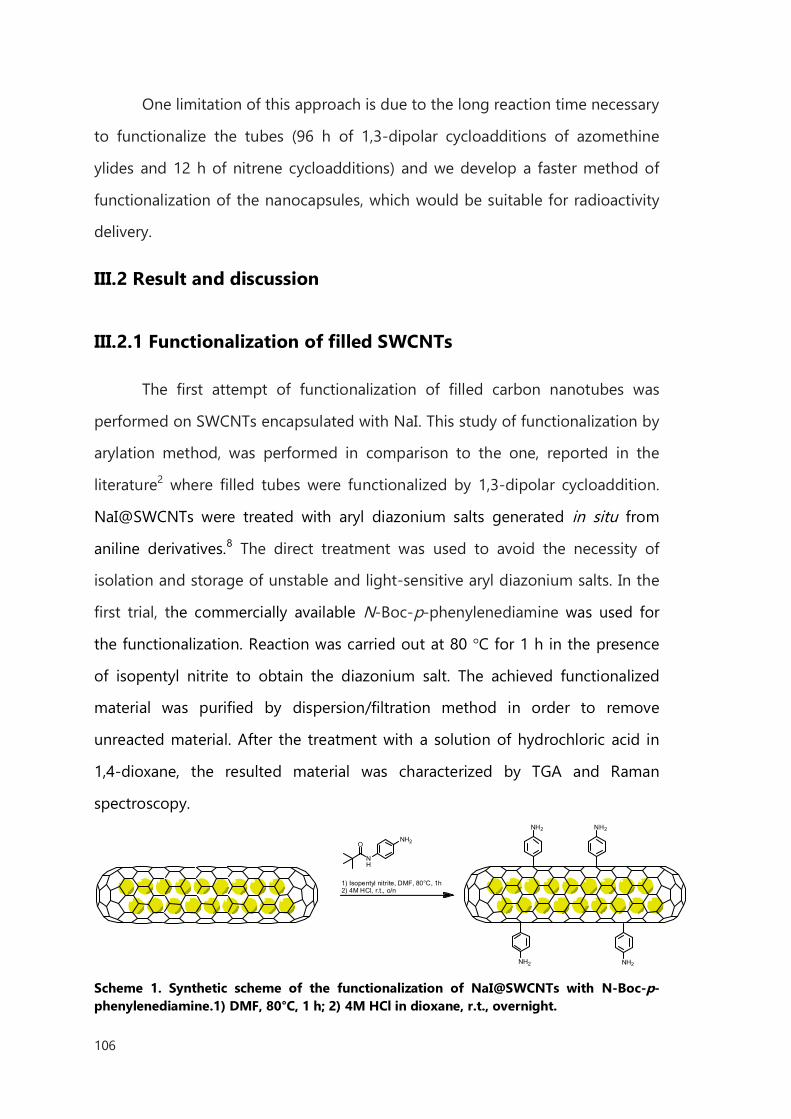
106
One limitation of this approach is due to the long reaction time necessary
to functionalize the tubes (96 h of 1,3-dipolar cycloadditions of azomethine
ylides and 12 h of nitrene cycloadditions) and we develop a faster method of
functionalization of the nanocapsules, which would be suitable for radioactivity
delivery.
III.2 Result and discussion
III.2.1 Functionalization of filled SWCNTs
The first attempt of functionalization of filled carbon nanotubes was
performed on SWCNTs encapsulated with NaI. This study of functionalization by
arylation method, was performed in comparison to the one, reported in the
literature2 where filled tubes were functionalized by 1,3-dipolar cycloaddition.
NaI@SWCNTs were treated with aryl diazonium salts generated in situ from
aniline derivatives.8 The direct treatment was used to avoid the necessity of
isolation and storage of unstable and light-sensitive aryl diazonium salts. In the
first trial, the commercially available N-Boc-p-phenylenediamine was used for
the functionalization. Reaction was carried out at 80 °C for 1 h in the presence
of isopentyl nitrite to obtain the diazonium salt. The achieved functionalized
material was purified by dispersion/filtration method in order to remove
unreacted material. After the treatment with a solution of hydrochloric acid in
1,4-dioxane, the resulted material was characterized by TGA and Raman
spectroscopy.
NH
NH2
1) Isopentyl nitrite, DMF, 80°C, 1h2) 4M HCl, r.t., o/n
O
NH2NH2
NH2NH2
Scheme 1. Synthetic scheme of the functionalization of NaI@SWCNTs with N-Boc-p-phenylenediamine.1) DMF, 80°C, 1 h; 2) 4M HCl in dioxane, r.t., overnight.
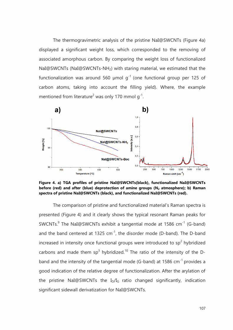
107
The thermogravimetric analysis of the pristine NaI@SWCNTs (Figure 4a)
displayed a significant weight loss, which corresponded to the removing of
associated amorphous carbon. By comparing the weight loss of functionalized
NaI@SWCNTs (NaI@SWCNTs-NH2) with staring material, we estimated that the
functionalization was around 560 µmol g-1 (one functional group per 125 of
carbon atoms, taking into account the filling yield). Where, the example
mentioned from literature2 was only 170 mmol g-1.
Figure 4. a) TGA profiles of pristine NaI@SWCNTs(black), functionalized NaI@SWCNTs before (red) and after (blue) deprotection of amine groups (N2 atmosphere); b) Raman spectra of pristine NaI@SWCNTs (black), and functionalized NaI@SWCNTs (red).
The comparison of pristine and functionalized material’s Raman spectra is
presented (Figure 4) and it clearly shows the typical resonant Raman peaks for
SWCNTs.9 The NaI@SWCNTs exhibit a tangential mode at 1586 cm-1 (G-band)
and the band centered at 1325 cm-1, the disorder mode (D-band). The D-band
increased in intensity once functional groups were introduced to sp2 hybridized
carbons and made them sp3 hybridized.10 The ratio of the intensity of the D-
band and the intensity of the tangential mode (G-band) at 1586 cm-1 provides a
good indication of the relative degree of functionalization. After the arylation of
the pristine NaI@SWCNTs the ID/IG ratio changed significantly, indication
significant sidewall derivatization for NaI@SWCNTs.
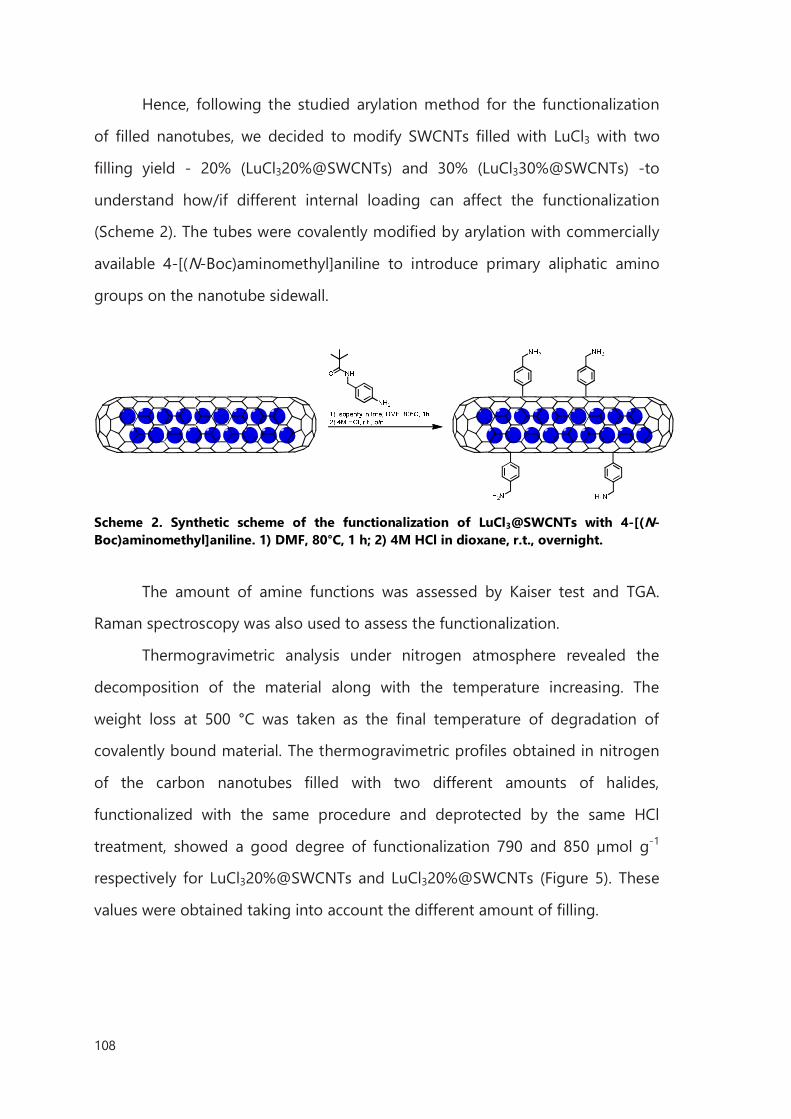
108
Hence, following the studied arylation method for the functionalization
of filled nanotubes, we decided to modify SWCNTs filled with LuCl3 with two
filling yield - 20% (LuCl320%@SWCNTs) and 30% (LuCl330%@SWCNTs) -to
understand how/if different internal loading can affect the functionalization
(Scheme 2). The tubes were covalently modified by arylation with commercially
available 4-[(N-Boc)aminomethyl]aniline to introduce primary aliphatic amino
groups on the nanotube sidewall.
Scheme 2. Synthetic scheme of the functionalization of LuCl3@SWCNTs with 4-[(N-Boc)aminomethyl]aniline. 1) DMF, 80°C, 1 h; 2) 4M HCl in dioxane, r.t., overnight.
The amount of amine functions was assessed by Kaiser test and TGA.
Raman spectroscopy was also used to assess the functionalization.
Thermogravimetric analysis under nitrogen atmosphere revealed the
decomposition of the material along with the temperature increasing. The
weight loss at 500 °C was taken as the final temperature of degradation of
covalently bound material. The thermogravimetric profiles obtained in nitrogen
of the carbon nanotubes filled with two different amounts of halides,
functionalized with the same procedure and deprotected by the same HCl
treatment, showed a good degree of functionalization 790 and 850 µmol g-1
respectively for LuCl320%@SWCNTs and LuCl320%@SWCNTs (Figure 5). These
values were obtained taking into account the different amount of filling.
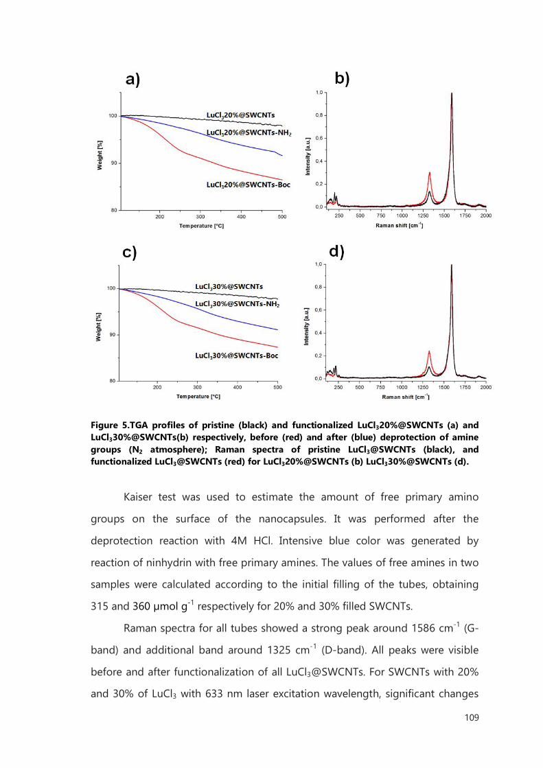
109
Figure 5.TGA profiles of pristine (black) and functionalized LuCl320%@SWCNTs (a) and LuCl330%@SWCNTs(b) respectively, before (red) and after (blue) deprotection of amine groups (N2 atmosphere); Raman spectra of pristine LuCl3@SWCNTs (black), and functionalized LuCl3@SWCNTs (red) for LuCl320%@SWCNTs (b) LuCl330%@SWCNTs (d).
Kaiser test was used to estimate the amount of free primary amino
groups on the surface of the nanocapsules. It was performed after the
deprotection reaction with 4M HCl. Intensive blue color was generated by
reaction of ninhydrin with free primary amines. The values of free amines in two
samples were calculated according to the initial filling of the tubes, obtaining
315 and 360 µmol g-1 respectively for 20% and 30% filled SWCNTs.
Raman spectra for all tubes showed a strong peak around 1586 cm-1 (G-
band) and additional band around 1325 cm-1 (D-band). All peaks were visible
before and after functionalization of all LuCl3@SWCNTs. For SWCNTs with 20%
and 30% of LuCl3 with 633 nm laser excitation wavelength, significant changes
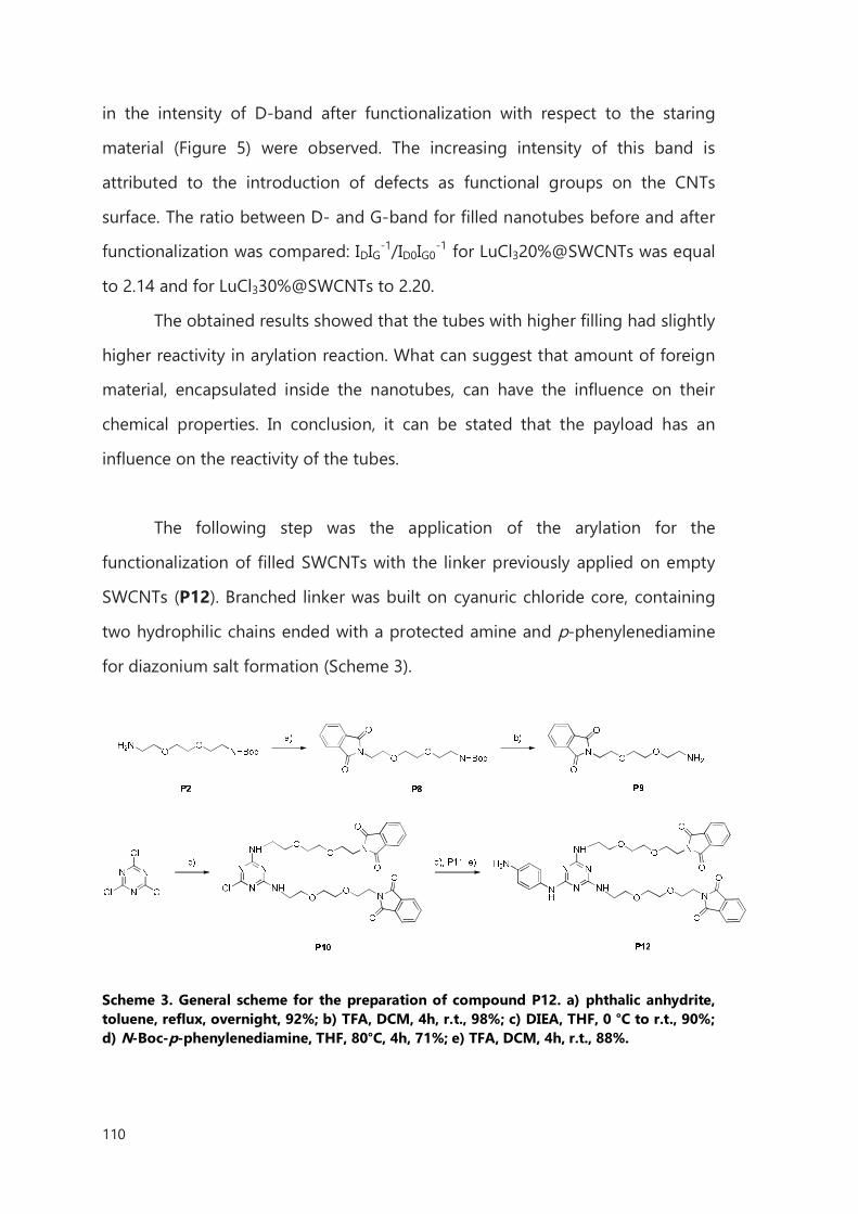
110
in the intensity of D-band after functionalization with respect to the staring
material (Figure 5) were observed. The increasing intensity of this band is
attributed to the introduction of defects as functional groups on the CNTs
surface. The ratio between D- and G-band for filled nanotubes before and after
functionalization was compared: IDIG-1/ID0IG0-1 for LuCl320%@SWCNTs was equal
to 2.14 and for LuCl330%@SWCNTs to 2.20.
The obtained results showed that the tubes with higher filling had slightly
higher reactivity in arylation reaction. What can suggest that amount of foreign
material, encapsulated inside the nanotubes, can have the influence on their
chemical properties. In conclusion, it can be stated that the payload has an
influence on the reactivity of the tubes.
The following step was the application of the arylation for the
functionalization of filled SWCNTs with the linker previously applied on empty
SWCNTs (P12). Branched linker was built on cyanuric chloride core, containing
two hydrophilic chains ended with a protected amine and p-phenylenediamine
for diazonium salt formation (Scheme 3).
Scheme 3. General scheme for the preparation of compound P12. a) phthalic anhydrite, toluene, reflux, overnight, 92%; b) TFA, DCM, 4h, r.t., 98%; c) DIEA, THF, 0 °C to r.t., 90%; d) N-Boc-p-phenylenediamine, THF, 80°C, 4h, 71%; e) TFA, DCM, 4h, r.t., 88%.
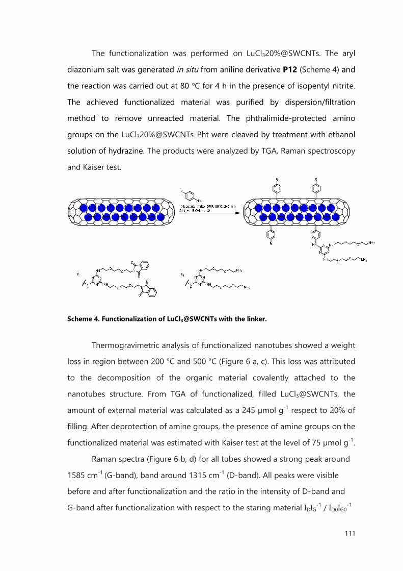
111
The functionalization was performed on LuCl320%@SWCNTs. The aryl
diazonium salt was generated in situ from aniline derivative P12 (Scheme 4) and
the reaction was carried out at 80 °C for 4 h in the presence of isopentyl nitrite.
The achieved functionalized material was purified by dispersion/filtration
method to remove unreacted material. The phthalimide-protected amino
groups on the LuCl320%@SWCNTs-Pht were cleaved by treatment with ethanol
solution of hydrazine. The products were analyzed by TGA, Raman spectroscopy
and Kaiser test.
Scheme 4. Functionalization of LuCl3@SWCNTs with the linker.
Thermogravimetric analysis of functionalized nanotubes showed a weight
loss in region between 200 °C and 500 °C (Figure 6 a, c). This loss was attributed
to the decomposition of the organic material covalently attached to the
nanotubes structure. From TGA of functionalized, filled LuCl3@SWCNTs, the
amount of external material was calculated as a 245 µmol g-1 respect to 20% of
filling. After deprotection of amine groups, the presence of amine groups on the
functionalized material was estimated with Kaiser test at the level of 75 µmol g-1.
Raman spectra (Figure 6 b, d) for all tubes showed a strong peak around
1585 cm-1 (G-band), band around 1315 cm-1 (D-band). All peaks were visible
before and after functionalization and the ratio in the intensity of D-band and
G-band after functionalization with respect to the staring material IDIG-1 / ID0IG0-1

112
(Figure 6c) was calculated as a 1.21.
Figure 6.TGA profiles of pristine (black) and functionalized filled SWCNTs, before and after deprotection of amine groups for a)LuCl3@SWCNTs c) SmCl3@SWCNTs (N2 atmosphere); Raman spectra of pristine (black), and functionalized (red) filled SWCNTs with b) LuCl3, d) SmCl3;.
The same experiment was performed also on SWCNTs filled with
samarium (III) chloride (SmCl3@SWCNTs). TGA and Kaiser test values were
processed to obtain the degree of functionalization for every filled and
functionalized SWCNTs, showing a good agreement between the two filling
(Table 1).
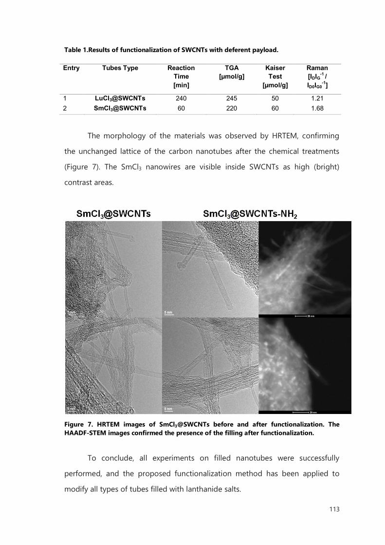
113
Table 1.Results of functionalization of SWCNTs with deferent payload.
Entry Tubes Type Reaction
Time [min]
TGA [µmol/g]
Kaiser Test
[µmol/g]
Raman [IDIG-1 / ID0IG0
-1]
1 LuCl3@SWCNTs 240 245 50 1.21 2 SmCl3@SWCNTs 60 220 60 1.68
The morphology of the materials was observed by HRTEM, confirming
the unchanged lattice of the carbon nanotubes after the chemical treatments
(Figure 7). The SmCl3 nanowires are visible inside SWCNTs as high (bright)
contrast areas.
Figure 7. HRTEM images of SmCl3@SWCNTs before and after functionalization. The HAADF-STEM images confirmed the presence of the filling after functionalization.
To conclude, all experiments on filled nanotubes were successfully
performed, and the proposed functionalization method has been applied to
modify all types of tubes filled with lanthanide salts.
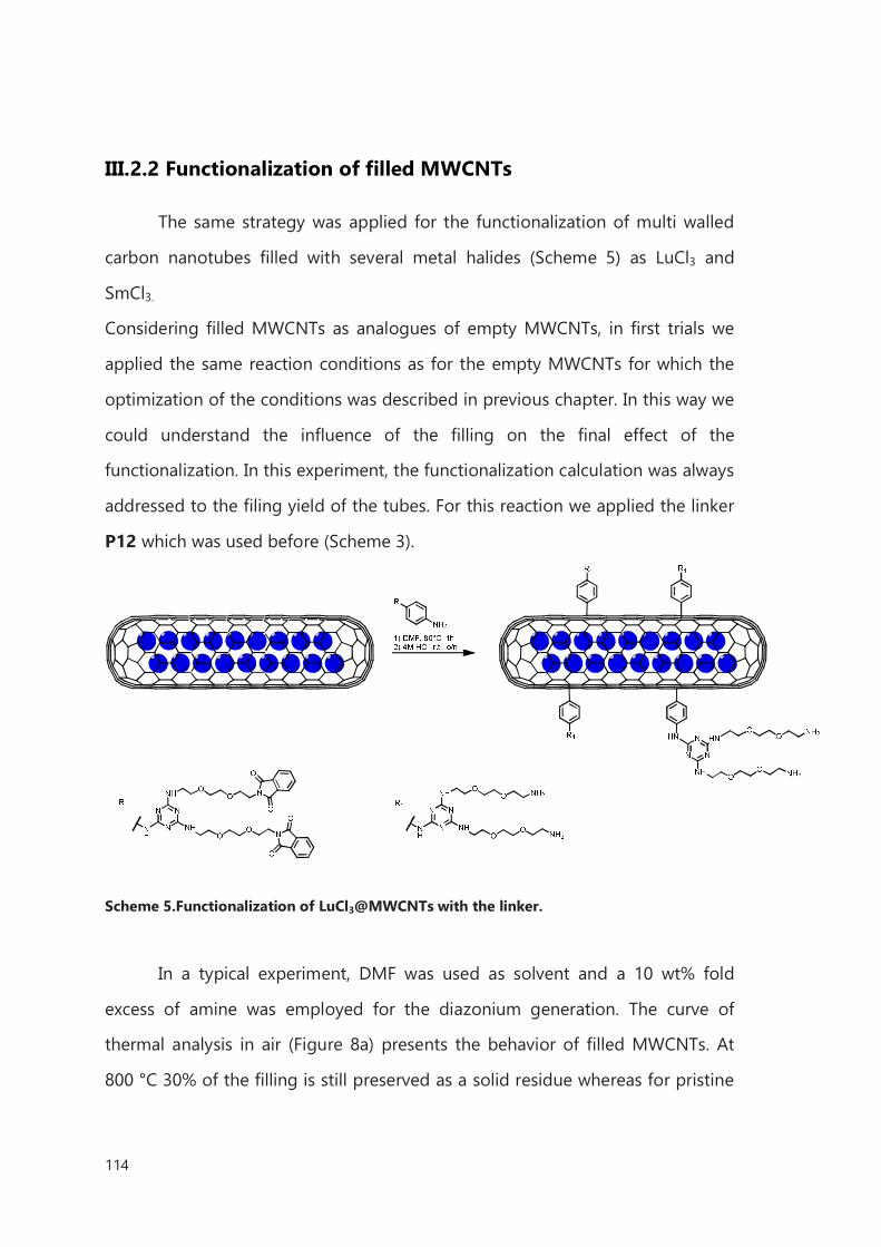
114
III.2.2 Functionalization of filled MWCNTs
The same strategy was applied for the functionalization of multi walled
carbon nanotubes filled with several metal halides (Scheme 5) as LuCl3 and
SmCl3.
Considering filled MWCNTs as analogues of empty MWCNTs, in first trials we
applied the same reaction conditions as for the empty MWCNTs for which the
optimization of the conditions was described in previous chapter. In this way we
could understand the influence of the filling on the final effect of the
functionalization. In this experiment, the functionalization calculation was always
addressed to the filing yield of the tubes. For this reaction we applied the linker
P12 which was used before (Scheme 3).
Scheme 5.Functionalization of LuCl3@MWCNTs with the linker.
In a typical experiment, DMF was used as solvent and a 10 wt% fold
excess of amine was employed for the diazonium generation. The curve of
thermal analysis in air (Figure 8a) presents the behavior of filled MWCNTs. At
800 °C 30% of the filling is still preserved as a solid residue whereas for pristine
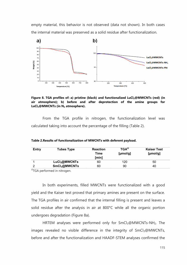
115
empty material, this behavior is not observed (data not shown). In both cases
the internal material was preserved as a solid residue after functionalization.
Figure 8. TGA profiles of: a) pristine (black) and functionalized LuCl3@MWCNTs (red) (in air atmosphere); b) before and after deprotection of the amine groups for LuCl3@MWCNTs (in N2 atmosphere).
From the TGA profile in nitrogen, the functionalization level was
calculated taking into account the percentage of the filling (Table 2).
Table 2.Results of functionalization of MWCNTs with deferent payload.
Entry Tubes Type Reaction
Time [min]
TGAa)
[µmol/g] Kaiser Test
[µmol/g]
1 LuCl3@MWCNTs 60 120 50 2 SmCl3@MWCNTs 60 90 40
a)TGA performed in nitrogen.
In both experiments, filled MWCNTs were functionalized with a good
yield and the Kaiser test proved that primary amines are present on the surface.
The TGA profiles in air confirmed that the internal filling is present and leaves a
solid residue after the analysis in air at 800°C while all the organic portion
undergoes degradation (Figure 8a).
HRTEM analyses were performed only for SmCl3@MWCNTs-NH2. The
images revealed no visible difference in the integrity of SmCl3@MWCNTs,
before and after the functionalization and HAADF-STEM analyses confirmed the
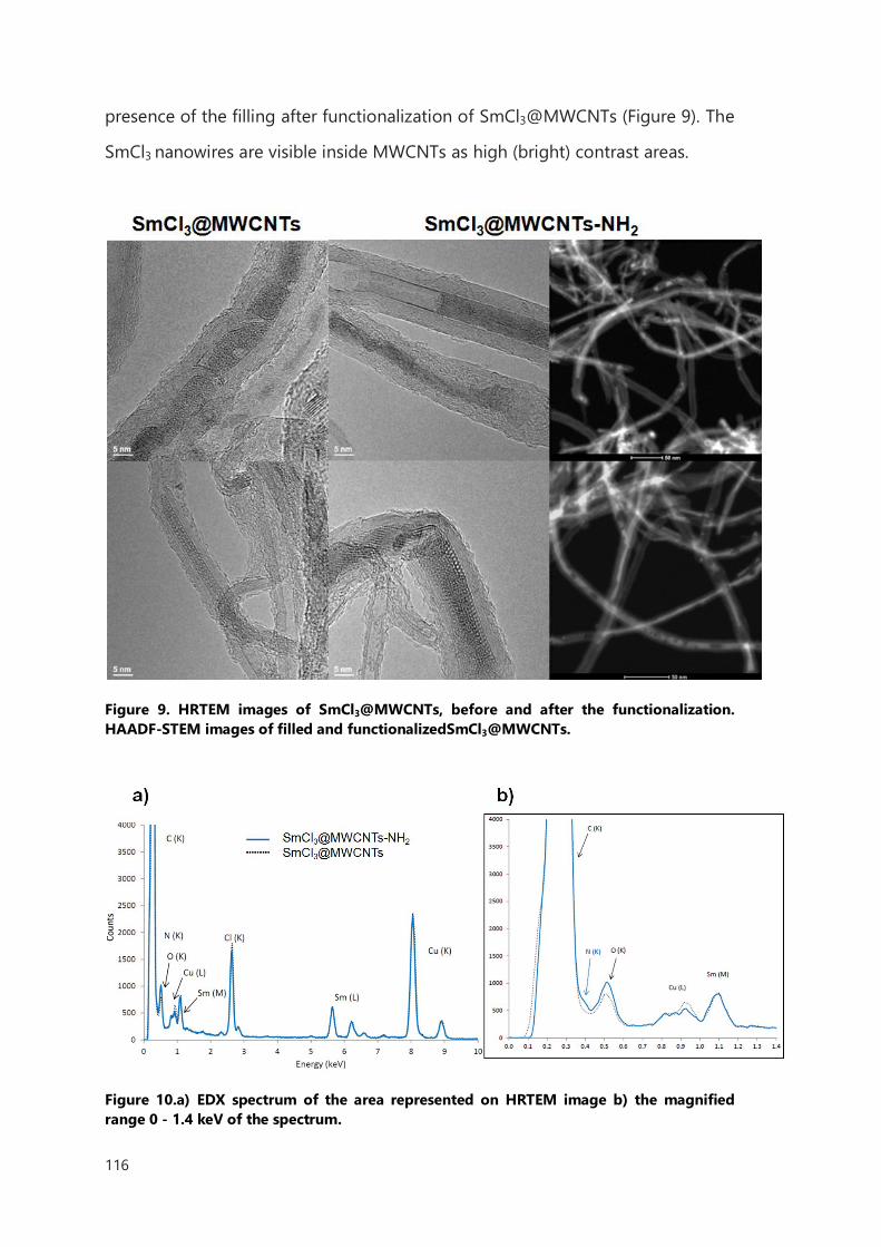
116
presence of the filling after functionalization of SmCl3@MWCNTs (Figure 9). The
SmCl3 nanowires are visible inside MWCNTs as high (bright) contrast areas.
Figure 9. HRTEM images of SmCl3@MWCNTs, before and after the functionalization. HAADF-STEM images of filled and functionalizedSmCl3@MWCNTs.
Figure 10.a) EDX spectrum of the area represented on HRTEM image b) the magnified range 0 - 1.4 keV of the spectrum.
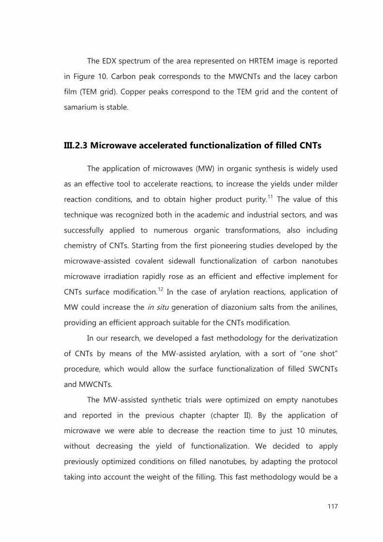
117
The EDX spectrum of the area represented on HRTEM image is reported
in Figure 10. Carbon peak corresponds to the MWCNTs and the lacey carbon
film (TEM grid). Copper peaks correspond to the TEM grid and the content of
samarium is stable.
III.2.3 Microwave accelerated functionalization of filled CNTs
The application of microwaves (MW) in organic synthesis is widely used
as an effective tool to accelerate reactions, to increase the yields under milder
reaction conditions, and to obtain higher product purity.11 The value of this
technique was recognized both in the academic and industrial sectors, and was
successfully applied to numerous organic transformations, also including
chemistry of CNTs. Starting from the first pioneering studies developed by the
microwave-assisted covalent sidewall functionalization of carbon nanotubes
microwave irradiation rapidly rose as an efficient and effective implement for
CNTs surface modification.12 In the case of arylation reactions, application of
MW could increase the in situ generation of diazonium salts from the anilines,
providing an efficient approach suitable for the CNTs modification.
In our research, we developed a fast methodology for the derivatization
of CNTs by means of the MW-assisted arylation, with a sort of “one shot”
procedure, which would allow the surface functionalization of filled SWCNTs
and MWCNTs.
The MW-assisted synthetic trials were optimized on empty nanotubes
and reported in the previous chapter (chapter II). By the application of
microwave we were able to decrease the reaction time to just 10 minutes,
without decreasing the yield of functionalization. We decided to apply
previously optimized conditions on filled nanotubes, by adapting the protocol
taking into account the weight of the filling. This fast methodology would be a
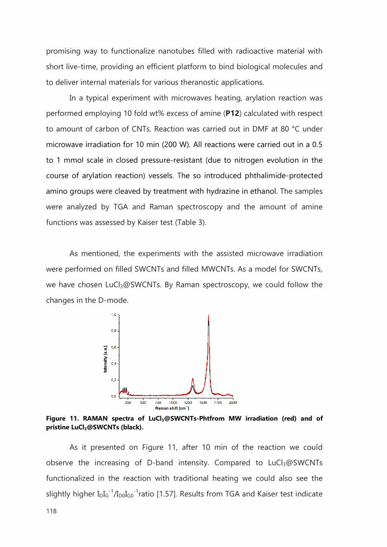
118
promising way to functionalize nanotubes filled with radioactive material with
short live-time, providing an efficient platform to bind biological molecules and
to deliver internal materials for various theranostic applications.
In a typical experiment with microwaves heating, arylation reaction was
performed employing 10 fold wt% excess of amine (P12) calculated with respect
to amount of carbon of CNTs. Reaction was carried out in DMF at 80 °C under
microwave irradiation for 10 min (200 W). All reactions were carried out in a 0.5
to 1 mmol scale in closed pressure-resistant (due to nitrogen evolution in the
course of arylation reaction) vessels. The so introduced phthalimide-protected
amino groups were cleaved by treatment with hydrazine in ethanol. The samples
were analyzed by TGA and Raman spectroscopy and the amount of amine
functions was assessed by Kaiser test (Table 3).
As mentioned, the experiments with the assisted microwave irradiation
were performed on filled SWCNTs and filled MWCNTs. As a model for SWCNTs,
we have chosen LuCl3@SWCNTs. By Raman spectroscopy, we could follow the
changes in the D-mode.
Figure 11. RAMAN spectra of LuCl3@SWCNTs-Phtfrom MW irradiation (red) and of pristine LuCl3@SWCNTs (black).
As it presented on Figure 11, after 10 min of the reaction we could
observe the increasing of D-band intensity. Compared to LuCl3@SWCNTs
functionalized in the reaction with traditional heating we could also see the
slightly higher IDIG-1/ID0IG0-1ratio [1.57]. Results from TGA and Kaiser test indicate
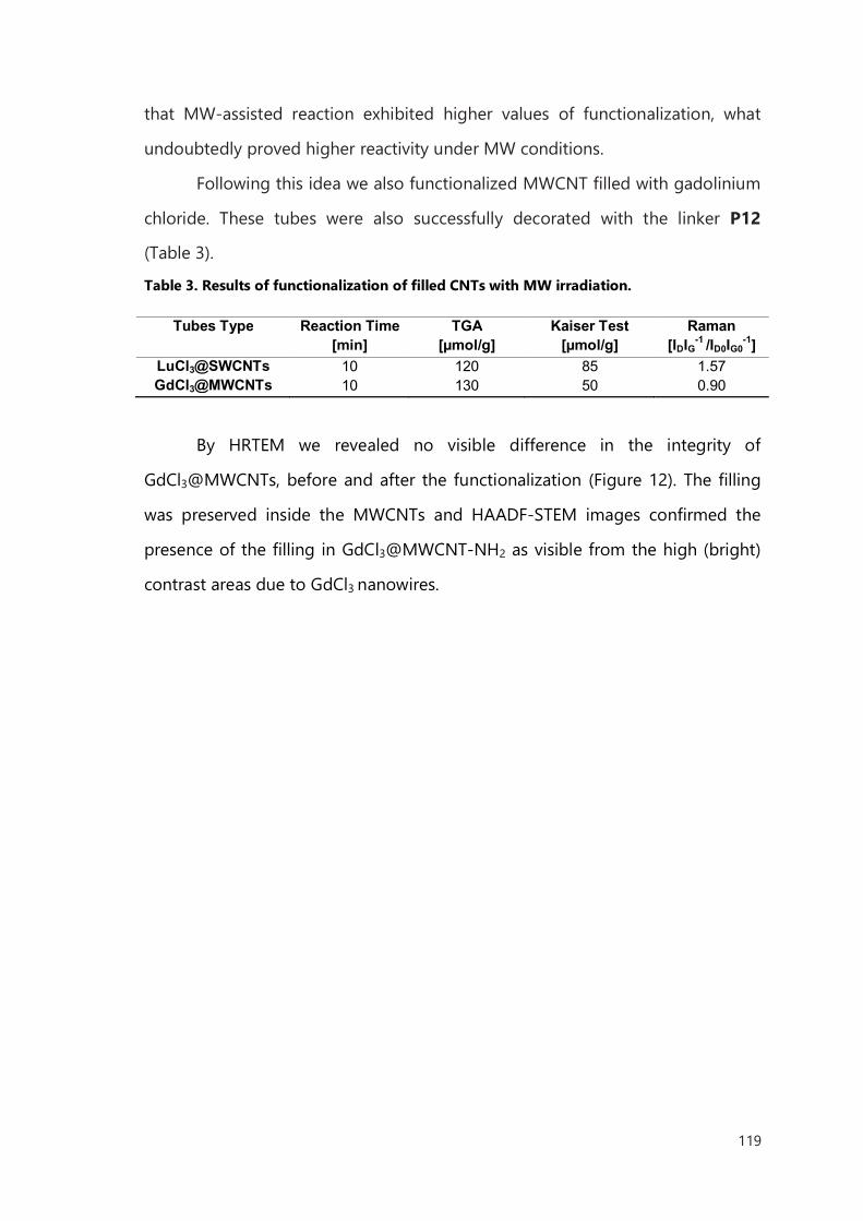
119
that MW-assisted reaction exhibited higher values of functionalization, what
undoubtedly proved higher reactivity under MW conditions.
Following this idea we also functionalized MWCNT filled with gadolinium
chloride. These tubes were also successfully decorated with the linker P12
(Table 3).
Table 3. Results of functionalization of filled CNTs with MW irradiation.
Tubes Type Reaction Time
[min] TGA
[µmol/g] Kaiser Test
[µmol/g] Raman
[IDIG-1 /ID0IG0-1]
LuCl3@SWCNTs 10 120 85 1.57 GdCl3@MWCNTs 10 130 50 0.90
By HRTEM we revealed no visible difference in the integrity of
GdCl3@MWCNTs, before and after the functionalization (Figure 12). The filling
was preserved inside the MWCNTs and HAADF-STEM images confirmed the
presence of the filling in GdCl3@MWCNT-NH2 as visible from the high (bright)
contrast areas due to GdCl3 nanowires.
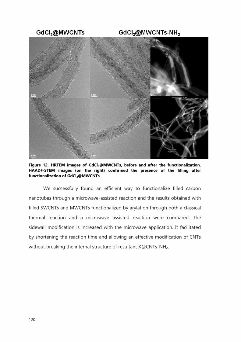
120
Figure 12. HRTEM images of GdCl3@MWCNTs, before and after the functionalization. HAADF-STEM images (on the right) confirmed the presence of the filling after functionalization of GdCl3@MWCNTs.
We successfully found an efficient way to functionalize filled carbon
nanotubes through a microwave-assisted reaction and the results obtained with
filled SWCNTs and MWCNTs functionalized by arylation through both a classical
thermal reaction and a microwave assisted reaction were compared. The
sidewall modification is increased with the microwave application. It facilitated
by shortening the reaction time and allowing an effective modification of CNTs
without breaking the internal structure of resultant X@CNTs-NH2.
121
III.3 Conclusions
The increasing application of nanotechnology in the biomedical field and
the current development of nanoscience and biomedicine are encouraging to
search for new nanoscale drug delivery systems with multiple functions and
advanced applications. The main advantage of nanocarriers is the possibility of
their versatile modification with different functionality. Indeed, multimodal
nanosystems with targeting, imaging, and drug delivery capabilities within a
single nanoscale construct, hold incredible promise in the treatment of different
types of diseases and in particular for cancer therapy. Theranostic, which
combines simultaneous diagnostics and therapeutics, represents a new modality
that utilizes therapy and real-time non-invasive in vivo imaging. This specific
combination of functions can provide biodistribution information, opportunities
to study therapeutic mechanisms, and strategies for improving therapeutic
efficacy and reducing side effects. The unique structural properties of carbon
nanotubes make this material a promising tool for nanoscience, in particular
nanomedicine.
The exploitation of different covalent functionalization reactions permits
the surface modification, which will be available for binding several cargos such
as bioactive compounds (small molecules or proteins), and the increase of CNTs
dispersibility. Following our research in this area, CNTs can be further
conjugated with therapeutic or tracking molecules. In particularly this short time
of reaction and convenience of covalent functionalization is suitable for delivery
of radioisotopes which could be closed inside the tubes. The biological study,
toxicity and first trials in vivo of such a system will be described in next chapter.
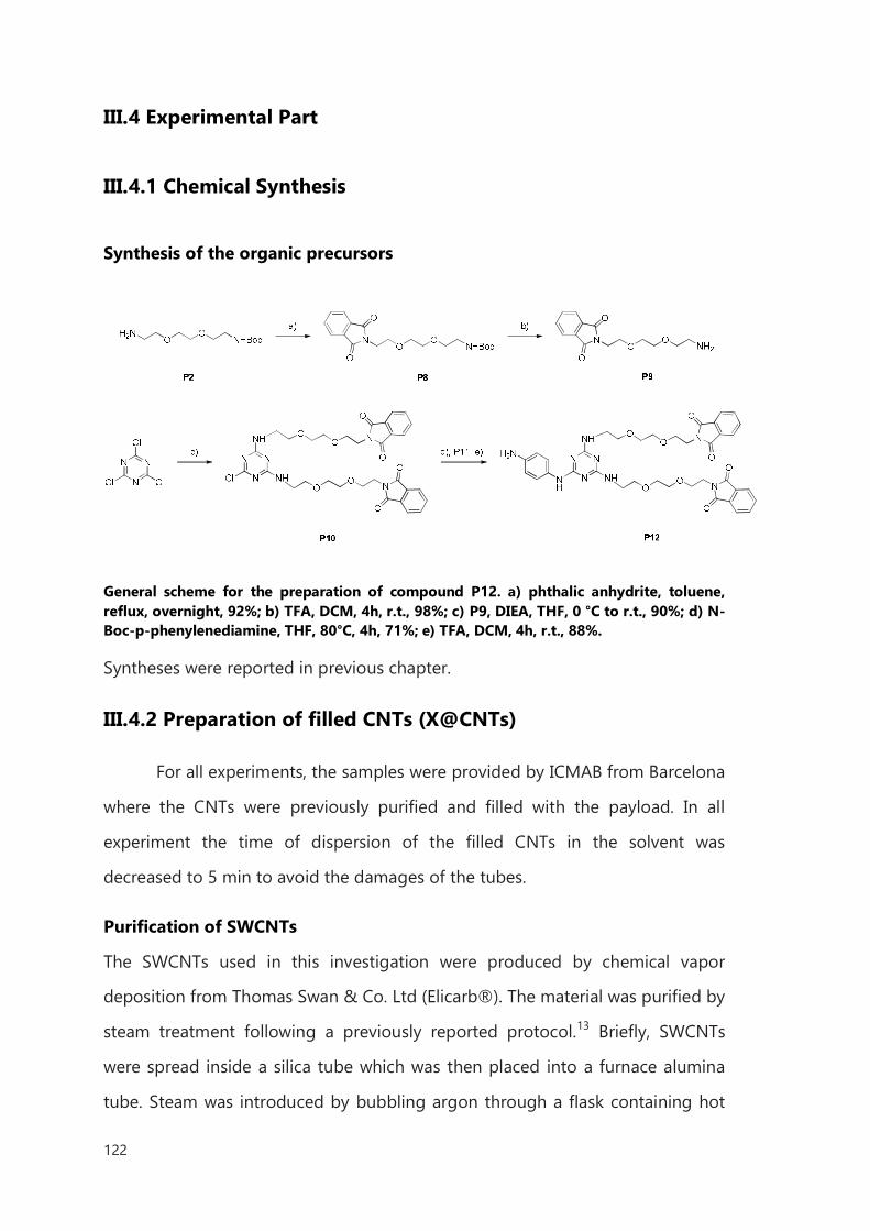
122
III.4 Experimental Part
III.4.1 Chemical Synthesis
Synthesis of the organic precursors
General scheme for the preparation of compound P12. a) phthalic anhydrite, toluene, reflux, overnight, 92%; b) TFA, DCM, 4h, r.t., 98%; c) P9, DIEA, THF, 0 °C to r.t., 90%; d) N-Boc-p-phenylenediamine, THF, 80°C, 4h, 71%; e) TFA, DCM, 4h, r.t., 88%.
Syntheses were reported in previous chapter.
III.4.2 Preparation of filled CNTs (X@CNTs)
For all experiments, the samples were provided by ICMAB from Barcelona
where the CNTs were previously purified and filled with the payload. In all
experiment the time of dispersion of the filled CNTs in the solvent was
decreased to 5 min to avoid the damages of the tubes.
Purification of SWCNTs
The SWCNTs used in this investigation were produced by chemical vapor
deposition from Thomas Swan & Co. Ltd (Elicarb®). The material was purified by
steam treatment following a previously reported protocol.13 Briefly, SWCNTs
were spread inside a silica tube which was then placed into a furnace alumina
tube. Steam was introduced by bubbling argon through a flask containing hot
123
distilled water. The whole system was initially purged with argon, to remove
atmospheric air, before heating the furnace to 900 °C during 4 h to allow the
purification of the material. The solid powder was collected and treated with 6
M HCl (Panreac) to dissolve the exposed metal nanoparticles. The purified
SWCNTs were collected by filtration through a 0.2 µm polycarbonate membrane
and thoroughly washed with water until neutral pH. The sample was oven-dried
at 80 °C overnight.
Purification of MWCNTs
The MWCNTs were purchased from Thomas Swan & Co. Ltd. As-received
MWCNTs were dispersed in fresh piranha solution (H2SO4/H2O2, 3:1) at a
concentration of 1 mg mL−1. The solution was stirred for 2 h at r.t. then the
reaction was quenched with distilled water. The pH of the solution was adjusted
until neutral. The sample was filtered on a polycarbonate membrane and dried
in the oven at 80 °C overnight. Next, the MWCNTs were treated with steam for 1
h at 900 °C and refluxed in 6 M HCl at 110 °C overnight. The solid sample was
collected by filtration and rinsed with distilled water until neutral pH.
Filling of CNTs
NaI@SWCNTs
Purified SWCNTs and aq. solution of sodium iodide were transferred into a silica
ampoule and heated to 70 °C overnight to remove water. The ampoule was
sealed under vacuum and annealed at 900 °C for 4 h. The mixture was then
washed with water to remove external material. Sample was filtered and dried to
afford NaI@SWCNTs.
LuCl3@CNTs
LuCl3 mixed with SWCNTs and grinded with an agate mortar in an argon-filled
glovebox. The mixture was sealed under vacuum inside a silica tube. The sample
was then annealed in a horizontal furnace at 960 °C during 12 h. The resulting
124
sample contained filled closed-ended nanotubes and external crystals of LuCl3.
The non-encapsulated material was removed by washing the sample in hot
water. From the performed TGA in air it was possible to determine the filling
yield of the encapsulated materials employing a reported formula (Appendix). In
the presented case, the two samples were prepared, one with about the 20 % of
filling yield (LuCl320%@SWCNTs) and 30 % (LuCl330%@SWCNTs).
SmCl3@CNTs
For encapsulation of samarium chloride, previously purified CNTs were mixed
with samarium (III) chloride (w/w 1:10 respectively) and grinded with an agate
mortar and pestle inside a glovebox until the mixture presented a
homogeneous color. The resulting mixture was then vacuum sealed inside a
silica ampoule and annealed at 900 °C for 12 h. After cooling down the sample,
the silica tube was opened and the material was stirred in water at 80 °C to
remove the non-encapsulated SmCl3. The filling yield of SmCl3@SWCNTs was
18.1 wt% and 19.5 wt% for SmCl3@MWCNTs.
GdCl3@MWCNTs
For encapsulation of gadolinium chloride previously purified MWCNTs were
then mixed with gadolinium (III) chloride (w/w 1:10 respectively) and grinded
with an agate mortar and pestle inside a glovebox until the mixture presented a
homogeneous color. The resulting mixture was then vacuum sealed inside a
silica ampoule and annealed at 900 °C for 12 h. After cooling down the sample,
the silica tube was opened and the material was stirred in water at 80 °C to
remove the non-encapsulated GdCl3 external to the CNTs.
125
III.4.3 Functionalization of CNTs
General functionalization using traditional heating functionalization
10 mg of CNTs in 5mL of DMF were dispersed by sonication for 10 min. Then
the corresponding aniline derivative was added in 25 fold wt% excess respect to
CNTs to in 5 mL of DMF was added, dispersed for another 5 min and cooled to
0°C. Isopentyl nitrite (5 fold molar excess with respect to the amine) was added
and reaction mixture was heated up to 80 °C and stirred for 1-4 hours. Then the
mixture was cooled to room temperature. The CNTs were filtered (membrane
MILIPORE, type: JHWP, pore size 0.45 µm) and washed with DMF until the
solvent eluted colorless, washed with DMF, water, MeOH, EtOAc, Et2O and dried.
General microwave-assisted functionalization
In a typical reaction of arylation, DMF dispersion of pristine filled CNTs were
loaded into a microwave vessel, along with DMF solution of amine P12 (25 fold
wt% excess respect to CNTs), cooled to 0°C and injected with isopentyl nitrite (5
fold molar excess respect to amine) for the generation of diazonium salt. The
microwave power was set to 200 W, the pressure at 125 psi, and the reaction
was carried out in ten cycles from 75 to 80 °C for 10 min in total. After cooling
to room temperature, the mixture was filtered (membrane MILIPORE, type:
JHWP, pore size 0.45 µm), washed with DMF, re-dispersed/filtered with water,
MeOH, dried with EtOAc and Et2O.
General procedure for deprotection of -Boc
For the deprotection, X@CNTs-Boc (10 mg) were dispersed in 4 M HCl solution
of dioxane (10mL) by sonication for 5 min. The dispersion was stirred at r.t. for
overnight, and then diluted with dioxane (50 mL) and filtered. After filtration, the
functionalized X@CNTs-NH2 were re-precipitated in water, filtered (membrane
MILIPORE, type: JHWP, pore size 0.45 µm), washed with water, MeOH, diethyl
126
ether finally dried under vacuum to afford functionalized material (X@CNTs-
NH2. The free amine loading was estimated by Kaiser test.
General procedure for deprotection of -NPht
For the deprotection, X@CNTs-Pht (30 mg) were dispersed in EtOH (30mL) by
sonicating for 5 min, and afterwards treated with hydrazine hydrate (3 mL). The
dispersion was stirred at r.t. for 2 h, and then diluted with EtOH (15 mL) and
filtered. After filtration, the functionalized SmCl3@MWCNTs-NH2 were re-
precipitated in EtOH, filtered(membrane MILIPORE, type: JHWP, pore size 0.45
µm), washed with 0.1 M HCl solution, water, MeOH, diethyl ether finally dried
under vacuum to afford functionalized X@CNTs (X@CNTs-NH2). The free amine
loading was estimated by Kaiser test.
127
Bibliography
1 R. Klingeler, R. B. Sim “Carbon Nanotubes for Biomedical Applications”; Springer, Berlin,
Heidelberg, 2011. 2 S. Y. Hong, G. Tobias, K. T. Al-Jamal, B. Ballesteros, H. Ali-Boucetta, S. Lozano-Perez, P. D.
Nellist, R. B. Sim, C. Finucane, S. J. Mather, M. L. H. Green, K. Kostarelos, B. G. Davis; Nature
materials 2010, 9, 485–490. 3 J. T. Wang, L. Cabana, M. Bourgognon, H. Kafa, A. Protti, K. Venner, A. M. Shah, J. Sosabowski, S.
J. Mather, A. Roig, X. Ke, G. V. Tendeloo, R. T. de Rosales, G. Tobias, K. T. Al-Jamal; Adv. Funct.
Mater. 2014, 24, 1880–1894. 4 M. K. Bakht, M. Sadeghi, M. Pourbaghi-Masouleh, C. Tenreiro; Current Cancer Drug Targets
2012, 12, 998–1015. 5 C. Spinato, A. P. Ruiz de Garibay, M. Kierkowicz, E. Pach, M. Martincic, R. Klippstein, M.
Bourgognon, J. Tzu-Wen Wang, C. Ménard-Moyon, K. T. Al-Jamal, B. Ballesteros, G. Tobias, A.
Bianco; Nanoscale 2016, Doi:10.1039/C5NR07923C. 6 M. Martincic, E. Pach, B. Ballesteros, G. Tobias; Phys. Chem. Chem. Phys. 2015, 17, 31662–
31669. 7 L. Lacerda, J. Russier, G. Pastorin, M. A. Herrero, E. Venturelli, H. Dumortier, K. T. Al-Jamal, M.
Prato, K. Kostarelos, A. Bianco; Biomaterials, 2012, 33, 3334–3343. 8 J. L. Bahr, J. M. Tour; Chem Mater 2001,13, 3823–3824. 9 R. Graupner; Journal of Raman Spectroscopy, 2007, 38, 673–683. 10 R. Sharma, J. Hyun B. Chrisantha, J. Perera, M. S. Strano; Nano Letters, 2010, 10, 398–405.
11 V. Polshettiwar, R. S. Varma; Pure Appl. Chem. 2008, 80, 777–790. 12 Y. Wang, Z. Iqbal, S. Mitra; Carbon 2005, 43, 1015–1020. 13 B. Ballesteros,G. Tobias, L. Shao, E. Pellicer, J. Nogués, E. Mendoza, M. L.H. Green; Small, 2008,
4, 1501–1506.

128
129
IV. Biomedical application of functionalized, filled
CNTs for radioactivity delivery
IV.1 Introduction
Up to now, the hypothesis of CNTs clinical administration is strongly
limited by multiple factors such as low solubility, impurity content,
reproducibility in the preparation of the pristine materials, toxicity and
pharmacokinetics, among the others.
The conjugation of pharmacological agents with nanoparticles has met
great interest from the medical point of view1 and there are many examples of
current application of these systems in clinical practice and also at various
stages of clinical development.2,3 Versatile agents attached to the particles show
improved pharmacological and toxicological properties against their non-
conjugated form. The circulating half-life of the conjugates, maximal tolerated
dose (MTD), and target selectivity are the important factors affecting the high
therapeutic index. Typically conjugates are attached to the surface of
nanoparticles, or encapsulated and protected inside the core. The nanoparticle
130
can also be designed to provide either controlled or triggered release of the
therapeutic molecule.4 Then, the surface of the particle can be properly
functionalized by various methods to increase the circulating half-life, and by
reducing nonspecific distribution in some cases by targeting tissue with specific
cell surface antigens with a specific ligand (peptide, aptamer, antibody, small
molecule). Surface functionalization can address the major limiting factor of
long-circulating nanoparticles, notably protein absorption. Proteins adsorbed on
the surface of the nanoparticle promote opsonization, leading to aggregation
with subsequent rapid clearance from the bloodstream.5 The resultant rapid
clearance is due to phagocytosis by the mononuclear phagocyte system (MPS)
in conjunction with the liver and spleen filtration network. Typically, the majority
of opsonized particles are cleared by a receptor-mediated mechanism within
minutes due to the high concentration of phagocytic cells in the liver and spleen
or alternatively they may be excreted.6 Several biological barriers are applied to
defend the human body from invasion by foreign particles. The biodistribution
of any nanoparticle is primarily ruled by their ability to penetrate biological
barriers.7 These barriers consist of: - the reticuloendothelial system (RES), -
endothelial/epithelial membranes, - complex networks of blood vessels, -
abnormal flow of blood and interstitial gradients. Endothelia composing the
blood vessels have been classified as continuous, fenestrated, or discontinuous,
depending on the morphological features and organ location.
The first in vivo study on functionalized CNTs biodistribution was
reported by Wang et al. using mice treated with intraperitoneally (i.p.)
administered short hydroxylated SWCNTs. The tubes were shown to accumulate
mainly in the liver and kidney, and, in a lower degree, in the spleen and lung,
and excreted mainly by the kidney within 18 days.8 Functionalized SWCNTs,
labeled with indium [111In] thanks to the presence of a chelating
molecule(diethylenetriaminepentaaceticacid- DTPA) on the surface were
demonstrated to not been retained in the reticuloendothelial system (liver or
131
spleen) after intravenous administration (i.v.), and were rapidly cleared from the
systemic blood circulation again via renal excretion.9 This studied was
performed tracing the indium radioactivity by gamma scintigraphy. It was also
possible to compare the biodistribution of two types of functionalization: the
first with no free amino groups and a second one with 40% free amino groups
resulting in charged surface. Both derivatized SWCNTs were found in kidney,
muscle, skin, and blood after 30 min. However the charged SWCNTs led to a
higher affinity for kidney, muscle, skin, and lung, leading to their rapid clearance
from all tissues. In 3 h the nanotubes were cleared from all organs down to
levels of 1–2% (relative to the 30 min time point). TEM analysis of urine samples
indicated high levels of intact functionalized CNTs, demonstrating that they are
rapidly cleared from the systemic circulation via the kidney.9 Later on, Lacerda et
al. presented an elimination mechanism for CNTs, using [111In] DTPA-MWCNTs.
After tail vein injection, CNTs fast distributed in the systemic blood followed by
rapid urinary clearance through via the kidney glomerular filtration (Fig. 3).10 The
reason for the more rapid elimination observed herein, in comparison to
derivatives functionalized with surfactants, is that in the latter case once in the
blood these surfactants desorbed from the CNTs with subsequent circulation of
bundles and accumulation in the liver tissue. It is important to note that [111In]
DTPA-MWCNTs complexes used in this study were considerably longer than the
dimensions of the glomerular capillary wall. Hence, the length does not appear
to be a critical parameter in their renal clearance. The mechanism by which
CNTs pass through the glomerular filtration system is believed to involve the
acquisition of a conformation in which the longitudinal dimension of the
nanotube is perpendicular to the glomerular fenestrations (cross section is
between 20 and 30 nm) and small enough to allow permeation through the
glomerular pores.10 This hypothesis was later confirmed by TEM imaging, where
individualized, well-dispersed MWCNTs were observed in the renal capillary
lumen. During their translocation through the glomerular filtration barrier, their
132
longitudinal axis was shown to be vertically oriented to the endothelial
fenestrations.11 Moreover, histological examination of the different tissues
confirmed that those MWCNTs complexes did not induce any physiological
abnormality 24 h after the injection.12
The findings described above have domostrated that CNTs can be
designed in many ways to form system, which allow them to enter blood
circulation, target cells, deliver pay-loads, be exocytosed and finally eliminated
from the body. As a nanoparticle for medical applications, the carbon nanotubes
need to achieve good biodistribution and good target specificity in order to
have therapeutic efficacy.
In a previous chapter, we reported functionalization on two types of
CNTs encapsulated with several metal halides like NaI, SmCl3, LuCl3, GaCl3. Filled
CNTs were successfully functionalized by covalent bounding with solubilizing
linkers. In all steps of processing the leakage of the internal cavities was not
observed.
In this chapter we showed the interactions between functionalized
SmCl3@CNTs and in vitro cellular models in order to understand their potential
mechanisms of toxicity.
We also used isotope of 153Sm encapsulated in functionalized MWCNTs
to follow exposure via tail vein injection, to discuss their biodistribution as
models for imaging and theranostic application.
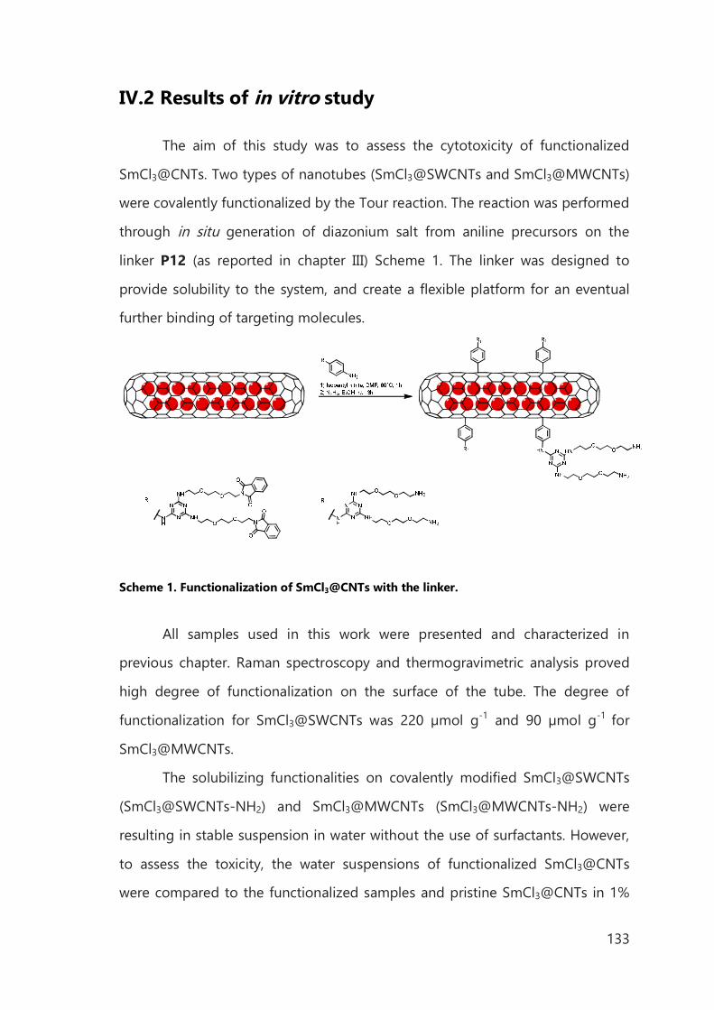
133
IV.2 Results of in vitro study
The aim of this study was to assess the cytotoxicity of functionalized
SmCl3@CNTs. Two types of nanotubes (SmCl3@SWCNTs and SmCl3@MWCNTs)
were covalently functionalized by the Tour reaction. The reaction was performed
through in situ generation of diazonium salt from aniline precursors on the
linker P12 (as reported in chapter III) Scheme 1. The linker was designed to
provide solubility to the system, and create a flexible platform for an eventual
further binding of targeting molecules.
Scheme 1. Functionalization of SmCl3@CNTs with the linker.
All samples used in this work were presented and characterized in
previous chapter. Raman spectroscopy and thermogravimetric analysis proved
high degree of functionalization on the surface of the tube. The degree of
functionalization for SmCl3@SWCNTs was 220 µmol g-1 and 90 µmol g-1 for
SmCl3@MWCNTs.
The solubilizing functionalities on covalently modified SmCl3@SWCNTs
(SmCl3@SWCNTs-NH2) and SmCl3@MWCNTs (SmCl3@MWCNTs-NH2) were
resulting in stable suspension in water without the use of surfactants. However,
to assess the toxicity, the water suspensions of functionalized SmCl3@CNTs
were compared to the functionalized samples and pristine SmCl3@CNTs in 1%
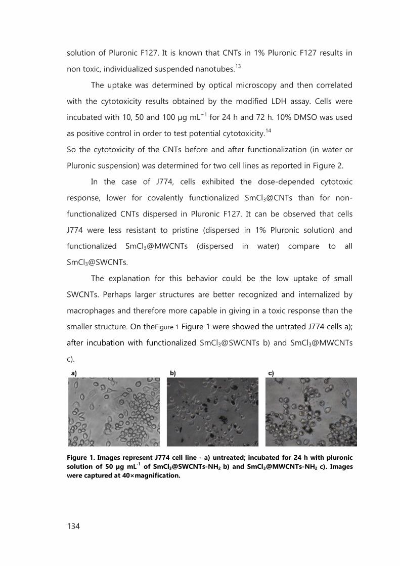
134
solution of Pluronic F127. It is known that CNTs in 1% Pluronic F127 results in
non toxic, individualized suspended nanotubes.13
The uptake was determined by optical microscopy and then correlated
with the cytotoxicity results obtained by the modified LDH assay. Cells were
incubated with 10, 50 and 100 µg mL−1 for 24 h and 72 h. 10% DMSO was used
as positive control in order to test potential cytotoxicity.14
So the cytotoxicity of the CNTs before and after functionalization (in water or
Pluronic suspension) was determined for two cell lines as reported in Figure 2.
In the case of J774, cells exhibited the dose-depended cytotoxic
response, lower for covalently functionalized SmCl3@CNTs than for non-
functionalized CNTs dispersed in Pluronic F127. It can be observed that cells
J774 were less resistant to pristine (dispersed in 1% Pluronic solution) and
functionalized SmCl3@MWCNTs (dispersed in water) compare to all
SmCl3@SWCNTs.
The explanation for this behavior could be the low uptake of small
SWCNTs. Perhaps larger structures are better recognized and internalized by
macrophages and therefore more capable in giving in a toxic response than the
smaller structure. On theFigure 1 Figure 1 were showed the untrated J774 cells a);
after incubation with functionalized SmCl3@SWCNTs b) and SmCl3@MWCNTs
c).
Figure 1. Images represent J774 cell line - a) untreated; incubated for 24 h with pluronic solution of 50 µg mL-1 of SmCl3@SWCNTs-NH2 b) and SmCl3@MWCNTs-NH2 c). Images were captured at 40×magnification.
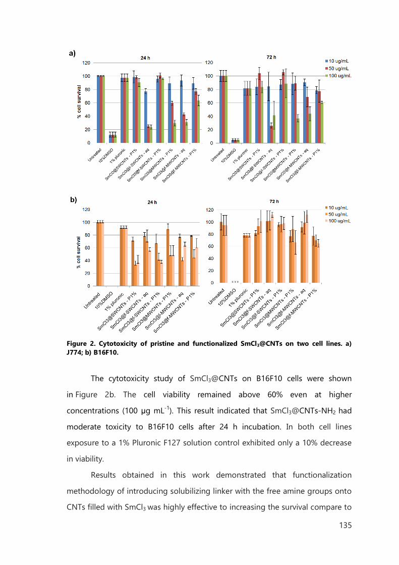
135
Figure 2. Cytotoxicity of pristine and functionalized SmCl3@CNTs on two cell lines. a) J774; b) B16F10.
The cytotoxicity study of SmCl3@CNTs on B16F10 cells were shown
in Figure 2b. The cell viability remained above 60% even at higher
concentrations (100 μg mL-1). This result indicated that SmCl3@CNTs-NH2 had
moderate toxicity to B16F10 cells after 24 h incubation. In both cell lines
exposure to a 1% Pluronic F127 solution control exhibited only a 10% decrease
in viability.
Results obtained in this work demonstrated that functionalization
methodology of introducing solubilizing linker with the free amine groups onto
CNTs filled with SmCl3 was highly effective to increasing the survival compare to

136
pristine, non-functionalized SmCl3@CNTs. The linker, successfully introduced on
SmCl3@SWCNTs and SmCl3@MWCNTs provide the best-possible dispersion of
the system. The high effective dose for to the in vitro system was showed
without causing reduced viability.
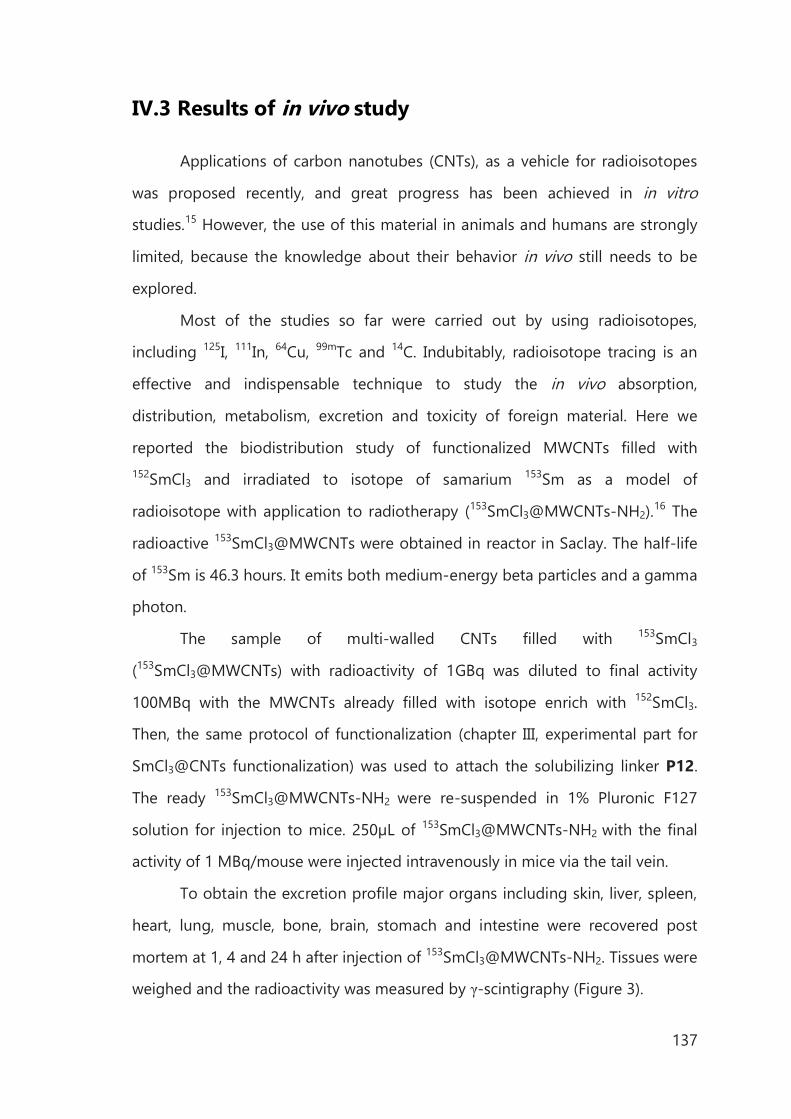
137
IV.3 Results of in vivo study
Applications of carbon nanotubes (CNTs), as a vehicle for radioisotopes
was proposed recently, and great progress has been achieved in in vitro
studies.15 However, the use of this material in animals and humans are strongly
limited, because the knowledge about their behavior in vivo still needs to be
explored.
Most of the studies so far were carried out by using radioisotopes,
including 125I, 111In, 64Cu, 99mTc and 14C. Indubitably, radioisotope tracing is an
effective and indispensable technique to study the in vivo absorption,
distribution, metabolism, excretion and toxicity of foreign material. Here we
reported the biodistribution study of functionalized MWCNTs filled with
152SmCl3 and irradiated to isotope of samarium 153Sm as a model of
radioisotope with application to radiotherapy (153SmCl3@MWCNTs-NH2).16 The
radioactive 153SmCl3@MWCNTs were obtained in reactor in Saclay. The half-life
of 153Sm is 46.3 hours. It emits both medium-energy beta particles and a gamma
photon.
The sample of multi-walled CNTs filled with 153SmCl3
(153SmCl3@MWCNTs) with radioactivity of 1GBq was diluted to final activity
100MBq with the MWCNTs already filled with isotope enrich with 152SmCl3.
Then, the same protocol of functionalization (chapter III, experimental part for
SmCl3@CNTs functionalization) was used to attach the solubilizing linker P12.
The ready 153SmCl3@MWCNTs-NH2 were re-suspended in 1% Pluronic F127
solution for injection to mice. 250µL of 153SmCl3@MWCNTs-NH2 with the final
activity of 1 MBq/mouse were injected intravenously in mice via the tail vein.
To obtain the excretion profile major organs including skin, liver, spleen,
heart, lung, muscle, bone, brain, stomach and intestine were recovered post
mortem at 1, 4 and 24 h after injection of 153SmCl3@MWCNTs-NH2. Tissues were
weighed and the radioactivity was measured by γ-scintigraphy (Figure 3).
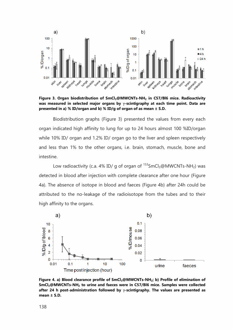
138
Figure 3. Organ biodistribution of SmCl3@MWCNTs-NH2 in C57/Bl6 mice. Radioactivity was measured in selected major organs by γ-scintigraphy at each time point. Data are presented in a) % ID/organ and b) % ID/g of organ of as mean ± S.D.
Biodistribution graphs (Figure 3) presented the values from every each
organ indicated high affinity to lung for up to 24 hours almost 100 %ID/organ
while 10% ID/ organ and 1.2% ID/ organ go to the liver and spleen respectively
and less than 1% to the other organs, i.e. brain, stomach, muscle, bone and
intestine.
Low radioactivity (c.a. 4% ID/ g of organ of 153SmCl3@MWCNTs-NH2) was
detected in blood after injection with complete clearance after one hour (Figure
4a). The absence of isotope in blood and faeces (Figure 4b) after 24h could be
attributed to the no-leakage of the radioisotope from the tubes and to their
high affinity to the organs.
Figure 4. a) Blood clearance profile of SmCl3@MWCNTs-NH2; b) Profile of elimination of SmCl3@MWCNTs-NH2 to urine and faeces were in C57/Bl6 mice. Samples were collected after 24 h post-administration followed by γ-scintigraphy. The values are presented as mean ± S.D.
139
The similar results for biodistribution of MWCNTs with free amino groups
were found in the work of Wang et al. where investigated the biodistribution
profile of functionalized MWCNTs with two different size and diameter.17 In that
work the biodistribution studies were evaluated for tubes functionalized with
1,3-dipolar cycloaddition, with the chain terminated with free amino groups and
then bounded to an antibody. However, it can be assumed that free amine
groups, present on the surface of MWCNTs could affect interaction with
biological membranes. Also the diameter can be the main factor that impacts
organ biodistribution in vivo.
140
IV.4 Conclusions
In concussions, SWCNTs and MWCNTs were functionalized by
endohedral and exohedral method and used as a model for radioactivity
delivery. No significant reduction of viability was observed in in vitro tests, for
two different cell lines at different compounds concentration: 10, 50 and 100 µg
mL−1.
Then, we have developed a convenient and general method to use 153Sm
inside the MWCNTs for in vivo biodistribution study. Among the radioisotopes
used in the tracing studies, samarium is the most convenient and it can be
sealed in MWCNTs easily and directly irradiated to samarium-153 (153Sm). It
emits beta particles what makes it highly therapeutic component and the
gamma rays for straightforward tracing.
By molten phase high temperature method, MWCNTs were filled with
metal halide (SmCl3). The larger diameter of MWCNTs allowed us to increase the
yield of filling respect to already reported work on SWCNTs.1 Thanks to this fact,
the final specific activity of the irradiated system reached almost 1 GBq. After
fast functionalization via alternative aryl diazonium chemistry (1 hour of reaction
suitable for half-life time of isotope 153Sm) an adequate level of functionalization
was achieved. In spite of the modification of the sp2 carbon network of
SmCl3@CNTs by the covalent attachment of the aniline modified with triazine
derivative, the internal part of the tubes was intact and the leakage of the
internal material was not observed after the functionalization or during the
biological study.
Our results suggest that the application of diazonium based arylation of
filled MWCNTs can be generally adoptable as an efficient and convenient
technique for functionalization, without breaking and opening the tubes. The
functionalized nanocapsules filled with 153Sm can be in this way, in vivo delivery
for long-term tracing studies.
141
IV.5 Experimental Part
IV.5.1 Preparation of samarium filled CNTs (SmCl3@CNTs) was described in Chapter III
IV.5.2 Preparation of radioactive 153SmCl3@MWCNTs
For encapsulation of samarium chloride, previously purified MWCNTs were
mixed with enrich 152-samarium (III) chloride (w/w 1:10 respectively) and
grinded with an agate mortar and pestle inside a glovebox until the mixture
presented a homogeneous color. The resulting mixture was then vacuum sealed
inside a silica ampoule and annealed at 900 °C for 12 h. After cooling down the
sample, the silica tube was opened and the material was stirred in water at 80 °C
to remove the non-encapsulated SmCl3. The filling yield of SmCl3@MWCNTs
was 19.5 wt%.
Then the radioactive 152SmCl3@MWCNTs was irradiated in reactor in Saclay and
provided as 153SmCl3@MWCNTs in a solution of DMF with final activity of 1 GBq.
IV.5.3 Functionalization of SmCl3@CNTs for in vitro study was the same as in Chapter III for filled nanotubes.
IV.5.4 Functionalization of 153SmCl3@MWCNTs
6 mg of 152SmCl3@MWCNTs in 4 mL of DMF were dispersed by sonication for
10 min. Then the 100 µL of 153SmCl3@MWCNTs dispersion in DMF, with the
activity 100 MBq, were injected. Then the P12 was added in excess (25 times in
weight) with respect to CNTs in 2 mL of DMF, dispersed for another 5 min and
cooled to 0°C. Isopentyl nitrite (5 fold molar excess with respect to amine) was
added and reaction mixture was heated up to 80 °C and stirred was continued
for one hours. Then the mixture was cooled to room temperature. The CNTs
were filtered (membrane MILIPORE, type: JHWP, pore size 0.1 µm) and washed
142
with DMF until the solvent eluted colorless, washed with DMF, water, MeOH,
EtOAc, Et2O and dried to afford 153SmCl3@MWNTs-Pht.
Deprotection of -NPht
For the deprotection, 153SmCl3@MWNTs-Pht (6 mg) were dispersed in EtOH (6
mL) by sonicating for 5 min, and afterwards treated with hydrazine hydrate (1
mL). The dispersion was stirred at r.t. for 3 h, and then diluted with EtOH (50 mL)
and filtered. After filtration, the functionalized SmCl3@MWCNTs-NH2 were re-
precipitated in EtOH, filtered (membrane MILIPORE, type: JHWP, pore size 0.45
m), washed with 0.1 M HCl solution, water, MeOH, diethyl ether finally dried μ
under vacuum to afford functionalized SmCl3@MWCNTs (SmCl3@MWCNTs-
NH2).
IV.5.5 Cell toxicity assays
Reagents
RPMI-1640 media, fetal bovine serum (FBS), penicillin/streptomycin,
Trypsin/EDTA, and phosphate buffered saline (PBS) were obtained from Gibco,
Invitrogen (UK). Cytotoxicity assay (LDH kit) was purchased from Promega UK
Ltd. B16F10 melanoma cells were cultivated in DMEM supplemented with 10%
fetal bovine serum, 100 IU/mL penicillin, and100 μg/mL streptomycin at 37 °C in
20% CO2 atmosphere. Culture of J774 macrophage cells were cultivated in
DMEM-HG supplemented with 10% heat-inactivated bovine fetal serum, 100
IU/mL of penicillin, 100 µm/mL of streptomycin, and 2 mM L-glutamine at 37° C
in 20% CO2.
Cell culture
Cells were seeded at a density of 8000 cells per well in flat-bottomed 96-well
plates and left to adhere overnight at 37° C and 20% CO2. Cells were then
143
treated with the CNTs dispersed in 1% of Pluronic F127 and or in water for 24 h
and 72 h. Dispersions were diluted in complete media at three concentrations:
10, 50, 100 g/mL. Healthy control cells were incubated with complete media μ
while positive controls were treated with 10% DMSO.
%Cell Survival=A490nm of treated cells
A490nm of untreated cells×100
IV.5.6 Biodistribution study of 153SmCl3@MWCNTs-NH2 in mice by gamma scintigraphy
Tissue biodistribution of SmCl3@MWCNTs-NH2 in mice was performed by
gamma scintigraphy. All in vivo experiments were conducted under the
authority of project and personal licences granted by the UK Home Office and
the UKCCCR Guidelines (1998). The blood circulation and the excretion profiles
of radio-labeled SmCl3@MWCNTs-NH2, as well as the biodistribution in major
organs were carried out using normal C57/Bl6 mice. The 153SmCl3@MWCNTs-
NH2 were centrifuged and re-suspended in 1% Pluronic F127 solution for
injection. Mice were injected intravenously via the tail vein with 250µL of 0.5 mg
mL-1 solution of 153SmCl3@MWCNTs-NH2 presenting the final activity of 1
MBq/mouse. To obtain the excretion profile, mice were housed singly in
metabolic cages in which animals had free access to water and food. After 24 h,
urine and faeces were collected from individual cages and counted by γ-
scintigraphy. For tissue biodistribution study, blood samples were collected in
heparinised capillaries from 4 min up to 24 h after injection and counted by γ-
scintigraphy (LKB Wallac 1282 Compugamma, PerkinElmer). Then the animal
was flushed with 10 mL of normal saline via the heart to clear any blood
remaining in the organs. Major organs including skin, liver, spleen, heart, lung,
muscle, bone, brain, stomach and intestine were obtained post mortem at 1, 4
and 24 h after injection of 153SmCl3@MWCNTs-NH2. Tissues were weighed and

144
the radioactivity was measured by γ-scintigraphy. The percentage injected dose
per gram of tissue was calculated for each organ. Four animals were used for
each time point for every compound.
145
Bibliography
1 L. Zhang, F. X. Gu, J. M. Chan, A. Z. Wang, R. S. Langer, O. C. Farokhzad; Clin. Pharmacol. Ther.
2008, 83, 761–769. 2 Hua He, L. Ai Pham-Huy, P. Dramou, D. Xiao, P. Zuo, C. Pham-Huy; Biomed. Res. Int. 2013,
578290–578302.
3 G. Seeta Rama Raju, L. Benton, E. Pavitraa, J. S. Yu; Chem. Commun. 2015, 51, 13248–13259. 4 Z. Liu, S. Tabakman, K. Welsher, H. Dai; Nano Research 2009, 2, 85-120. 5 N. Kotagiri, J.-W. Kim; International Journal of Nanomedicine 2014, 9, 85–105. 6 S. M. Moghimi, A. C. Hunter, J. C. Murray; Pharmacol. Rev. 2001, 53, 283–318. 7 M. Ferrari; Nat. Rev. Cancer 2005, 5, 161–171. 8 H. Wang, J. Wang, X. Deng, H. Sun, Z. Shi, Z. Gu, Y. Liu, Y. Zhaoc; J. Nanosci. Nanotechnol. 2004,
4, 1019–1024.
9 R. Singh. Proc. Natl. Acad. Sci. USA 2006,103, 3357–3362. 10 L. Lacerda, et al.; Adv. Mater. 2008, 20, 225–230. 11 L. Lacerda, M. A. Herrero, K. Venner, A. Bianco, M. Prato, K. Kostarelos; Small 2008, 4, 1130–
1132. 12 L. Lacerda, H. Ali-Boucetta, M. A. Herrero, G. Pastorin, A. Bianco, M. Prato, K. Kostarelos;
Nanomedicine 2008, 3, 149–161. 13 M. S. Strano, V. Moore, M. K. Miller, M. J. Allen, E. H. Haroz, C. Kittrell, R. H.Hauge; Smalley, R.
E.; J. Nanosci. Nanotechnol. 2003, 3, 81–86.
14 H. Ali-Boucetta, K. T. Al-Jamal, K. H. Müller, S. Li, A. E. Porter, A. Eddaoudi, M. Prato, A. Bianco,
K. Kostarelos; Small 2011, 7, 3230–3238. 15 C. Spinato, A. P. Ruiz de Garibay, M. Kierkowicz, E. Pach, M. Martincic, R. Klippstein, M.
Bourgognon, J. Tzu-Wen Wang, C. Ménard-Moyon, K. T. Al-Jamal, B. Ballesteros, G. Tobias, A.
Bianco; Nanoscale 2016, Doi:10.1039/C5NR07923C. 16 M. K. Bakht, M. Sadeghi, M. Pourbaghi-Masouleh, C. Tenreiro; Current Cancer Drug Targets,
2012, 12, 998–1015. 17 J. T.-W. Wang, C. Fabbro, E. Venturelli, C. Ménard-Moyon, O. Chaloin, T. Da Ros, L. Methven, A.
Nunes, J. K. Sosabowski, S. J. Mather, M. K. Robinson, J. Amadou, M. Prato, A. Bianco, K.
Kostarelos, K. T. Al-Jamal; Biomaterials 2014, 35, 9517–9528.

146
147
V. Appendix
V.1 Chemicals and Solvents
Chemicals were purchased from, Aldrich, Acros, Alfa Aesar and used as received.
Solvents were purchased from Aldrich, Acros, and Alfa Aesar. Deuterated
solvents from Aldrich and Cambridge Isotope Laboratories. All solvents used for
synthesis were analytical grade. When anhydrous conditions were required, high
quality commercial solvents were used (THF, DCM, o-DCB, toluene, DMF). Water
was purified using a Millipore filter system MilliQ®.
V.2 Characterization Methods and Instrumentation
Nuclear magnetic resonance spectroscopy (NMR) - NMR spectra were
recorded on a Bruker 400 or on a Varian 500 spectrometer (500 MHz for 1H, 125
MHz for 13C). All the spectra were run at ambient temperature and chemical
shifts were reported in ppm according to tetramethylsilane using as internal
reference. Deuterated solvents (d-chlorophorm: H = 7.26 ppm, C = 77.16 ppm;
d-methanol: H = 3.31). Resonance multiplicity was described as s (singlet), d
(doublet), t (triplet), m (multiplet) and br (broad signal).
Thermogravimetric analysis (TGA) - All TGAs were performed using Q500 (TA
Instruments) to record TGA analysis under N2 and air, by equilibrating from
room temperature to 100°C, and following a thermal ramp 10 °C/min up to 800
°C with a flow rate of 90 mL/min. The loss of weight was extracted as a
difference by the weight % and evaluated at the plateau of the curve for each
derivative. About 0.7 mg of sample per each analysis was required. Reported
graphs are an average of at least two separate measurements.
Determination of the filling yield:
The filling yield (FY) was calculated consider the amount of TGA residues in air:
from empty nanotubes (R1), clean filled nanotubes (R2) and bulk material (RA):
148
FY (wt%)=100∙(R2-R1)
RA-R1 (1)
RA was calculated basing on stoichiometry of the oxidative reaction which was
taking place during TGA analysis. Where MA and MB are molecular weights of A
and B; x and y are stoichiometric coefficients of reaction. Thus residue was
calculated according to following formula (2):
RA=100∙y∙MWB
x∙MWA (2)
High Resolution Transmission Electron Microscopy (HRTEM) - TEM analyses
were performed on Hitachi H7500 microscope (Tokyo, Japan) with an
accelerating voltage of 80 kV, equipped with an AMT Hamamatsu camera
(Tokyo, Japan). HAADF-STEM images were acquired at20 kV on a FEI Magellan
XHR 400L SEM equipped with a dedicated STEM detector. HRTEM micrographs
and EDX spectra were acquired on a FEI Tecnai G2 F20 operated at 200 kV and
equipped with an EDAX super ultra-thin window (SUTW) X-ray detector.
Samples were dispersed in ethanol and deposited on lacey carbon Cu TEM grids
(Agar).
Thin Layer Chromatography (TLC) - was conducted on pre-coated aluminium
plateswith 0.25 mm Macherey-Nagel silica gel with fluorescent indicator UV254.
Column Chromatography - Chromatographic purifications were carried out
with silicagel (Merck Kieselgel 60 - 200 mesh ASTM).
Infrared spectra (IR) were recorded on Varian 660-R, FT-IR spectrometer using
KBrplates or on a Perkin Elmer Spectrum One ATR-FT-IR spectrometer.
Mass spectra Both ESI and MALDI HR were performed on a Waters Synapt G2-
Si
UV-VIS-NIR spectroscopy - Spectra were recorded on Cary 5000
spectrophotometer (Varian) using 1 cm path quartz cuvettes.
149
Microwave - Microwave assisted reactions were carried out in a CEM Discover.
Kaiser test - Kaiser test (Kaiser test kit, Sigma Aldrich) was performed to
determine the degree of amine groups on the synthesized CNTs constructs. In a
typical test, 0.3-0.5 mg of CNTs were dispersed in a mixture of phenol (75 µL,
80% in ethanol) and KCN (100 µL in H2O/pyridine) solution and sonicated for 2
min in an ultrasonic bath. Subsequently, 75 µL of ninhydrin solution (6% in
ethanol) were added, and the mixture was heated at 120 °C for 10 min. It was
then cooled and diluted with 60% ethanol in water to a final volume of 3 mL.
After centrifugation (3000 rpm, 10 min), the absorption spectrum of the
supernatant was measured, using as blank a solution obtained in the same way
but without CNTs. The absorbance maximum at 570 nm was used to calculate
the amine loading in the CNTs samples (molar extinction coefficient =15000 M-
1 cm-1). Reported values are an average of at least two separated measurements.
µmolg =
[Abssample-Absblanc
]∙dilution (mL)∙106
ε ∙sample weight (mg)