UNIVERSITA’ DEGLI STUDI DI PADOVA -...
136
UNIVERSITA’ DEGLI STUDI DI PADOVA Dipartimento di Biologia SCUOLA DI DOTTORATO DI RICERCA IN BIOCHIMICA E BIOTECNOLOGIE INDIRIZZO BIOTECNOLOGIE XIII CICLO Molecular Characterization of Mitochondria Interactions with other Organelles Direttore della Scuola: Ch.mo Prof. Giuseppe Zanotti Coordinatore dell’Indirizzo: Ch.mo Prof. Giorgio Valle Supervisore: Ch.mo Prof. Fiorella Lo Schiavo Dottoranda: Cristina Ruberti
Transcript of UNIVERSITA’ DEGLI STUDI DI PADOVA -...
UNIVERSITA’ DEGLI STUDI DI PADOVA
Dipartimento di Biologia
SCUOLA DI DOTTORATO DI RICERCA
IN BIOCHIMICA E BIOTECNOLOGIE
INDIRIZZO BIOTECNOLOGIE
XIII CICLO
Molecular Characterization of Mitochondria
Interactions with other Organelles
Direttore della Scuola: Ch.mo Prof. Giuseppe Zanotti
Coordinatore dell’Indirizzo: Ch.mo Prof. Giorgio Valle
Supervisore: Ch.mo Prof. Fiorella Lo Schiavo
Dottoranda: Cristina Ruberti

2
INDICE
ABSTRACT pag. 1
RIASSUNTO pag. 4
INTRODUCTION
Mitochondria pag. 7
Mitochondrial fission machinery pag. 9
Roles of mitochondrial fission pag. 11
Matrixules pag. 11
Chloroplasts pag. 12
Chloroplast fission machinery pag. 13
Stromules pag. 14
Peroxisomes pag. 14
Peroxisomal fission machinery pag. 15
Peroxules pag. 16
Mitochondria, peroxisomes and chloroplasts:
metabolic, functional and physical inter-connections pag. 16
Leaf senescence: a physiological process
where mitochondria and chloroplasts presumably co-operate pag. 17
Topics of PhD project pag. 18
REFERENCES pag. 19
CHAPTER 1
Changes in mitochondrial morphology associated with cell aging during
grapevine leaf spontaneous senescence
INTRODUCTION pag. 27
II
RESULTS
Analyses of mitochondrial morphology during spontaneous
senescence in grapevine suspension cell cultures pag. 29
Grapevine plants transformed with -GFP-targeted
to mitochondria pag. 29
Analyses of mitochondrial morphology during leaf senescence pag. 30
Physiological and molecular characterization of
grapevine leaf senescence pag. 31
DISCUSSION pag. 33
MATERIAL AND METHODS
Suspension cell cultures and TMRM treatment pag. 35
Cell viability assay pag. 36
Cell cultures and plant material pag. 36
Semi-quantitative RT-CR analysis in pBIGYIN::GUS plants pag. 39
REFERENCES pag. 40
CHAPTER 2
BIGYN, a tail anchored protein, recruits cytosolic ELM1 protein at
mitochondria and chloroplast level
INTRODUCTION pag. 45
RESULTS
Subcellular localization of ELM1::YFP pag. 48
Subcellular localization of YFP::BIGYIN pag. 50
Co-localization analysis of BIGYIN and ELM1 pag. 53
Bimolecular fluorescence complementation assay to test
BIGYIN-ELM1 in vivo interaction pag. 53
Subcellular localization of BIGYIN and ELM1-interction sites pag. 56
Expression pattern of ELM1 and BIGYIN in seedlings pag. 57
DISCUSSION
YFP::BIGYIN localizes to mitochondria, peroxisomes
and chloroplasts pag. 60
III
BIGYIN and ELM1 co-localize to mitochondria and chloroplasts pag. 61
BIGYIN and ELM1 interact in vivo pag. 62
BIGYIN and ELM1 interact in vivo on mitochondria
and chloroplasts pag. 63
BIGYIN and ELM1 are expressed in the same
plant tissues in Arabidopsis seedlings pag. 63
MATERIAL AND METHODS
Plant materials and growth condition pag. 65
Genetic materials pag. 65
Protoplasts isolation pag. 68
Protoplasts transfection assay pag. 69
Agrobacterium tumefaciens strain pag. 69
Tobacco leaf agroinfiltration pag. 70
Confocal analyses pag. 70
BiFC fluorescence quantification pag. 71
-glucoronidase (GUS) histochemical analyses pag. 71
Statistic pag. 71
REFERENCES pag. 72
CHAPTER 3
The subcellular localization of BIGYIN, an Arabidopsis protein involved in
mitochondrial and peroxisomal division, unveils a dynamic network of
tubules and organelles
INTRODUCTION pag. 77
RESULTS
Structure of Arabidopsis FIS1-type proteins pag. 81
BIGYIN expression pattern in germinating seeds,
young seedlings, adult rosettes and mature flowering plants pag. 82
Localization pattern of BIGYIN protein in germinating seeds,
young seedlings, adult rosettes and mature flowering plants pag. 84
Subcellular localization of YFP::BIGYIN in Arabidopsis plants pag. 85
IV
Network of „bigyinules‟ pag. 90
Analysis of the anti-BIGYIN antibody specificity pag. 92
Effects of an actin-inhibitor on movement of
organelles labelled with YFP::BIGYIN and bigyinules pag. 94
Effects of an actin inhibitor on movement of
YFP::BIGYIN within organelle membranes pag. 94
Effects of an ER-Golgi vesicle trafficking
inhibitor on movement of YFP::BIGYIN pag. 96
Characterization of a T-DNA insertional mutant for BIGYIN pag. 96
Analyses in BIGYIN protein structure of domains
involved in multiple targeting pag. 98
DISCUSSION
BIGYIN is a tail-anchored protein belonging to the FIS1-type protein pag. 103
Expression and localization pattern of BIGYIN in Arabidopsis pag. 103
BIGYIN localizes to mitochondria, peroxisomes, chloroplasts
and to membranous protrusions extending from these organelles pag. 104
C-terminal of BIGYIN is necessary and sufficient for BIGYIN targeting pag. 105
Anti-BIGYIN antibody is specific for BIGYIN pag. 106
BIGYIN unveils a dynamic network of organelles
and tubular protrusions pag. 107
BIGYIN may play a role also in chloroplast division pag. 109
MATERIAL AND METHODS
Plant materials and growth condition pag. 111
DNA constructs pag. 111
Semi-quantitative RT-PCR analysis in pBIGYIN::GUS plants pag. 114
-glucoronidase (GUS) histochemical analysis pag. 115
Inhibitor treatment pag. 115
Antibody pag. 115
Protein extraction and Western blot analyses pag. 116
BIGYIN insertion mutant pag. 116
Analysis of mitochondria pag. 117
Transient expression experiments pag. 117
Confocal analyses pag. 119

V
Fluorescence recovery after photobleaching pag. 119
Accession Numbers pag. 119
Statistic pag. 120
REFERENCES pag. 121
CONCLUSIONS pag.127

VI
1
ABSTRACT
The topic of this PhD thesis was to study organelles in plant cells, analysing in
particular the mitochondrial morphology. Recent papers have, in fact, reported
that regulation of mitochondrial morphology is important not only for maintaining
mitochondrial number during cell division, and mitochondrial distribution within
each cell (Westermann, 2010; Logan, 2003), but also for executing many
biological functions. In animals, a shift in mitochondrial morphology towards the
organelle fragmentation is an early event during apoptosis (Suen et al., 2008). In
plants, a link between mitochondrial morphology and senescence-associated cell
death (i.e. a genetically controlled programme of self destruction) has been
reported in Medicago truncatula cultured cells (Zottini et al., 2006).
The understanding of molecular mechanisms, used by plants to regulate
senescence, might allow biotechnological applications through genetic
manipulations of key elements affecting senescence and provide interesting
applicative outputs, especially in agronomically relevant species.
In light of these findings, I focused on the study of mitochondrial morphology
during senescence in grapevine (Vitis spp.) plants, one of the most important crops
in Mediterranean area that has also the potentiality of becoming a model organism
for fruit trees due to the knowledge of its genome sequence (Jaillon et al., 2007;
Velasco et al., 2007).
Suspension cell cultures of Vitis spp. were chosen as an initial model system
to study mitochondrial morphology during senescence/PCD, because they are an
accessible and not complex experimental system, sharing some of fundamental
regulatory mechanisms with PCD processes in plant. Then, in order to study
changes in mitochondrial morphology during different stages of leaf development
and senescence, transgenic grapevine plants, expressing GFP (green fluorescent
protein) fluorescent marker targeted to mitochondria, were generated. In the
grapevine leaves the senescence/PCD process was characterized by analyzing the
chlorophyll degradation and the expression of several VvSAGs genes (i.e. Vitis
homologues of Arabidopsis senescence associated genes). Analyses performed in
grapevine senescent leaves showed mitochondria prefunding altered in their
morphology. Then, we moved to investigate to the molecular aspects of these
2
morphological changes. Indeed, it is known that in eukaryotic cells, the number,
size and distribution of mitochondria are regulated by continuous cycles of
mitochondrial fusions and fission events (Logan, 2003) but so far in plants, no
genes involved in mitochondrial fusion machinery have been identified, while
several genes involved in mitochondrial fission have been recently described in
Arabidopsis thaliana, a plant model organism. Hence, I moved to Arabidopsis
plant and I focused on the study of the subcellular localization, expression and
interaction of two proteins (ELM1 and BIGYIN), known to be involved in
mitochondrial fission. ELM1 and BIGYIN subcellular localization was carefully
analyzed, by using several organelle fluorescent markers and adopting different
methods of transient expression in leaf cells, combined with confocal microscope
analyses. Yet, I investigated whether in vivo interactions between ELM1 and
BIGYIN occurred in plant cells. Then, I analyzed ELM1 and BIGYIN expression
pattern in plant to verify if their presence in the same tissues was indeed detected.
The analysis was carried out through histochemical GUS assay in transgenic
plants, stable expressing the ELM1 and BIGYIN promoter region fused to the -
glucuronidase (GUS) reporter.
In order to define the physiological role of BIGYIN, a detailed subcellular
localization of BIGYIN was performed in the whole plant, stably expressing
BIGYIN under control of its own promoter, in order to mimic physiological
conditions. These analyses allowed us to establish an association between
BIGYIN and chloroplasts/plastids and, in particular, to identify protrusions,
marked by YFP::BIGYIN, extruding from chloroplast, mitochondria and
peroxisomes. Such kind of tubular protrusions has already been observed and
termed matrixules, peroxules stromules respectively, and it has been proposed to
play a role in physical inter-organellar interactions, by increasing the transfer
efficiency of metabolites/molecules among organelles (Scott et al., 2007). The
presence of BIGYIN on these protrusions is indeed an unexpected finding that led
us to investigate more in detail this peculiar BIGYIN localization. I focused in
detail on the dynamics and behaviour of these YFP::BIGYIN marked tubules, and
investigated, in different tissues and different developmental stages, whether a
protein trafficking through these organelles indeed occurred.
3
The understanding of physical inter-organellar interactions is connected
with the importance of elucidating relationships and cross-talks among organelles,
key events to understand the mechanisms of interactions between plant and its
environment that can be of particular importance for the coordination of the
senescent program.
4
RIASSUNTO
In questa tesi di dottorato, è stata condotta un‟analisi degli organelli in cellule
vegetali, studiando, in particolare, i mitocondri e la loro morfologia in diverse
condizioni fisiologiche. La regolazione della morfologia mitocondriale è
importante sotto molteplici aspetti, infatti controlla il numero dei mitocondri
durante la divisione cellulare e la loro distribuzione all‟interno di ogni cellula
(Wastermann, 2010; Logan, 2003), controlla infine alcune funzioni biologiche.
Nelle cellule animali, per esempio, la frammentazione dei mitocondri è un evento
precoce che caratterizza il processo di apoptosi (Suen et al., 2008).
Un‟associazione tra morfologia mitocondriale e senescenza/morte cellulare
programmata (PCD) è stata riportata anche in colture cellulare vegetali (Zottini et
al., 2006).
La comprensione dei meccanismi molecolari, utilizzati dalla pianta per
regolare il processo di senescenza/PCD, può essere importante da un punto di
vista biotecnologico, in quanto potrebbe permettere una modulazione dei
parametri di crescita e di sviluppo della pianta attraverso miglioramenti genetici e
manipolazioni di fattori ambientali che regolano il processo di senescenza. I
risvolti applicativi di tale approcio sono molto interessanti, soprattutto per le
specie di rilevanza agronomica.
Alla luce di tali premesse, la morfologia mitocondriale è stata dettagliatamente
analizzata durante il processo di senescenza/PCD in vite (Vitis spp). La vite
rapprensenta una delle più importanti piante coltivate della zona del Mediterraneo,
ed a seguito del sequenziamento del suo genoma (Jaillon et al., 2007; Velasco et
al., 2007), è ritenuta un organismo modello negli studi sugli alberi da frutto.
Inizialmente l‟analisi della morfologia mitocondriale durante il processo di
senescenza/morte cellulare programmata è stata condotta in colture cellulari di
Vitis spp.. In seguito, lo studio è stato continuato in pianta, in particolare è stata
portata avanti un‟analisi dettagliata della morfologia mitocondriale durante i
differenti stadi di sviluppo e di senescenza della foglia. A tal fine, sono state
prodotte ed analizzate piante transgeniche, esprimenti un marcatore fluorescente
mitocondriale. Le analisi condotte hanno dimostrato che in foglie senescenti i
mitocondri presentano delle morfologie caratteristiche. Questi risultati ci hanno
5
indirizzato ad uno studio degli aspetti molecolari coinvolti nella regolazione della
morfologia mitocondriale.
In numerose cellule eucariotiche, la morfologia mitocondriale è regolata
dal continuo alternarsi di eventi di fusione e fissione mitocondriale (Logan, 2003).
Nelle piante, i componenti molecolari coinvolti nel meccanismo di fusione
mitocondriale non sono stati ancora individuati, mentre i componenti proteici
implicati nella fissione sono stati recentemente descritti in Arabidopsis. A tuttoggi
non è stato ancora compreso nè il preciso ruolo svolto da tali proteine, nè le loro
precise interazioni fisiche, responsabili del processo di fissione mitocondriale.
Durante il mio dottorato, ho quindi analizzato, nel sistema modello Arabidopsis
thaliana, la localizzazione subcellulare, l‟espressione e l‟interazione di due
proteine (ELM1 ed BIGYIN), coinvolte nel processo di divisione mitocondriale.
La localizzazione subcellulare di ELM1 e BIGYIN è stata determinata in cellule
vegetali, utilizzando differenti metodi di espressione transiente combinati con
analisi di microscopia confocale. Sono state poi eseguite analisi per verificare
l‟interazione di queste due proteine in vivo in cellule vegetali. Successivamente è
stata condotta in pianta un‟analisi del pattern di espressione di BIGYIN ed ELM1,
in modo tale da verificarne l‟espressione di queste due proteine negli stessi tessuti,
prerequisito fondamentale per una loro eventuale interazione. A tal fine, sono state
prodotte ed analizzate mediante saggio istochimico-colorimetrico piante
transgeniche di Arabidopsis thaliana stabilmente esprimenti il promotore di tali
geni fuso al gene che codifica per l‟enzima -glucuronidasi (GUS).
Per definire il ruolo fisiologico di BIGYIN, una dettagliata localizzazione
subcellulare di questa proteina è stata eseguita in piante di Arabidopsis,
stabilmente esprimenti il costrutto YFP::BIGYIN sotto il controllo del proprio
promotore. Queste analisi hanno messa in evidenza un‟associazione tra BIGYIN e
cloroplasti/plastidi ed ha portato ad individuare particolari protrusioni, marcate
con la proteina di fusione YFP::BIGYIN, che si estondono dai cloroplasti,
mitocondri e perossisomi. Protrusioni simili erano già state riportate in letteratura
e prendono il nome di „stromuli‟, „matrixuli‟, „peroxuli‟ a seconda che si
estendano rispettivamente da cloroplasti, mitocondri o perossisomi.
Recentemente, è stato ipotizzato che tali le protrusioni abbiano un ruolo nelle
interazioni fisiche tra i differenti organelli, aumentando i contatti fisici tra gli

6
organuli e migliorando l‟efficienza di scambio di metaboliti/molecule (Scott et al.,
2007). Tuttavia nessun dato è stato riportato a conferma di tale ipotesi. L‟inattesa
presenza di BIGYIN su tali protrusioni, ci ha permesso di studiare in dettaglio la
loro dinamicità e di analizzare la presenza di interazioni fisiche tra gli organelli.
La comprensione delle interazioni fisiche tra gli organelli si colloca nel
campo di indagine delle relazioni tra tali compartimenti subcellulari che viene ora
considerato un campo fondamentale per la conoscenza dei meccanismi di base
della biologia cellulare vegetale.
7
INTRODUCTION
Mitochondria, peroxisomes and plastids are essential and ubiquitous subcellular
organelles in plants. Each of these organelles are specialized compartments
playing specific functions. Extensive metabolic exchanges are well known
(Schrader and Yoon, 2007; Bauwe et al., 2010), instead, physical and functional
signalling interactions among them are now emerging form recent data
(Sweetlove et al., 2007; Rhoads and Subbaiah, 2007).
Mitochondria
Mitochondria are essential bio-energetic subcellular organelles in most eukaryotic
cells. Their major role is to synthesize ATP, by coupling substrate oxidation to
electron transport and the generation of a proton electrochemical gradient.
Mitochondria are also involved in a range of other processes, i.e. in controlling
cellular redox state (Noctor et al., 2006), in Ca2+
homeostasis (Vandecasteele et
al., 2001; Logan and Knight, 2003) and in programmed cell death (PCD) (Lam,
2004) both in animals and in plants. Moreover mitochondria are not independent
and autonomous organelles, but they are involved in metabolic and functional
connection with other subcellular compartments. For example, in higher plants,
mitochondria, peroxisomes, chloroplasts and cytosol are involved together in
photorespiration process (Bauwe et al., 2010).
Each mitochondrion is composed of compartments that carry out
specialized functions. These compartments include the outer membrane, the inner
membrane, the intermembrane space (the space between the outer and inner
membranes), the matrix (space within the inner membrane), the crystal membrane
(infolding of the inner membranes) and the intercrystal space (Logan, 2006). The
outer membranes act as a barrier to large molecules (>10 kDa), that can only enter
into mitochondria through specific pores located within the lipid bilayer. The
inner membrane is characterized by a highly complex structure. Components of
the electron transport system and the ATP synthetase are an integral part of the
inner membranes. The matrix contains enzymes responsible for the citric acid

8
cycle reaction and also several metabolites (i.e. NAD, NADP, ADP, and ATP)
(Bowsher and Tobin, 2001).
It is widely accepted that mitochondria originate from a common ancestral
free-living α-proteobacterium, that colonised pro-eukaryotic cell around two
billion years ago (Gray et al., 1999). During evolution, the engulfed
proteobacterium transferred the majority of its genes to the nucleus. As result of
this process, several mitochondrial processes (i.e. biochemistry pathways) are now
under nuclear control. In addition, the mitochondria lost also components of their
bacterial origin (i.e. genes originally involved in its division as prokaryotic cell);
and it co-opted other components from the host (i.e. the main components
involved in division machinery of the outer membrane, originated in consequence
of incorporation into the host eukaryotic cell) (Schrader, 2006).
Mitochondria are not created de novo, but they arise by fission events of
pre-exiting mitochondria in the cytosol. In various eukaryotic cells, mitochondria
undergo continuous cycles of fission and fusion and these concerted activities
control the maintenance of mitochondrial number during cell division, and the
mitochondrial distribution and morphology within a single cell (Logan, 2003). As
result of these fission-fusion events, mitochondrial shape and size are continually
changing within the living cells. This great heterogeneity in the dynamics and
morphology of mitochondria has been reported within a single cell, in different
cell types of the same organism, and also in different organisms.
In yeasts and in most animal cell types, mitochondria are usually described
as an „interconnected reticular network‟, because the chondriome (i.e. all the
mitochondria in a cell collectively) is organized into long tubules or reticula,
characterized by dynamical shifting between a fragmentized state and a tubular
„reticulum continuum‟ (Logan, 2006). By contrast, in higher plants, the
chondriome is described as a „discontinuous whole‟, because it is a highly
dynamic structure composed mainly of discrete organelles (Logan, 2006).
Yeasts, mammals and higher plants show a common molecular mechanism
involved in mitochondrial fission events (Delille et al., 2009). Concerning,
instead, the molecular mechanisms involved in mitochondrial fusion event, in
yeasts and mammals the molecular players have been described (Westermann,
9
2010), while in plants none molecular component has yet been identified (Logan,
2003).
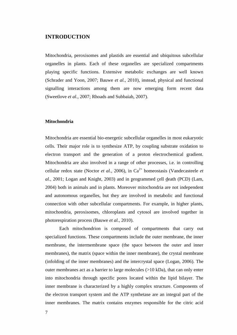
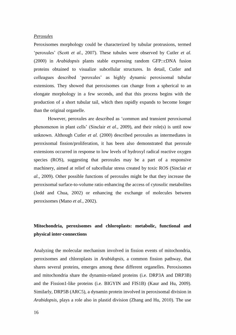
Mitochondrial fission machinery
Molecular mechanisms involved in mitochondrial fission events have been
studied extensively in yeast Saccharomyces cerevisiae. Mitochondrial fission
machinery in yeast is composed by four proteins: Fission1 (Fis1), Dynamin1
(Dnm1), Mitochondrial division1 (Mdv1) and Caf4. Fis1 is an integral membrane
protein, located to the mitochondrial outer membranes. Its N-terminal domain is
exposed to the cytoplasmatic side and forms a tetratricopeptide (TRP)-like bundle
helix, involved in protein-protein interaction (Suzuki et al., 2003), while the C-
terminal tail contains a single transmembrane domain. For this topology, Fis1 is
considered a member of tail-anchored (TA) (Nout-Cin) family of membrane
proteins (Borghese et al., 2007). Dnm1 is a yeast dynamin-related protein (DRP),
characterized by a N-terminal GTPase domain and a C-terminal GTPase effector.
During mitochondrial fission, Fis1 recruits to mitochondrial fission sites the
cytosolic molecular adapter Mdv1 (and its paralog Caf4) and Dnm1. Dnm1 and
Mdv1 are thought to form higher-order multimer complexes, named fission
complexes, that surround and pinch off mitochondria (Fig.1).
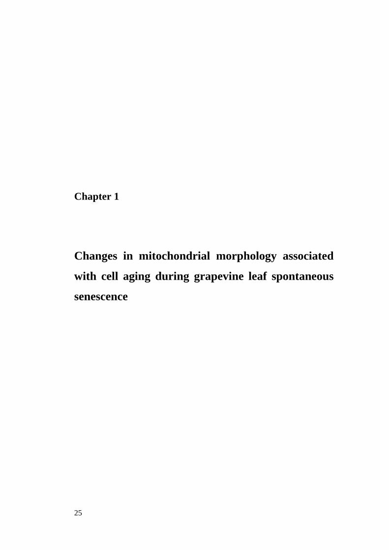
Fig.1. Molecular model of mitochondrial fission in Yeast: interaction between Dnm1, Mdv1 and
Fis1. MOM stands for the mitochondrial outer membrane.
10
In higher plants, studies on molecular mechanism, involved in
mitochondrial fission events, have just begun. Recently, in Arabidopsis thaliana,
several genes involved in remodelling of mitochondria have been described: two
dynamin-related proteins (DRPs), termed DRP3A (formerly, Arabidopsis
dynamin-like protein 2A [ADL2a] and ADL2b, respectively; (Zhang and Hu,
2008a); two fission-like protein, homologue of yeasts Fis1, termed BIGYIN and
FIS1B (Scott et al., 2007); a plant specific protein, named ELM1, which does not
present sequence similarity with the yeast Mdv1/Caf4 (Arimura et al., 2008).
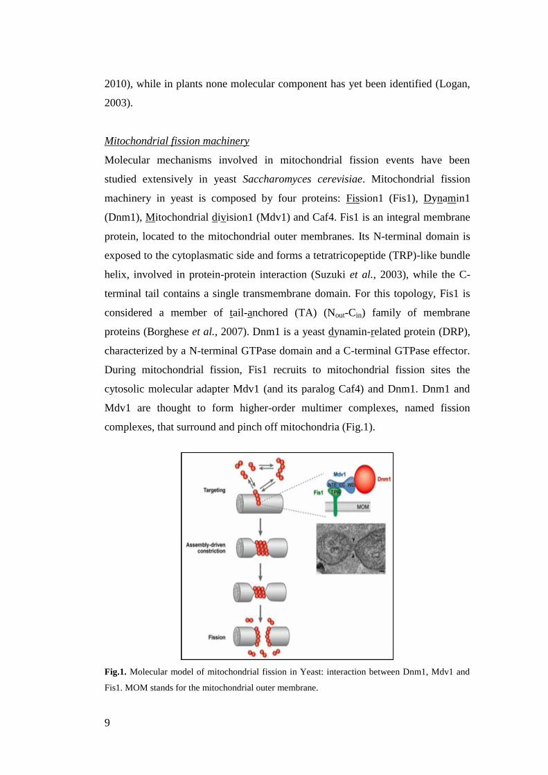
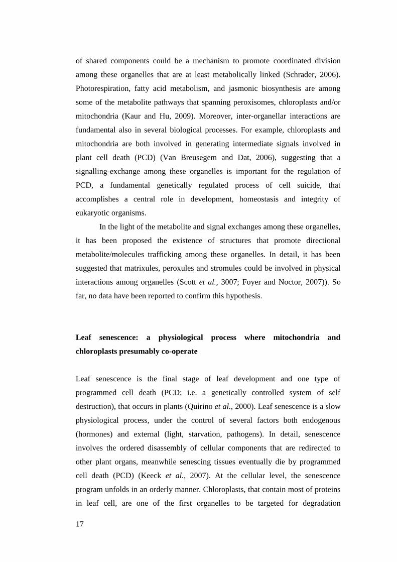
How these proteins specifically interact among them and what is the
precise role of each of these proteins during mitochondrial fission events is yet
unclear. So far, it is known that DRP3A and BIGYIN do not interact each other,
while DRP3A, DRP3B and ELM1 interact among them (Arimura et al., 2008;
Zhang and Hu, 2008a) (Fig. 2). Moreover, ELM1 is required for the subcellular
transfer of DRP3A from the cytosol to mitochondrial fission sites (Arimura et al.,
2008). Since ELM1 protein structure does not present transmembrane domain or
other predicted membrane-anchoring domains, it has been supposed that its
mitochondrial localization could depend on the interaction with protein(s) located
to mitochondrial surfaces. BIGYIN is one of the proteins that have been proposed
to interact with ELM1, but results showing this interaction have not yet reported.
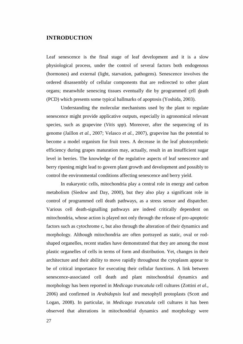
Fig.2 Comparison of Mitochondrial fission Factors in Yeas and in Plant. In Yeast, Dnm1,
a dynamin-related protein, is recruited from the cytosol to mitochondria in a manner
dependent on Fis1 and an adaptor protein (Mdv1). In the Plant Arabidopsis, dynamin
related protein, DRP3A and DRP3B, interact with ELM1 for their localization to
mitochondria. A black bar in Fis1 shows putative transmembrane domains. OM, outer
membrane; IM, inner membrane (Arimura et al., 2008)
11
Roles of mitochondrial fission
Mitochondrial fission is important not only for the maintenance of mitochondrial
number during cell division, or for mitochondrial morphology within a single cell
(Westermann, 2010; Logan, 2003), but mitochondrial architecture is also
important for executing many biological functions. For example, mitochondrial
fission and subsequent mitochondrial fragmentation are an early event during
apoptosis in yeasts and mammals (Suen et al., 2008). By contrast, a decrease in
mitochondrial fission and the subsequent giant elongated mitochondria are
protective features in old human endothelial cells cultivated in vitro (Mai et al.,
2010). Elongation of mitochondria allows both a decrease of the energy-
consuming processes of mitochondrial dynamics, and a fast distribution and
exchange of molecules into the mitochondrial matrix. As result, these elongation
of mitochondria rendered cells more resistant against apoptotic stimuli (Mai et al.,
2010). In plants, similar giant mitochondria during senescence-associated cell
death have been reported in Medicago truncatula cultured cells (Zottini et al.,
2006) and in Arabidopsis leaf and mesophyll protoplasts (Scott and Logan, 2008),
suggesting a link between plant mitochondrial morphology and senescence-
associated cell death.
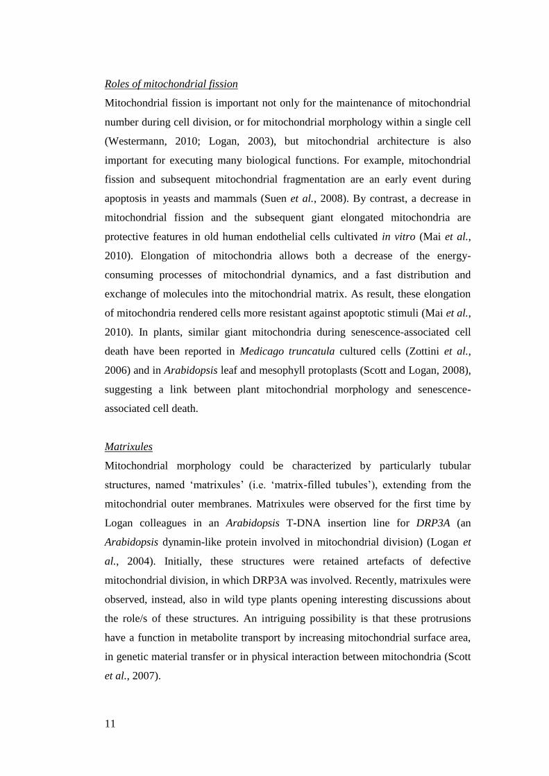
Matrixules
Mitochondrial morphology could be characterized by particularly tubular
structures, named „matrixules‟ (i.e. „matrix-filled tubules‟), extending from the
mitochondrial outer membranes. Matrixules were observed for the first time by
Logan colleagues in an Arabidopsis T-DNA insertion line for DRP3A (an
Arabidopsis dynamin-like protein involved in mitochondrial division) (Logan et
al., 2004). Initially, these structures were retained artefacts of defective
mitochondrial division, in which DRP3A was involved. Recently, matrixules were
observed, instead, also in wild type plants opening interesting discussions about
the role/s of these structures. An intriguing possibility is that these protrusions
have a function in metabolite transport by increasing mitochondrial surface area,
in genetic material transfer or in physical interaction between mitochondria (Scott
et al., 2007).
12
Chloroplasts
Plant cell contains plastids, which represent one of the principal hallmarks that
differentiate plant cell from other eukaryotic cells. „Plastid‟ is a general term
applied to an important and wide group of functionally distinct subcellular
organelles. Plastids develop from small undifferentiated proplastids in dividing
meristematic cells. These undifferentiated proplastids differentiate into a variety
of specific plastid type, depending on the particular cell type in which they are
located and the stage of cellular development. „Leucoplasts‟, lacking of
pigmentation, are specialized for bulk storage („amyloplasts‟), or lipids
(„elaioplasts‟), or protein („proteinoplasts‟) (Bowsher and Tobin, 2001).
„Chloroplasts‟, instead, contain chlorophyll, they are involved in photosynthesis
and presumably for this important role they are also the best studied among the
different plastidial type.
Chloroplasts arose from an endosymbiotic event between a prokaryote and
a photosynthetic prokaryote (Gray, 1999). During evolution numerous plastid
genes have been lost or transferred to the nuclear genome. As result, the correct
plastid development and function are now under nuclear control.
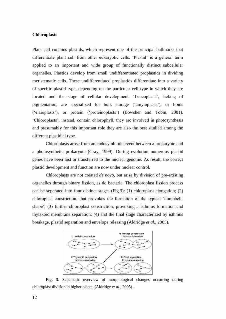
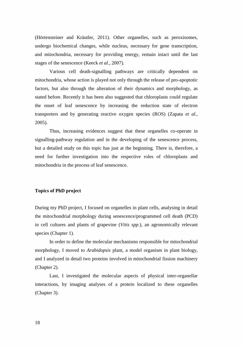
Chloroplasts are not created de novo, but arise by division of pre-existing
organelles through binary fission, as do bacteria. The chloroplast fission process
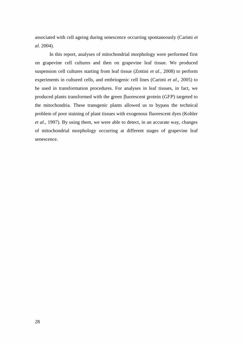
can be separated into four distinct stages (Fig.3): (1) chloroplast elongation; (2)
chloroplast constriction, that provokes the formation of the typical „dumbbell-
shape‟; (3) further chloroplast constriction, provoking a isthmus formation and
thylakoid membrane separation; (4) and the final stage characterized by isthmus
breakage, plastid separation and envelope releasing (Aldridge et al., 2005).
Fig. 3. Schematic overview of morphological changes occurring during
chloroplast division in higher plants. (Aldridge et al., 2005).
13
Chloroplast fission machinery
In most organisms, the chloroplast division apparatus consist of a double ring
structure, with one ring on the cytosolic face of the outer membrane (outer ring),
and one on the stromal face of the inner envelope (inner ring) (Maple and Møller,
2007). The plastid division is controlled by a combination of prokaryote-derived
and host eukaryote-derived proteins, but the precise roles of these components in
chloroplast division machinery and how these components are coordinated is still
unclear.
In Arabidopsis, homologues of the bacterial cell division FtsZ have been
identify in the nuclear genome (i.e. AtFtsZ1-1 and AtFtsZ2-1). These proteins are
imported into chloroplasts and formed a ring (Z-ring) on the stromal side of the
inner membrane. In detail, the Z-ring probably determines the site of division,
where successively molecular components of eukaryotic origin are assembled to
form the inner and outer ring (Aldridge et al., 2005). The correct position of the
Z-ring at the chloroplast centre could be, at least in part, mediated by the
AtMinD1 and AtMinE1, homologues of the bacterial MinD and MinE. The Z-ring
is probably stabilized and anchored to the inner membrane by the interaction
between AtFtsZ2-1 and the transmembrane protein ARC6, protein of prokaryotic
origin localized to the stromal side of the inner membrane (Maple and Møller,
2007).
Unlike the Z-ring, the inner and the outer rings have been probably
recruited from the eukaryotic cells, and, so far, the composition of both rings is
unknown. Several component of eukaryotic origin involved in plastid division
have been recently identified in Arabidopsis: DRP5B (i.e. dynamin-related
protein5B, named also ARC5), PDV1 and PDV2 (i.e. plastid division1 and plastid
division2). PDV1 and PDV2 are transmembrane proteins integrated into the
chloroplast outer membrane, with the N-terminal region exposed to the cytosol.
PDV1 and PDV2 are required for the localization of DRP5B at the division site
(Miyagishima et al., 2006). DRP5B is a member of the dynamin-like superfamily
of eukaryotic membrane-remodelling GTPases. Since members of the dynamin-
like superfamily are involved in membrane pinching events in eukaryotes and also
in mitochondrial membrane scissions in yeast, mammals and plants, it is
14
reasonable that DRP5B may play a role to pinch off the outer chloroplast
membrane at late stages of the division process (Gao et al., 2003).
Stromules
In higher plants, all plastid types are characterized by highly dynamic protrusions,
named „stromules‟ (i.e. „stroma-filled tubules), extending from plastidial surfaces
(Köhler and Hanson, 2000). Stromules could protrude from a single plastid or
connect two o more plastids among them. It has been reported that stromules are
involved in exchange of endogenous stromal proteins (i.e. RubisCO and aspartate
aminotransferase), between connected chloroplasts (Kwok and Hanson, 2004a).
However, this is not the primary function of stromules, given that stromules can
freely extend from a single chloroplast without necessarily to be linked to another
chloroplast. Stromules could have a function in metabolite/molecules exchange
also with cytosol, and, since a close apposition of stromules and nuclei has been
frequently observed, it has also been suggested that stromules could play a role in
interactions between plastids and nuclei, such as allowing the rapid transit of
molecules and signals between these subcellular compartments (Kwok and
Hanson, 2004b).
Peroxisomes
Peroxisomes are single-membrane organelles ubiquitously present in all
eukaryotic cells. In plants, peroxisomes mediate several metabolic functions, i.e.
fatty acid -oxidation or hydrogen peroxide degradation.
During evolution, these organelles arose from cellular membrane system,
probably the endoplasmic reticulum, as an invention of eukaryotic cells (Michels
et al., 2005).
Peroxisomes are highly dynamic organelles, capable of adapting to a
variety of environmental and developmental cues by altering their abundance and
morphology. Peroxisomes generated mainly from pre-existing peroxisomes
through a constitutive division (named also „peroxisomal division‟) or through an
induced many-fold increment in their number (termed „peroxisomal
15
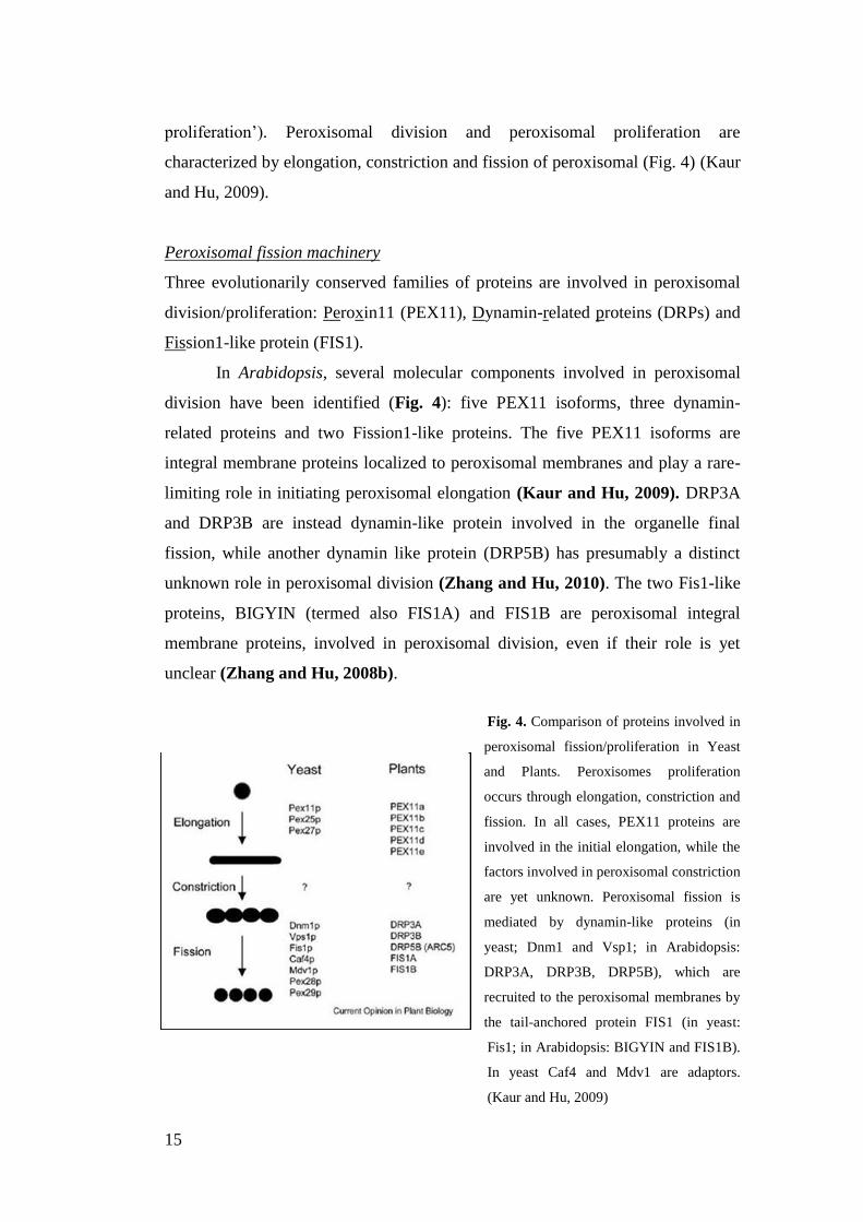
proliferation‟). Peroxisomal division and peroxisomal proliferation are
characterized by elongation, constriction and fission of peroxisomal (Fig. 4) (Kaur
and Hu, 2009).
Peroxisomal fission machinery
Three evolutionarily conserved families of proteins are involved in peroxisomal
division/proliferation: Peroxin11 (PEX11), Dynamin-related proteins (DRPs) and
Fission1-like protein (FIS1).
In Arabidopsis, several molecular components involved in peroxisomal
division have been identified (Fig. 4): five PEX11 isoforms, three dynamin-
related proteins and two Fission1-like proteins. The five PEX11 isoforms are
integral membrane proteins localized to peroxisomal membranes and play a rare-
limiting role in initiating peroxisomal elongation (Kaur and Hu, 2009). DRP3A
and DRP3B are instead dynamin-like protein involved in the organelle final
fission, while another dynamin like protein (DRP5B) has presumably a distinct
unknown role in peroxisomal division (Zhang and Hu, 2010). The two Fis1-like
proteins, BIGYIN (termed also FIS1A) and FIS1B are peroxisomal integral
membrane proteins, involved in peroxisomal division, even if their role is yet
unclear (Zhang and Hu, 2008b).
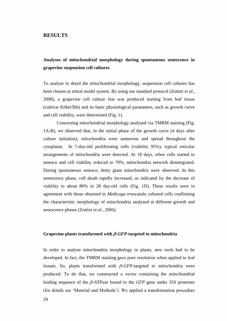
Fig. 4. Comparison of proteins involved in
peroxisomal fission/proliferation in Yeast
and Plants. Peroxisomes proliferation
occurs through elongation, constriction and
fission. In all cases, PEX11 proteins are
involved in the initial elongation, while the
factors involved in peroxisomal constriction
are yet unknown. Peroxisomal fission is
mediated by dynamin-like proteins (in
yeast; Dnm1 and Vsp1; in Arabidopsis:
DRP3A, DRP3B, DRP5B), which are
recruited to the peroxisomal membranes by
the tail-anchored protein FIS1 (in yeast:
Fis1; in Arabidopsis: BIGYIN and FIS1B).
In yeast Caf4 and Mdv1 are adaptors.
(Kaur and Hu, 2009)
16
Peroxules
Peroxisomes morphology could be characterized by tubular protrusions, termed
„peroxules‟ (Scott et al., 2007). These tubules were observed by Cutler et al.
(2000) in Arabidopsis plants stable expressing random GFP::cDNA fusion
proteins obtained to visualize subcellular structures. In detail, Cutler and
colleagues described „peroxules‟ as highly dynamic peroxisomal tubular
extensions. They showed that peroxisomes can change from a spherical to an
elongate morphology in a few seconds, and that this process begins with the
production of a short tubular tail, which then rapidly expands to become longer
than the original organelle.
However, peroxules are described as „common and transient peroxisomal
phenomenon in plant cells‟ (Sinclair et al., 2009), and their role(s) is until now
unknown. Although Cutler et al. (2000) described peroxules as intermediates in
peroxisomal fission/proliferation, it has been also demonstrated that peroxule
extensions occurred in response to low levels of hydroxyl radical reactive oxygen
species (ROS), suggesting that peroxules may be a part of a responsive
machinery, aimed at relief of subcellular stress created by toxic ROS (Sinclair et
al., 2009). Other possible functions of peroxules might be that they increase the
peroxisomal surface-to-volume ratio enhancing the access of cytosolic metabolites
(Jedd and Chua, 2002) or enhancing the exchange of molecules between
peroxisomes (Mano et al., 2002).
Mitochondria, peroxisomes and chloroplasts: metabolic, functional and
physical inter-connections
Analyzing the molecular mechanism involved in fission events of mitochondria,
peroxisomes and chloroplasts in Arabidopsis, a common fission pathway, that
shares several proteins, emerges among these different organelles. Peroxisomes
and mitochondria share the dynamin-related proteins (i.e. DRP3A and DRP3B)
and the Fission1-like proteins (i.e. BIGYIN and FIS1B) (Kaur and Hu, 2009).
Similarly, DRP5B (ARC5), a dynamin protein involved in peroxisomal division in
Arabidopsis, plays a role also in plastid division (Zhang and Hu, 2010). The use
17
of shared components could be a mechanism to promote coordinated division
among these organelles that are at least metabolically linked (Schrader, 2006).
Photorespiration, fatty acid metabolism, and jasmonic biosynthesis are among
some of the metabolite pathways that spanning peroxisomes, chloroplasts and/or
mitochondria (Kaur and Hu, 2009). Moreover, inter-organellar interactions are
fundamental also in several biological processes. For example, chloroplasts and
mitochondria are both involved in generating intermediate signals involved in
plant cell death (PCD) (Van Breusegem and Dat, 2006), suggesting that a
signalling-exchange among these organelles is important for the regulation of
PCD, a fundamental genetically regulated process of cell suicide, that
accomplishes a central role in development, homeostasis and integrity of
eukaryotic organisms.
In the light of the metabolite and signal exchanges among these organelles,
it has been proposed the existence of structures that promote directional
metabolite/molecules trafficking among these organelles. In detail, it has been
suggested that matrixules, peroxules and stromules could be involved in physical
interactions among organelles (Scott et al., 3007; Foyer and Noctor, 2007)). So
far, no data have been reported to confirm this hypothesis.
Leaf senescence: a physiological process where mitochondria and
chloroplasts presumably co-operate
Leaf senescence is the final stage of leaf development and one type of
programmed cell death (PCD; i.e. a genetically controlled system of self
destruction), that occurs in plants (Quirino et al., 2000). Leaf senescence is a slow
physiological process, under the control of several factors both endogenous
(hormones) and external (light, starvation, pathogens). In detail, senescence
involves the ordered disassembly of cellular components that are redirected to
other plant organs, meanwhile senescing tissues eventually die by programmed
cell death (PCD) (Keeck et al., 2007). At the cellular level, the senescence
program unfolds in an orderly manner. Chloroplasts, that contain most of proteins
in leaf cell, are one of the first organelles to be targeted for degradation
18
(Hörtensteiner and Kräutler, 2011). Other organelles, such as peroxisomes,
undergo biochemical changes, while nucleus, necessary for gene transcription,
and mitochondria, necessary for providing energy, remain intact until the last
stages of the senescence (Keeck et al., 2007).
Various cell death-signalling pathways are critically dependent on
mitochondria, whose action is played not only through the release of pro-apoptotic
factors, but also through the alteration of their dynamics and morphology, as
stated before. Recently it has been also suggested that chloroplasts could regulate
the onset of leaf senescence by increasing the reduction state of electron
transporters and by generating reactive oxygen species (ROS) (Zapata et al.,
2005).
Thus, increasing evidences suggest that these organelles co-operate in
signalling-pathway regulation and in the developing of the senescence process,
but a detailed study on this topic has just at the beginning. There is, therefore, a
need for further investigation into the respective roles of chloroplasts and
mitochondria in the process of leaf senescence.
Topics of PhD project
During my PhD project, I focused on organelles in plant cells, analysing in detail
the mitochondrial morphology during senescence/programmed cell death (PCD)
in cell cultures and plants of grapevine (Vitis spp.), an agronomically relevant
species (Chapter 1).
In order to define the molecular mechanisms responsible for mitochondrial
morphology, I moved to Arabidopsis plant, a model organism in plant biology,
and I analyzed in detail two proteins involved in mitochondrial fission machinery
(Chapter 2).
Last, I investigated the molecular aspects of physical inter-organellar
interactions, by imaging analyses of a protein localized to these organelles
(Chapter 3).
19
REFERENCE
- Aldridge C, Maple J, Møller SG (2005).The molecular biology and
plastid division in higher plant. Journal of Experimental Botany, 56
(414):1016-1077
- Arimura SI, Fujimoto M, Doniwa Y, Kadoya N, Nakazono M, Sakamoto
W, Tsutsumi N (2008). Arabidopsis ELONGATED MITOCHONDRIA1
is required for the localization of DYNAMIN-RELATED PROTEIN3A to
mitochondrial fission sites. The Plant Cell, 20(6):1555-66
- Bauwe H, Hagemann M, Ferine AR (2010). Photorespiration: players,
partners and origin. Trends in Plant Science, 15 (6):330-336
- Borghese N, Brambilasca S, Colombo S (2007). How tails guide tail-
anchored proteins to their destinations. Current Opinion in Cell Biology,
19:368–375
- Bowsher CG, Tobin AK (2001). Compartmentation of metabolism within
mitochondria and plastids. Journal of Experimental Botany, 52 (356):513-
518
- Cutler SR, Ehrhardt DW, Griffitts JS, Sommerville CR (2000). Random
GFP::cDNA fusions enable visualization of subcellular structures in cells
of Arabidopsis at high frequency. PNAS, 97 (7):3718-3723
- Delille HK, Alves R, Schrader M (2009). Biogenesis of peroxisomes and
mitochondria: linked by division. Histochem Cell Biol, 131: 441-446
- Foyer CH, Noctor G (2007). Shape-shifters building bridges? Stromules,
matrixules and metabolite channelling in photorespiration. TRENDS in
Plant Science, 12 (9):381-382
- Gao H, Kadirjan-Kalbach D, Froehlich JE, Osteryoung W (2003). ARC5,a
cytosolic dynamin-like protein from plants, is part of the chloroplast
division machinery. PNAS, 100 (7): 4328-4333
- Gray MV (1999). Evolution of organellar genomes. Current Opinion in
Genetics and Development, 9 (6):678-687
- Gray MW, Burger G, Lang BF (1999). Mitochondrial evolution. Science,
283 (5407):1476-1481
20
- Hörtensteiner S and Kräutler (2011). Chlorophyll breakdown in higher
plants. Biochimica et Biophysica Acta, doi:10.1016/j.bbabio.2010.12.007
- Kaur N, Hu J (2009). Dynamic of peroxisomes abundance: a tale of
division and proliferation. Current Opinion in Plant Biology, 12: 781-
788
- Keeck O, Pesquet E, Ahad A, Askne A, Nordvall D, Vodnala SM,
Tuominen H, Hurry V, Dizengremel P, Gardeström P (2007). The
different fates of mitochondrial and chloroplasts during dark-induced
senescence in Arabidopsis leaves. Plant, Cell and Environment, 30:1523-
1534
- Köhler RH, Hanson MR (2000). Plastid tubules of higher plants are
tissue-specific and developmentally regulated. Journal of Cell Science,
113 (1):81-89
- Kwok EY, Hanson MR (2004a). GFP-labelled Rubisco and aspartate
aminotransferase are present in plastid stromules and traffic between
plastids. Journal of Experimental Botany, 55(397):595-604
- Kwok EY, Hanson MR (2004b). Stromules and dynamic nature of plastid
morphology. Journal of Microscopy, 214 (2):124-137
- Jaillon O, Aury JM, Noel B, Policriti A, Clepet C, Casagrande A, Choisne
N, Aubourg S, Vitulo N, Jubin C, Vezzi A, Legeai F, Hugueney P, Dasilva
C, Horner D, Mica E, Jublot D, Poulain J, Bruyère C, Billault A, Segurens
B, Gouyvenoux M, Ugarte E, Cattonaro F, Anthouard V, Vico V, Del
Fabbro C, Alaux M, Di Gaspero G, Dumas V, Felice N, Paillard S, Juman
I, Moroldo M, Scalabrin S, Canaguier A, Le Clainche I, Malacrida G,
Durand E, Pesole G, Laucou V, Chatelet P, Merdinoglu D, Delledonne M,
Pezzotti M, Lecharny A, Scarpelli C, Artiguenave F, Pè ME, Valle G,
Morgante M, Caboche M, Adam-Blondon AF, Weissenbach J, Quétier F,
Wincker P; French-Italian Public Consortium for Grapevine Genome
Characterization. (2007) The grapevine genome sequence suggests
ancestral hexaploidization in major angiosperm phyla. Nature 449: 463-
467
21
- Jedd G, Chua NH (2002). Visualization of peroxisomes in living plant
cells reveals acto-myosin dependent cytoplasmic streaming and
peroxisome budding. Plant Cell Physiology, 43 (4):384-392
- Lam E (2004). Controlled cell death, plant survival and development.
Molecular Cell Biology, 4:305-315
- Logan DC (2006). The mitochondrial compartment. Journal of
Experimental Botany, 57 (6): 1225-1243
- Logan DC, Scott I, Tobin AK (2004).AL2a, like ADL2b, is involved in
the control of higher plant mitochondrial morphology. Journal of
Experimental Botany, 55(397):783-785
- Logan DC, Knight MR (2003). Mitochondrial and cytosolic calcium
dynamics are differentially regulated in plants. Plant Physiology, 133: 21-
24
- Logan DC (2003). Mitochondrial dynamics. New Phytologist, 160 (3):
463–478
- Michels PA, Moyersoen J, Krazy H, Galland N, Herman M, Hannaert V
(2005). Peroxisomes, glyoxysomes and glycosomes. Molecular membrane
biology, 22(1-2):133-145
- Mai S, Klinkenberg M, Auburger G, Bereiter-Hahn J, Jendrach M (2010).
Decreased expression of Drp1 and Fis1 mediates mitochondrial elongation
in senescent cells and enhances resistance to oxidative stress through
PINK1. Journal of Cell Science, 123: 917-926
- Mano S, Nakamori C, Hayashi M, Kato A, Kondo M, Nishimura M
(2002). Distribution and characterization of peroxisomes in Arabidopsis
by visualization with GFP: dynamic morphology and actin-dependent
movement. Plant Cell Physiology, 43 (3):331-341
- Maple J and Møller SG (2007). Plastid division coordination across a
double-membraned structure. FEBS Letter, 581 (11):2162-2167.
- Miyagishima SY, Froehlich JE, Osteryoung KW (2006). PDV1 and
PDV2 mediate recruitment of the dynamin-related protein ARC5 to the
plastid division site. The Plant Cell, 18 (10):2517-2530
- Noctor G, De Paepe R, Foyer CH (2007). Mitochondrial redox biology
and homeostasis in plants. TRENDS in Plant Science, 12 (3):125-134
22
- Quirino BF, Noh YS, Himelblau E, Amasino RM (2000). Molecular
aspects of leaf senescence. 5(7):278-82.
- Rhoads DM, Subbaiah C (2007). Mitochondrial retrograde regulation in
plants. Mitochonrion, 7: 177-194
- Schrader M, Yoon Y (2007). Mitochondria and peroxisomes: are the „Big
Brother‟ and the „Little Sister‟ closer than assumed? BioEssay, 29: 1105-
1114
- Schrader M (2006). Shared components of mitochondrial and
peroxisomal division. Biochimica et Biophysica Acta, 1763(5-6):531-541
- Scott, I and Logan, D.C. (2008) Mitochondrial morphology transition is
an early indicator of subsequent cell death in Arabidopsis. New
Phytologist 177, 90-101
- Scott I, Sparkes IA, Logan DC (2007). The missing link: inter-organellar
connection in mitochondria and peroxisomes? TRENDS in Plant Science,
12 (9):380-381
- Scott I, Tobin AK, Logan DC (2006). BIGYIN, an orthologue of human
and yeast FIS1 genes functions in the control of mitochondrial size and
number in Arabidopsis thaliana. Journal of Experimental Botany, 57
(6):1275-1280
- Sinclair AM, Trobacher CP, Mathur N, Greenwood JS, Mathur J (2009).
Peroxule extension over ER-defined paths constitutes a rapid subcellular
response to hydroxyl stress. The Plant Journal, 59:231-247
- Suen DF, Norris KL, Youle RJ (2008). Mitochondrial dynamics and
apoptosis, Genes and Development, 22: 1577-1590
- Suzuki M, Jeong SY, Karbowski M, Youle RJ, Tjandra N (2003). The
solution structure of human mitochondria fission protein Fis1 reveals a
novel TPR-like helix bundle. Journal of Molecular Biology, 334 (3):445-
58.
- Sweetlove L, Fait A, Nunes-Nesi A, Williams T, Fernie AR (2007). The
mitochondrion: an integration point of cellular metabolism and signalling.
Critical Reviews in Plant Sciences, 26:17-43
- Van Breusegem F, Dat JF (2006). Reactive oxygen species in plant cell
death. Plant Physiology, 141 (2):384-90
23
- Vandecasteele G, Szabadkai G, Rizzuto R (2001). Mitochondrial calcium
homeostasis: mechanisms and molecules. IUBMB Life, 52: 213-219
- Velasco R, Zharkikh A, Troggio M, Cartwright DA, Cestaro A, Pruss D,
Pindo M, FitzGerald LM, Vezzulli S, Reid ., Malacarne G, Iliev D,
Coppola G, Wardell B, Micheletti D, Macalma TM, Facci M,. Mitchell JT,
Perazzolli M, Eldredge G, Gatto P, Oyzerski R, Moretto M, Gutin N,
Stefanini M, Chen Y, Segala C, Davenport C, Demattè L, Mraz A,
Battilana J, Stormo K, Costa F, Tao Q, Si-Ammour A, Harkins T, Lackey
A, Perbost C, Taillon B, Stella A, Solovyev V, Fawcett JA, Sterck L,
Vandepoele K, Grando MS, Toppo S, Moser C, Lanchbury J, Bogden R,
Skolnick M, Sgaramella V,. Bhatnagar SK, Fontana P, Gutin A, Van de
Peer Y, Salamini F, Viola RA, (2007) High quality draft consensus
sequence of the genome of a heterozygous grapevine variety, PLoS ONE ,
2 (12), p. e1326
- Westermann B, (2010). Mitochondrial fusion and fission in cell life and
death. Molecular Cell Biology, 11: 872-884
- Zapata JM, Guera A, Esteban-Charrasco A, Martin M, Sebater B (2005).
Chloroplast regulate leaf senescence: delayed senescence in transgenic
ndhF-defective tobacco
- Zhang XC, Hu JP (2010). The Arabidopsis chloroplast division protein
DYNAMIN-RELATED PROTEIN5B also mediates peroxisomes division.
The Plant Cell, 22 (2):431-42.
- Zhang XC, Hu JP (2008a).Two small protein families, DYNAMIN-
RELATED PROTEIN3 and FISSION1, are required for peroxisome
fission in Arabidopsis. The Plant Journal, 57(1):146-59
- Zhang XC, Hu JP (2008b). FISSION1A and FISSION1B proteins
mediate the fission of peroxisomes and mitochondria in Arabidopsis.
Molecular Plant, 1(6):1036-47
- Zottini M, Barizza E, Bastianelli F, Carimi F, Lo Schiavo F (2006).
Growth and senescence of Medicago truncatula cultured cells are
associated with characteristic mitochondrial morphology. New
Phytologist, 172 :239-247

24
25
Chapter 1
Changes in mitochondrial morphology associated
with cell aging during grapevine leaf spontaneous
senescence

26
27
INTRODUCTION
Leaf senescence is the final stage of leaf development and it is a slow
physiological process, under the control of several factors both endogenous
(hormones) and external (light, starvation, pathogens). Senescence involves the
ordered disassembly of cellular components that are redirected to other plant
organs; meanwhile senescing tissues eventually die by programmed cell death
(PCD) which presents some typical hallmarks of apoptosis (Yoshida, 2003).
Understanding the molecular mechanisms used by the plant to regulate
senescence might provide applicative outputs, especially in agronomical relevant
species, such as grapevine (Vitis spp). Moreover, after the sequencing of its
genome (Jaillon et al., 2007; Velasco et al., 2007), grapevine has the potential to
become a model organism for fruit trees. A decrease in the leaf photosynthetic
efficiency during grapes maturation may, actually, result in an insufficient sugar
level in berries. The knowledge of the regulative aspects of leaf senescence and
berry ripening might lead to govern plant growth and development and possibly to
control the environmental conditions affecting senescence and berry yield.
In eukaryotic cells, mitochondria play a central role in energy and carbon
metabolism (Siedow and Day, 2000), but they also play a significant role in
control of programmed cell death pathways, as a stress sensor and dispatcher.
Various cell death-signalling pathways are indeed critically dependent on
mitochondria, whose action is played not only through the release of pro-apoptotic
factors such as cytochrome c, but also through the alteration of their dynamics and
morphology. Although mitochondria are often portrayed as static, oval or rod-
shaped organelles, recent studies have demonstrated that they are among the most
plastic organelles of cells in terms of form and distribution. Yet, changes in their
architecture and their ability to move rapidly throughout the cytoplasm appear to
be of critical importance for executing their cellular functions. A link between
senescence-associated cell death and plant mitochondrial dynamics and
morphology has been reported in Medicago truncatula cell cultures (Zottini et al.,
2006) and confirmed in Arabidopsis leaf and mesophyll protoplasts (Scott and
Logan, 2008). In particular, in Medicago truncatula cell cultures it has been
observed that alterations in mitochondrial dynamics and morphology were
28
associated with cell ageing during senescence occurring spontaneously (Carimi et
al. 2004).
In this report, analyses of mitochondrial morphology were performed first
on grapevine cell cultures and then on grapevine leaf tissue. We produced
suspension cell cultures starting from leaf tissue (Zottini et al., 2008) to perform
experiments in cultured cells, and embriogenic cell lines (Carimi et al., 2005) to
be used in transformation procedures. For analyses in leaf tissues, in fact, we
produced plants transformed with the green fluorescent protein (GFP) targeted to
the mitochondria. These transgenic plants allowed us to bypass the technical
problem of poor staining of plant tissues with exogenous fluorescent dyes (Kohler
et al., 1997). By using them, we were able to detect, in an accurate way, changes
of mitochondrial morphology occurring at different stages of grapevine leaf
senescence.
29
RESULTS
Analyses of mitochondrial morphology during spontaneous senescence in
grapevine suspension cell cultures
To analyse in detail the mitochondrial morphology, suspension cell cultures has
been chosen as initial model system. By using our standard protocol (Zottini et al.,
2008), a grapevine cell culture line was produced starting from leaf tissue
(cultivar Köber5bb) and its basic physiological parameters, such as growth curve
and cell viability, were determined (Fig. 1).
Concerning mitochondrial morphology analysed via TMRM staining (Fig.
1A-B), we observed that, in the initial phase of the growth curve (4 days after
culture initiation), mitochondria were numerous and spread throughout the
cytoplasm. In 7-day-old proliferating cells (viability 95%), typical reticular
arrangements of mitochondria were detected. At 18 days, when cells started to
senesce and cell viability reduced to 70%, mitochondria network disintegrated.
During spontaneous senesce, dotty giant mitochondria were observed. In this
senescence phase, cell death rapidly increased, as indicated by the decrease of
viability to about 80% in 28 day-old cells (Fig. 1D). These results were in
agreement with those obtained in Medicago truncatula cultured cells confirming
the characteristic morphology of mitochondria analysed at different growth and
senescence phases (Zottini et al., 2006).
Grapevine plants transformed with -GFP-targeted to mitochondria
In order to analyse mitochondria morphology in plants, new tools had to be
developed. In fact, the TMRM staining gave poor resolution when applied to leaf
tissues. So, plants transformed with -GFP-targeted to mitochondria were
produced. To do that, we constructed a vector containing the mitochondrial
leading sequence of the -ATPase bound to the GFP gene under 35S promoter
(for details see „Material and Methods‟). We applied a transformation procedure
30
using embryogenic cell cultures as starting material. Embryogenic cultures were
the starting material for transformation experiments. GFP-transformed embryos
were visualized by epi-fluorescence stereomicroscope: GFP expression was
detectable with a patchy distribution in ten days embryogenic callus (Fig. 2A; t1).
Three and four months later (Fig. 2A; t2 and t3), transformed globular and
torpedo embryos were clearly observed, respectively. In order to avoid chimeras,
secondary embryos were regenerated from primary transgenic plantlets (Fig. 2 B).
Adult plants obtained from those selected transgenic secondary embryos
maintained GFP fluorescence in leaf and root tissues (Fig. 2B) thus demonstrating
the absence of silencing phenomena eventually developed during plant growth.
Laser scanning confocal microscopy was employed to determine the distribution
of GFP-fluorescent mitochondria at the cellular level. In Fig. 2C mitochondria
were visualized at the embryo level, in the following paragraphs analyses at the
level of leaf tissues will be presented.
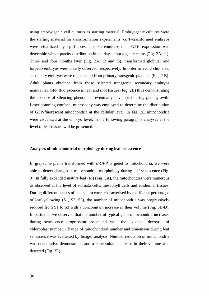
Analyses of mitochondrial morphology during leaf senescence
In grapevine plants transformed with -GFP targeted to mitochondria, we were
able to detect changes in mitochondrial morphology during leaf senescence (Fig.
3). In fully expanded mature leaf (M) (Fig. 3A), the mitochondria were numerous
as observed at the level of stomata cells, mesophyll cells and epidermal tissues.
During different phases of leaf senescence, characterised by a different percentage
of leaf yellowing (S1, S2, S3), the number of mitochondria was progressively
reduced from S1 to S3 with a concomitant increase in their volume (Fig. 3B-D).
In particular we observed that the number of typical giant mitochondria increases
during senescence progression associated with the expected decrease of
chloroplast number. Change of mitochondrial number and dimension during leaf
senescence was evaluated by ImageJ analysis. Number reduction of mitochondria
was quantitative demonstrated and a concomitant increase in their volume was
detected (Fig. 3E).
31
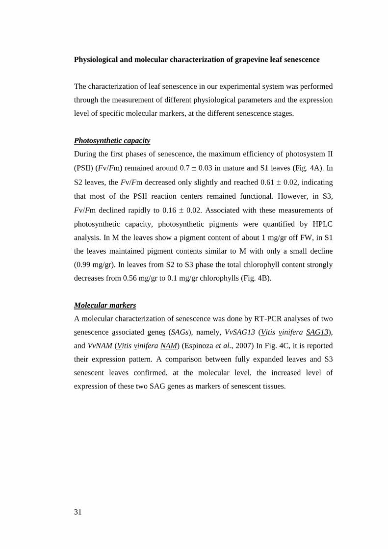
Physiological and molecular characterization of grapevine leaf senescence
The characterization of leaf senescence in our experimental system was performed
through the measurement of different physiological parameters and the expression
level of specific molecular markers, at the different senescence stages.
Photosynthetic capacity
During the first phases of senescence, the maximum efficiency of photosystem II
(PSII) (Fv/Fm) remained around 0.7 0.03 in mature and S1 leaves (Fig. 4A). In
S2 leaves, the Fv/Fm decreased only slightly and reached 0.61 0.02, indicating
that most of the PSII reaction centers remained functional. However, in S3,
Fv/Fm declined rapidly to 0.16 0.02. Associated with these measurements of
photosynthetic capacity, photosynthetic pigments were quantified by HPLC
analysis. In M the leaves show a pigment content of about 1 mg/gr off FW, in S1
the leaves maintained pigment contents similar to M with only a small decline
(0.99 mg/gr). In leaves from S2 to S3 phase the total chlorophyll content strongly
decreases from 0.56 mg/gr to 0.1 mg/gr chlorophylls (Fig. 4B).
Molecular markers
A molecular characterization of senescence was done by RT-PCR analyses of two
senescence associated genes (SAGs), namely, VvSAG13 (Vitis vinifera SAG13),
and VvNAM (Vitis vinifera NAM) (Espinoza et al., 2007) In Fig. 4C, it is reported
their expression pattern. A comparison between fully expanded leaves and S3
senescent leaves confirmed, at the molecular level, the increased level of
expression of these two SAG genes as markers of senescent tissues.

32
33
DISCUSSION
In this work we describe the mitochondria dynamics and morphology occurring in
Vitis vinifera leaves during natural senescence. The molecular aspects of the
various stages of senescence are not known in details; due to the slowness and
heterogeneity of the process. Being simple and homogeneous, cell cultures
represent an ideal system for studying some general aspects of senescence,
allowing defining the relationships between senescence and PCD (Carimi et al.
2004).
In order to perform an accurate analysis of mitochondria, during
senescence, at the cellular level, we used suspension cell cultures for our initial
experiments. So, a grapevine cell culture from leaf tissues was produced and its
growth curve established. The growth curve, as usual, identified different phases:
initial, in which cells condition their medium; log, in which cell division takes
place; and final, in which cells stop dividing, elongate and senesce. In fact, if cells
were not sub-cultured at the end of the growth period, they started senescing and
PCD ensued. The last phase of the curve is identified as spontaneous senescence
phase. Growing cultured cells showed typical reticular arrangements of fast
moving mitochondria; the networks disappear in ageing cells and giant, not
mobile mitochondria characterised this type of cells. These results are matching
the ones observed in Medicago truncatula cell cultures, where similar analyses
were done (Zottini, et al. 2006).
Then, we developed new tools to observe alterations of mitochondrial
morphology in plant. In fact, we produced grape plants transformed with GFP
targeted to mitochondria and we were able to detect in an accurate way
morphology and dynamics of mitochondria during leaf senescence. A significant
progress in producing transgenic grapevines was made when embryogenic cell
lines were used as target tissue (Vivier and Pretorius, 2002). Our transformation
protocol based, in fact, on long lasting embryogenic cell lines, that we produced
and maintained as such indefinitely. We were able to achieve that alternating
cycles of cell de-differentiation with cycles of embryo differentiation (see
„Material and Methods‟ for detail). The use of PEM combined with
Agrobacterium infection provided an efficient transformation procedure.
34
As reported in fig.4, we observed the evolution of mitochondrial
morphology at different leaf stages. In fully expanded green leaf, the
mitochondria visualised in different tissues (i.e. epidermis, stomata and mesophyll
cells), were numerous and mobile. In senescent leaf, their number reduced, their
volume increased and giant mitochondria are detected. The ageing cells of
senescent leaf tissue is therefore characterized by these mitochondrial
morphological alterations. These data are in agreement with the alterations of
mitochondrial morphology and motility observed in Arabidopsis protoplast and
leaf tissues during induced PCD (Yao et al.,2004; Scott and Logan, 2008).
It is important to underline the similarity of events observed in cell
cultured and in leaf tissue. The correspondence between the evolution of
dynamics and morphology of mitochondria from growth to ageing cells in
cultures and from greening to yellowing cells in leaf tissue confirms that analyses
performed in cell cultures may contribute positively in dissecting and understand
cellular mechanisms of important physiological processes, such as cell ageing and
senescence in plants. This is, in fact, a good example in which results from
experiments performed in cultured cells can be used as guidelines to perform
experiments in complex tissues.
Yet, the understanding of some of the cellular mechanisms occurring
during grapevine senescent events might lead to a controlled regulation of this
process with important potentials in improving quantity and quality of an
important crop production and its post-harvest shelf life.
35
MATERIAL AND METHODS
Suspension cell cultures and TMRM treatment
Suspension cell cultures preparation was according to Zottini et al. (2008).
Briefly, grapevine cultivar Köber5bb cell lines were obtained from leaf dish
explants incubated on selective B5 (Gamborg B5 medium, Duchefa; Gamborg et
al. 1968) solid (8 g l-1
agar) medium supplemented with 2.26 µM 2,4-
dichlorophenoxy-acetic acid (SIGMA) (B5F). After several subculture cycles
aliquots of callus were utilized for liquid cultures. For subculture cycles, 2 ml
were transferred to Erlenmeyer flasks (250 ml) filled with 50 ml liquid B5
medium supplemented with 2.26 µM 2,4-dichlorophenoxy-acetic acid . The
suspension cultures were subcultured in fresh medium every week and maintained
in a climate growth chamber at 25 °C on an orbital shaker (80 rpm) under a 16 h
day length. To determine dry weight, integer cells were separated from the culture
medium and cell debris through a vacuum filtration unit (Sartorius, Florence,
Italy).
A Nikon PCM2000 laser scanning confocal microscope (Nikon, Italy) was
used for analysis of mitochondrial morphology. The tetramethylrhodamine methyl
ester dye (TMRM) (Molecular Probes, Leiden, the Netherlands), a mitochondrial
membrane potential sensor, was used for visualizing mitochondria in cell culture
as described by Zottini et al. (2006). Cell suspensions (300 μl) were collected at
different times during their growth cycle, and incubated in 700 μl B5F medium
containing 1 μM TMRM for 15 min on a rotary shaker. Cells were centrifuged for
3 min at 10 000 xg, the supernatant was discarded and the pellet washed twice
with 700 μl B5F. Cells were then resuspended in 500 μl B5F. For microscope
analysis, 100 μl cell suspension was placed on a microscope slide and visualized
under a confocal microscope (excitation 548 nm, emission 573 nm). Images were
processed using Corel PHOTO -PAINT. For mitochondrial morphology
experiments, a randomized complete block design was used with three replicates
(individual Erlenmeyer flasks). Each experiment was repeated three times.
36
Cell viability assay
Cell viability was determined by fluorescein diacetate (FDA) assay according to
Amano et al. (2003). Immediately before each assay, a stock solution of FDA
(0.5% w/v in acetone) was diluted with distilled water to create a fresh 0.01% w/v
FDA working solution which was kept in the dark at 4°C. Cell suspensions of
grapevine was aliquot in 2 ml fractions on Poly-Prep Cromatography columns
(BioRad) then diluited 1/10 with PBS (2.7mM KCl, 137mM NaCl, 1.8mM
KH2PO4, 4.0mM Na2HPO4). 100 l of this solution was then mixed by gentle
stirring with 0.01% w/v FDA in a quartz cuvette. A spectrophotofluorimeter
(Perkin Elmer, UK) equipped with a stirrer was employed. Excitation and
emission wavelengths were selected at 493 and 510 nm, respectively. The
increase in fluorescence was recorded over a 120-s time period. The slope of the
fluorescence increase (between 60 to 90 s) was calculated for each cellular
suspension to determine the correlation between cell viability and the velocity of
FDA conversion. A standard cell viability curve was set up using several cellular
suspensions containing different known amounts of viable cells. To achieve this,
dead cells were prepared by boiling of viable cells. After, aliquots of these control
dead cells were added to several different quantities of healthy viable cells to
obtain suspensions whose cellular viabilities varied between 0 and 100% (in
increments of 20%). Cell suspension set was then used to determine the
correlation between FDA conversion and cell viability using a spectrofluorimetric
assay.
Cell cultures and plant material
Plasmid construction and Agrobacterium tumefaciens used for transformation
For the expression of GFP targeted to the mitochondria, the Agrobacterium
tumefaciens strain GV3101 harbouring the p BI121 binary vector, with a T-DNA
incorporating the Green Fluorescent Protein (GFP) gene targeted to mitochondria
(β::GFP) was obtained following the procedures as previously described by
Zottini et al. (2008). Briefly, the cDNA coding sequences of β::GFP were
subcloned from the β::GFP plasmid (Zhao et al., 2000; Duby et al., 2001) to the

37
pBI121 binary vector (Clontech Laboratories, USA) by replacing the β-
glucuronidase cDNA sequence. For sub-cloning, the β-GFP fusion construct into
the pBI121 binary vector the BglII/SacI restriction sites were used. The p BI121
binary vector contain the coding sequence for neomycin phosphotransferase II
(nptII) that allowed for selection of transgenic cells based on kanamycin
resistance.
Competent cells of Agrobacterium tumefaciens GV3101 strain were
prepared according to Sambrook et al. (1989) and the binary vectors were
introduced by electroporation as described by Zottini et al. (2008). The growth of
bacteria was optimized by growing in YEP medium (Bacto-Trypton, 10 gL-1
;
yeast extract, 10 gL-1
, NaCl, 5 gL-1
; pH 7.0) The media were supplemented with
the antibiotics rifampicin 100 mgL-1
, gentamycin 50 mgL-1
, kanamycin 50 mgL-1
.
Regeneration of embryogenic cell lines
Embryogenic cell lines of grapevine (cv Moscato giallo) were produced from
stigma/style cultures as described by Carimi et al. (2005). Briefly, explants were
dissected from unopened flowers and placed on Nitsch and Nitsch (1969) salts
and vitamins, 88 mM sucrose, 9 µM BA and 10 µM NOA. Medium pH was
adjusted to 5.7 before the addition of 8 g l-1
Plant agar (Duchefa) and autoclaving
at 121 °C for 20 min. Cultures were placed in an acclimatized cabinet at 25°C and
16 h light photoperiod, and subcultured at 30-day intervals. White embryogenic
globules, around 1-3 mm in size, were separated from the callus grown from the
original stigma/style explants and were cultured alternating, every 3 weeks, solid
MS growth regulator free medium and solid B5 2.26 µM 2-4D medium. The
alternation of such medium permitted us to maintain the embryogenic cell line for
long time.
Liquid suspensions for transformation were initiated from habituated
embryogenic cultures by transferring 1 g of PEM collected from solid MS growth
regulator free medium (MS-) to Erlenmeyer flasks (250 ml) filled with 50 ml
liquid MS- medium. The flasks were cultured for 3 days on an orbital shaker at 80
rpm and incubated at 25 °C in the dark.
38
Transformation of embryogenic cultures and selection of transgenic plants
Before transformation with Agrobacterium tumefaciens, 500 mg of PEM and
embryos were transferred into a petri disc contained 1 ml of liquid induction
medium (LIM = NN medium supplemented with 58 mM sucrose, 2.26 µM 2,4-
D) and were incised with a sharp razor blade. Bacteria suspension preparation was
according to Zottini et al. (2008). Agrobacterium tumafaciens suspension was
diluted to OD550 0.5 in LIM and added (5 mL) to the dissected embryos that were
previously transferred in bacteria-free LIM (1 mL). Embryo were incubated (room
temperature, dark) for 10 min after which the cultures were washed 5 times with
induction medium (3 min washing). Infected embryos were blotted dry on sterile
filter paper and plated on NN solid medium and incubated at 25 °C in the dark.
Two days later the cultures were transferred to NN solid medium supplemented
with 300 mgL-1
cefotaxime and maintained at 25°C in the dark. After 10 days the
cultures were transferred on NN solid medium supplemented with 20 mgL-1
kanamycin and 300 mgL-1
cefotaxime. After 20 days the cultures were transferred
on NN solid medium supplemented with 40 mgL-1
kanamycin and 300 mgL-
1cefotaxime and subcultured at 20 day-intervals.Embryo clusters differentiated at
the callus surface maintained on 40 mgL-1
kanamycin were collected and
transferred on NN hormone free solid medium for germination. Individual
germinated somatic embryos were transferred to Microbox Containers (Duchefa,
The Netherland) in half strength MS solid medium (0.8% plant agar Duchefa, The
Netherland) supplemented with 44 mM sucrose and were multiplied by clonal
propagation and maintained (30-day intervals). Plants were incubated in a growth
chamber at 25+1 °C under a 16 h day length, and a photosynthetic photon flux of
35 μmol m-2
s-1
Osram cool-white 18 W fluorescent lamps.
Hydroponic cultivation
Transformed plants grown in vitro were transferred with roots, after elimination of
agar by washing, to hydroponic conditions. The nutrient solution composition was
designed, tested and optimized for Vitis vinifera : 0.5 mM KH2PO4; 0.5 mM
K2SO4; 2 mM Ca(NO3)2.4H2O; 0.65 mM MgSO4; 0.5 μM H3BO3; 0.045 μM
CuSO4X 5H2O; 0.05 μM ZnSO4 X 7H2O; 0.02 μM (NH4)6Mo7O24 X 4H2O; 0.5
μM MnSO4; 10 μM Fe-EDDHA. The hydroponic system consists in a Microbox
39
Containers containing a floating polystyrene circle with a sponge placed in the
middle holding the plant.
Evaluation of gene expression trough fluorescent proteins
GFP-dependent fluorescence in leaves was analyzed using an epifluorescence
stereo microscope. Suspension cells and transformed leaves were analyzed using a
confocal microscopy Nikon PCM2000 (Bio-Rad, Germany) laser scanning
confocal imaging system. For GFP detection, excitation was at 488 nm and
emission between 515/530 nm. Image analysis was done with the ImageJ bundle
software (http://rsb.info.nih.gov/ij/).
Semi-quantitative RT-PCR analysis in pBIGYIN::GUS plants
Total RNA was extracted from leaved characterized by different phases of leaf
senescence (M, S1, S2, S4). RNA isolation was carried out using the 'Master Pure
Plant RNA Purification‟ Kit (EPICENTRE® Biotechnologies), according to
manufacturer‟s specification. After DNAse I treatment (Ambion Ltd, UK), first
strand synthesis and PCR were carried out starting from 1 g of total RNA,
according to the manufacturer‟s instructions (ImProm Reverse Transcriptase,
Promega). After first strand cDNA synthesis, samples were diluted 5 times and
used as templates for semi-quantitative RT-PCR. RT-PCR analyses were
performed using the follow specific primers: VvActin-1 (housekeeping gene, For:
5‟ -GACAATGGAACTGGAATGGTGAAG-3‟; Rev 5'-
TACGCCCACTGGCATATAGAGAAA-3‟), for VvSAG13 (For: 5‟-
GCTTCCTGCTCCAGATGC-3‟; Rev: 5‟- TGCCACCGTACACACCTG-3‟), for
VvNam For: 5‟-ATGCTCACAATCCGTAACCG-3‟; Rev 5‟-
CAGCCACAACATCAAGCATC-3‟).
RT-PCR reactions were performed using GoTaq®
DNA Polymerase (Promega), in
a total reaction volume of 50µL according to manufacturer's recommendations
containing 5µL of cDNA. PCR amplification cycle was performed with an initial
denaturation step at 94°C for 2min, followed by 32 cycles for avVvActin-1, 31
cycle for VvNam and 28 cycles for VvSAG13 (95°C for 20s; 61°C for 30s; 72°C
for 30s), and finally with an elongation step at 72°C for 5min.
40
REFERENCES
- Amano T, Hirasawa K, O‟Donohue MJ, Pernolle JC, Shioi Y (2003). A
versatile assay for the accurate, time-resolved determination of cellular
viability. Analytical Biochemistry, 314: 555-565
- Carimi F, Terzi M, De Michele R, Zottini M, Lo Schiavo F (2004). High
levels of cytokinin BAP induce PCD by accelerating senescence. Plant
Science, 166 : 963-969
- Carimi F, Barizza E, Gardiman M, Lo Schiavo F (2005). Somatic
embryogenesis from stigmas and styles of grapevine. In vitro cellular
development biology – plant, 41(3): 249-252
- Duby G, Oufattole M, Boutry M (2001) Hydrophobic residues within the
predicted N-terminal amphiphilic alpha-helix of a plant mitochondrial
targeting presequence play a major role in in vivo import. Plant Journal,
27:539–549
- Espinoza C, Medina C, Sommerville S, Arce-Johnson (2007).
Senescence-associated genes induced during compatible viral interactions
with grapevine and Arabidopsis. Journal of Experimental Botany, 58
(12):3197-3212
- Gamborg OL, Miller RA, Ojima K (1968). Nutrient requirements of
suspension cultures of soybean root cells. Experimental Cell Research
50:151-158.
- Jaillon O, Aury JM, Noel B, Policriti A, Clepet C, Casagrande A, Choisne
N, Aubourg S, Vitulo N, Jubin C, Vezzi A, Legeai F, Hugueney P, Dasilva
C, Horner D, Mica E, Jublot D, Poulain J, Bruyère C, Billault A, Segurens
B, Gouyvenoux M, Ugarte E, Cattonaro F, Anthouard V, Vico V, Del
Fabbro C, Alaux M, Di Gaspero G, Dumas V, Felice N, Paillard S, Juman
I, Moroldo M, Scalabrin S, Canaguier A, Le Clainche I, Malacrida G,
Durand E, Pesole G, Laucou V, Chatelet P, Merdinoglu D, Delledonne M,
Pezzotti M, Lecharny A, Scarpelli C, Artiguenave F, Pè ME, Valle G,
Morgante M, Caboche M, Adam-Blondon AF, Weissenbach J, Quétier F,
Wincker P; French-Italian Public Consortium for Grapevine Genome
Characterization (2007). The grapevine genome sequence suggests
41
ancestral hexaploidization in major angiosperm phyla. Nature, 449: 463-
467
- Kohler RH, Zipfel WR, Webb WW, Hanson MR (1997). The green
fluorescent protein as a marker to visualize plant mitochondria in vivo. The
Plant Journal, 11(3): 613-621
- Main GD, Reynolds S, Gartland JS (1995) Electroporation protocols in
Agrobacterium. In: Gartland KMA, Davey MR (eds) Methods in
molecular biology, vol 44: Agrobacterium protocols. Humana Press,
Totowa, pp 405–412
- Nitsch JP; Nitsch C (1969). Haploid plants from pollen grains. Science,
163:85-87.
- Sambrook J, Fritsch EF, Maniatis T (1989). Molecular Cloning: A
Laboratory Manual, 2nd edn. Cold Spring Harbor Laboratory Press, New
York.
- Siedow JN, Day DA (2000). Respiration and photorespiration, in
Biochemistry and Molecular Biology of Plants, edited by B. B. Buchanan,
W. Gruissem and R. L. Jones, Eds., American Society of Plant
Physiologists, pp. 676-728.
- Scott I, Logan DC (2008). Mitochondrial morphology transition is an
early indicator of subsequent cell death in Arabidopsis. New Phytologist,
177, 90-101
- Velasco R, Zharkikh A, Troggio M, Cartwright DA, Cestaro A, Pruss D,
Pindo M, FitzGerald LM, Vezzulli S, Reid ., Malacarne G, Iliev D,
Coppola G, Wardell B, Micheletti D, Macalma TM, Facci M,. Mitchell JT,
Perazzolli M, Eldredge G, Gatto P, Oyzerski R, Moretto M, Gutin N,
Stefanini M, Chen Y, Segala C, Davenport C, Demattè L, Mraz A,
Battilana J, Stormo K, Costa F, Tao Q, Si-Ammour A, Harkins T, Lackey
A, Perbost C, Taillon B, Stella A, Solovyev V, Fawcett JA, Sterck L,
Vandepoele K, Grando MS, Toppo S, Moser C, Lanchbury J, Bogden R,
Skolnick M, Sgaramella V,. Bhatnagar SK, Fontana P, Gutin A, Van de
Peer Y, Salamini F, Viola RA, (2007) High quality draft consensus
sequence of the genome of a heterozygous grapevine variety, PLoS ONE ,
2 (12), p. e1326
42
- Vivier MA, Pretorius IS (2002). Genetically tailored grapevines for the
wine industry. Trends in Biotechnology, 20, 472-478
- Yao N, Eisfelder BJ, Marvin J, Greenberg, JT (2004). The mitochondrion-
an organelle commonly involved in programmed cell death in Arabidopsis
thaliana. Plant Journal, 40:596-610
- Yoshida S (2003). Molecular regulation of leaf senescence. Current
Opinion of Plant Biology, 6: 79-84
- Zhao R, Dielen V, Kinet JM, Boutry M (2000) Cosuppression of a plasma
membrane H+-ATPase isoform impairs sucrose translocation, stomatal
opening, plant growth, and male fertility. Plant Cell, 12:535–546
- Zottini M, Barizza E, Bastianelli F, Carimi F, Lo Schiavo F (2006).
Growth and senescence of Medicago truncatula cultured cells are
associated with characteristic mitochondrial morphology. New
Phytologist, 172: 239-247
- Zottini M, Barizza E, Costa A, Formentin E, Ruberti C, Carimi F and Lo
Schiavo F (2008). Agroinfiltration of grapevine leaves for fast transient
assays of gene expression and for long-term production of stable
transformed cells. Plant Cell Report. 27 (5): 845-853
43
Chapter 2
BIGYIN, a tail anchored protein, recruits cytosolic
ELM1 protein at mitochondria and chloroplast
level

44
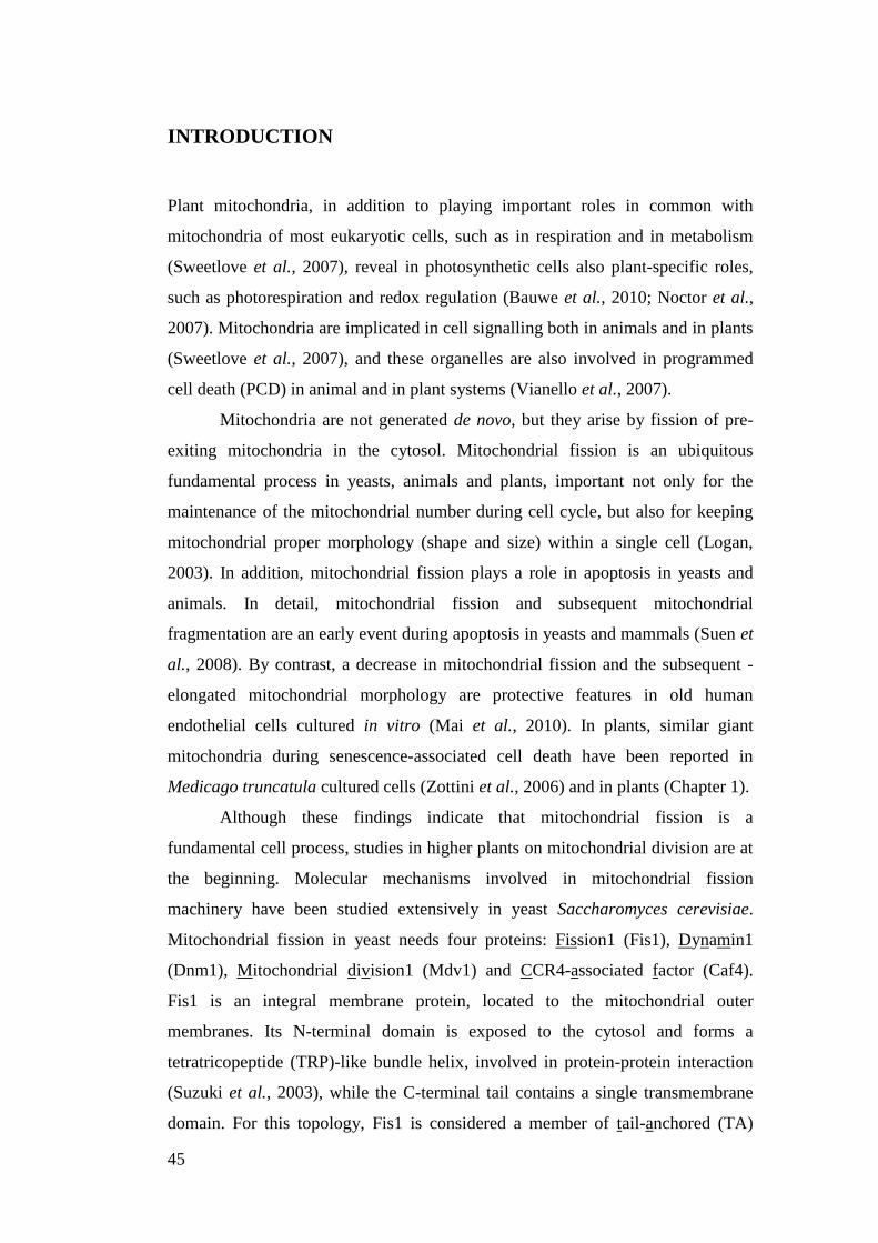
45
INTRODUCTION
Plant mitochondria, in addition to playing important roles in common with
mitochondria of most eukaryotic cells, such as in respiration and in metabolism
(Sweetlove et al., 2007), reveal in photosynthetic cells also plant-specific roles,
such as photorespiration and redox regulation (Bauwe et al., 2010; Noctor et al.,
2007). Mitochondria are implicated in cell signalling both in animals and in plants
(Sweetlove et al., 2007), and these organelles are also involved in programmed
cell death (PCD) in animal and in plant systems (Vianello et al., 2007).
Mitochondria are not generated de novo, but they arise by fission of pre-
exiting mitochondria in the cytosol. Mitochondrial fission is an ubiquitous
fundamental process in yeasts, animals and plants, important not only for the
maintenance of the mitochondrial number during cell cycle, but also for keeping
mitochondrial proper morphology (shape and size) within a single cell (Logan,
2003). In addition, mitochondrial fission plays a role in apoptosis in yeasts and
animals. In detail, mitochondrial fission and subsequent mitochondrial
fragmentation are an early event during apoptosis in yeasts and mammals (Suen et
al., 2008). By contrast, a decrease in mitochondrial fission and the subsequent -
elongated mitochondrial morphology are protective features in old human
endothelial cells cultured in vitro (Mai et al., 2010). In plants, similar giant
mitochondria during senescence-associated cell death have been reported in
Medicago truncatula cultured cells (Zottini et al., 2006) and in plants (Chapter 1).
Although these findings indicate that mitochondrial fission is a
fundamental cell process, studies in higher plants on mitochondrial division are at
the beginning. Molecular mechanisms involved in mitochondrial fission
machinery have been studied extensively in yeast Saccharomyces cerevisiae.
Mitochondrial fission in yeast needs four proteins: Fission1 (Fis1), Dynamin1
(Dnm1), Mitochondrial division1 (Mdv1) and CCR4-associated factor (Caf4).
Fis1 is an integral membrane protein, located to the mitochondrial outer
membranes. Its N-terminal domain is exposed to the cytosol and forms a
tetratricopeptide (TRP)-like bundle helix, involved in protein-protein interaction
(Suzuki et al., 2003), while the C-terminal tail contains a single transmembrane
domain. For this topology, Fis1 is considered a member of tail-anchored (TA)
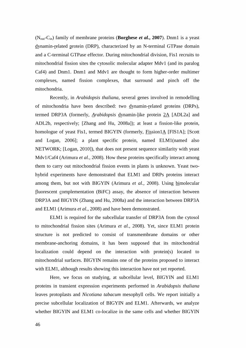
46
(Nout-Cin) family of membrane proteins (Borghese et al., 2007). Dnm1 is a yeast
dynamin-related protein (DRP), characterized by an N-terminal GTPase domain
and a C-terminal GTPase effector. During mitochondrial division, Fis1 recruits to
mitochondrial fission sites the cytosolic molecular adapter Mdv1 (and its paralog
Caf4) and Dnm1. Dnm1 and Mdv1 are thought to form higher-order multimer
complexes, named fission complexes, that surround and pinch off the
mitochondria.
Recently, in Arabidopsis thaliana, several genes involved in remodelling
of mitochondria have been described: two dynamin-related proteins (DRPs),
termed DRP3A (formerly, Arabidopsis dynamin-like protein 2A [ADL2a] and
ADL2b, respectively; [Zhang and Hu, 2008a]); at least a fission-like protein,
homologue of yeast Fis1, termed BIGYIN (formerly, Fission1A [FIS1A]; [Scott
and Logan, 2006]; a plant specific protein, named ELM1(named also
NETWORK; [Logan, 2010]), that does not present sequence similarity with yeast
Mdv1/Caf4 (Arimura et al., 2008). How these proteins specifically interact among
them to carry out mitochondrial fission events in plants is unknown. Yeast two-
hybrid experiments have demonstrated that ELM1 and DRPs proteins interact
among them, but not with BIGYIN (Arimura et al., 2008). Using bimolecular
fluorescent complementation (BiFC) assay, the absence of interaction between
DRP3A and BIGYIN (Zhang and Hu, 2008a) and the interaction between DRP3A
and ELM1 (Arimura et al., 2008) and have been demonstrated.
ELM1 is required for the subcellular transfer of DRP3A from the cytosol
to mitochondrial fission sites (Arimura et al., 2008). Yet, since ELM1 protein
structure is not predicted to consist of transmembrane domains or other
membrane-anchoring domains, it has been supposed that its mitochondrial
localization could depend on the interaction with protein(s) located to
mitochondrial surfaces. BIGYIN remains one of the proteins proposed to interact
with ELM1, although results showing this interaction have not yet reported.
Here, we focus on studying, at subcellular level, BIGYIN and ELM1
proteins in transient expression experiments performed in Arabidopsis thaliana
leaves protoplasts and Nicotiana tabacum mesophyll cells. We report initially a
precise subcellular localization of BIGYIN and ELM1. Afterwards, we analyze
whether BIGYIN and ELM1 co-localize in the same cells and whether BIGYIN

47
and ELM1 do interact in vivo in our experimental systems. Moreover, we analyze
whether BIGYIN and ELM1 are expressed in the same plant tissues in
Arabidopsis, a prerequisite to make possible their interactions.
48
RESULTS
Subcellular localization of ELM1::YFP
In order to investigate whether BIGYIN and ELM1 interact in vivo, we decided at
first to determine the precise subcellular localization pattern of these two proteins,
adopting different methods of transient expression, such as the transformation of
protoplasts obtained from Arabidopsis leaves and the agro-infiltration of
Nicotiana tabacum leaves, combined with confocal microscope analyses.
To investigate the intracellular localization of ELM1, we fused ELM1
coding sequence (CDS) to the N-termini of yellow fluorescent protein (YFP),
downstream the constitutive cauliflower mosaic virus 35S (CaMV 35S) promoter.
This fusion construct, termed ELM1::YFP, was subcloned into a plant pGreenII
binary vector (Hellens et al., 2000). The subcellular localization of ELM1::YFP
was performed using several organelle markers. To detect mitochondria, two
reporters were used: the red fluorescent protein (RFP) fused to Arabidopsis
cytochrome c oxidase-related (COX::RFP; obtained in our laboratory), and the
mCherry fluorescent protein fused to yeast cytochrome c oxidase IV pre-sequence
(COX4::mCherry; Nelson et al., 2007). To visualize peroxisomes, we fused RFP
to KSRM sequence tag (RFP::KSRM; obtained in our laboratory). To identify
chloroplasts, chlorophyll autofluorescence was analyzed in leaf mesophyll cells.
Subcellular localization of ELM1 to mitochondria
To test whether ELM1::YFP fusion protein was localized to mitochondria, the
pGreen-ELM1::YFP was introduced by polyethylene glycol transformation
method (Yoo et al., 2007) into protoplasts isolated from Arabidopsis leaves,
stably expressing the mitochondrial COX4::mCherry marker (Nelson et al., 2007).
Protoplasts were analyzed 24h after transfection. ELM1::YFP was present in
many discrete structures that mainly overlapped with mitochondrial COX::RFP
(Fig. 1A). In detail, ELM1::YFP was located to the periphery of mitochondria,
and it surrounded the mitochondrial RFP marker. Moreover, observing
mitochondrial distribution in protoplasts transformed with ELM1::YFP, we noted
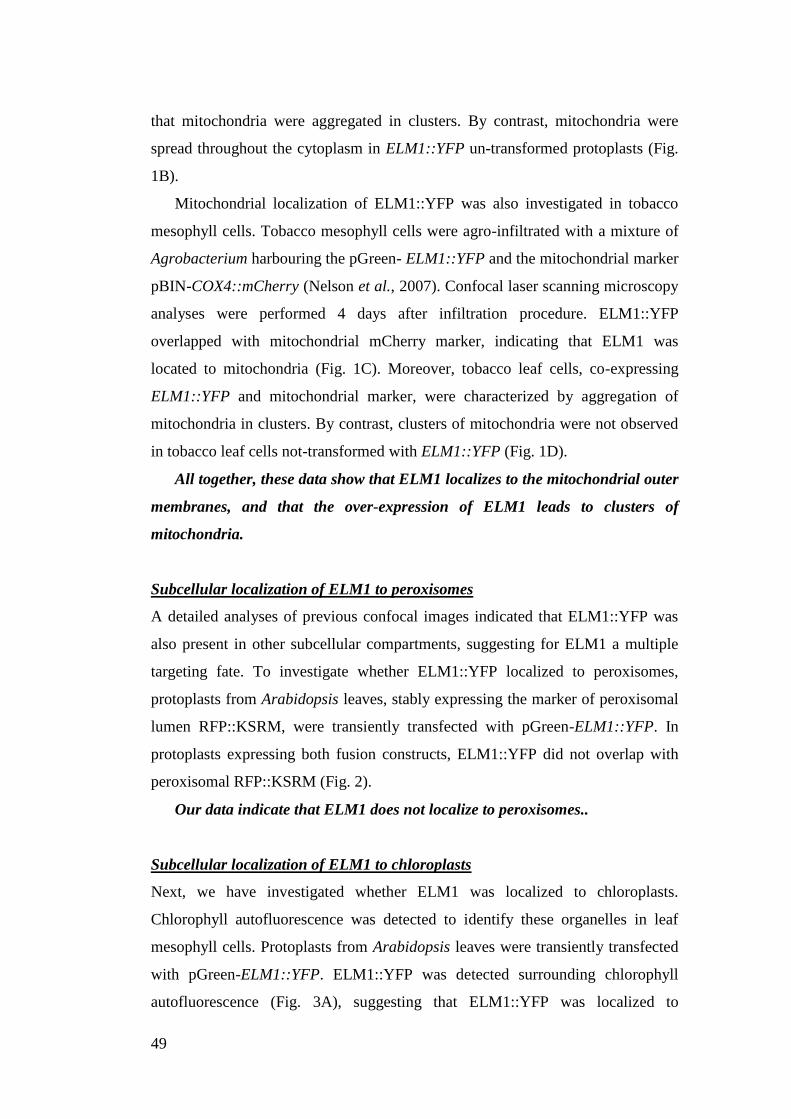
49
that mitochondria were aggregated in clusters. By contrast, mitochondria were
spread throughout the cytoplasm in ELM1::YFP un-transformed protoplasts (Fig.
1B).
Mitochondrial localization of ELM1::YFP was also investigated in tobacco
mesophyll cells. Tobacco mesophyll cells were agro-infiltrated with a mixture of
Agrobacterium harbouring the pGreen- ELM1::YFP and the mitochondrial marker
pBIN-COX4::mCherry (Nelson et al., 2007). Confocal laser scanning microscopy
analyses were performed 4 days after infiltration procedure. ELM1::YFP
overlapped with mitochondrial mCherry marker, indicating that ELM1 was
located to mitochondria (Fig. 1C). Moreover, tobacco leaf cells, co-expressing
ELM1::YFP and mitochondrial marker, were characterized by aggregation of
mitochondria in clusters. By contrast, clusters of mitochondria were not observed
in tobacco leaf cells not-transformed with ELM1::YFP (Fig. 1D).
All together, these data show that ELM1 localizes to the mitochondrial outer
membranes, and that the over-expression of ELM1 leads to clusters of
mitochondria.
Subcellular localization of ELM1 to peroxisomes
A detailed analyses of previous confocal images indicated that ELM1::YFP was
also present in other subcellular compartments, suggesting for ELM1 a multiple
targeting fate. To investigate whether ELM1::YFP localized to peroxisomes,
protoplasts from Arabidopsis leaves, stably expressing the marker of peroxisomal
lumen RFP::KSRM, were transiently transfected with pGreen-ELM1::YFP. In
protoplasts expressing both fusion constructs, ELM1::YFP did not overlap with
peroxisomal RFP::KSRM (Fig. 2).
Our data indicate that ELM1 does not localize to peroxisomes..
Subcellular localization of ELM1 to chloroplasts
Next, we have investigated whether ELM1 was localized to chloroplasts.
Chlorophyll autofluorescence was detected to identify these organelles in leaf
mesophyll cells. Protoplasts from Arabidopsis leaves were transiently transfected
with pGreen-ELM1::YFP. ELM1::YFP was detected surrounding chlorophyll
autofluorescence (Fig. 3A), suggesting that ELM1::YFP was localized to
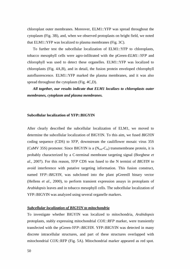
50
chloroplast outer membranes. Moreover, ELM1::YFP was spread throughout the
cytoplasm (Fig. 3B), and, when we observed protoplasts on bright field, we noted
that ELM1::YFP was localized to plasma membranes (Fig. 3C).
To further test the subcellular localization of ELM1::YFP to chloroplasts,
tobacco mesophyll cells were agro-infiltrated with the pGreen-ELM1::YFP and
chlorophyll was used to detect these organelles. ELM1::YFP was localized to
chloroplasts (Fig. 4A,B), and in detail, the fusion protein enveloped chlorophyll
autofluorescence. ELM1::YFP marked the plasma membranes, and it was also
spread throughout the cytoplasm (Fig. 4C,D).
All together, our results indicate that ELM1 localizes to chloroplasts outer
membranes, cytoplasm and plasma membranes.
Subcellular localization of YFP::BIGYIN
After clearly described the subcellular localization of ELM1, we moved to
determine the subcellular localization of BIGYIN. To this aim, we fused BIGYIN
coding sequence (CDS) to YFP, downstream the cauliflower mosaic virus 35S
(CaMV 35S) promoter. Since BIGYIN is a (Nout-Cin) transmembrane protein, it is
probably characterized by a C-terminal membrane targeting signal (Borghese et
al., 2007). For this reason, YFP CDS was fused to the N termini of BIGYIN to
avoid interference with putative targeting information. This fusion construct,
named YFP::BIGYIN, was subcloned into the plant pGreenII binary vector
(Hellens et al., 2000), to perform transient expression assays in protoplasts of
Arabidopsis leaves and in tobacco mesophyll cells. The subcellular localization of
YFP::BIGYIN was analyzed using several organelle markers.
Subcellular localization of BIGYIN to mitochondria
To investigate whether BIGYIN was localized to mitochondria, Arabidopsis
protoplasts, stably expressing mitochondrial COX::RFP marker, were transiently
transfected with the pGreen-YFP::BIGYIN. YFP::BIGYIN was detected in many
discrete intracellular structures, and part of these structures overlapped with
mitochondrial COX::RFP (Fig. 5A). Mitochondrial marker appeared as red spot.
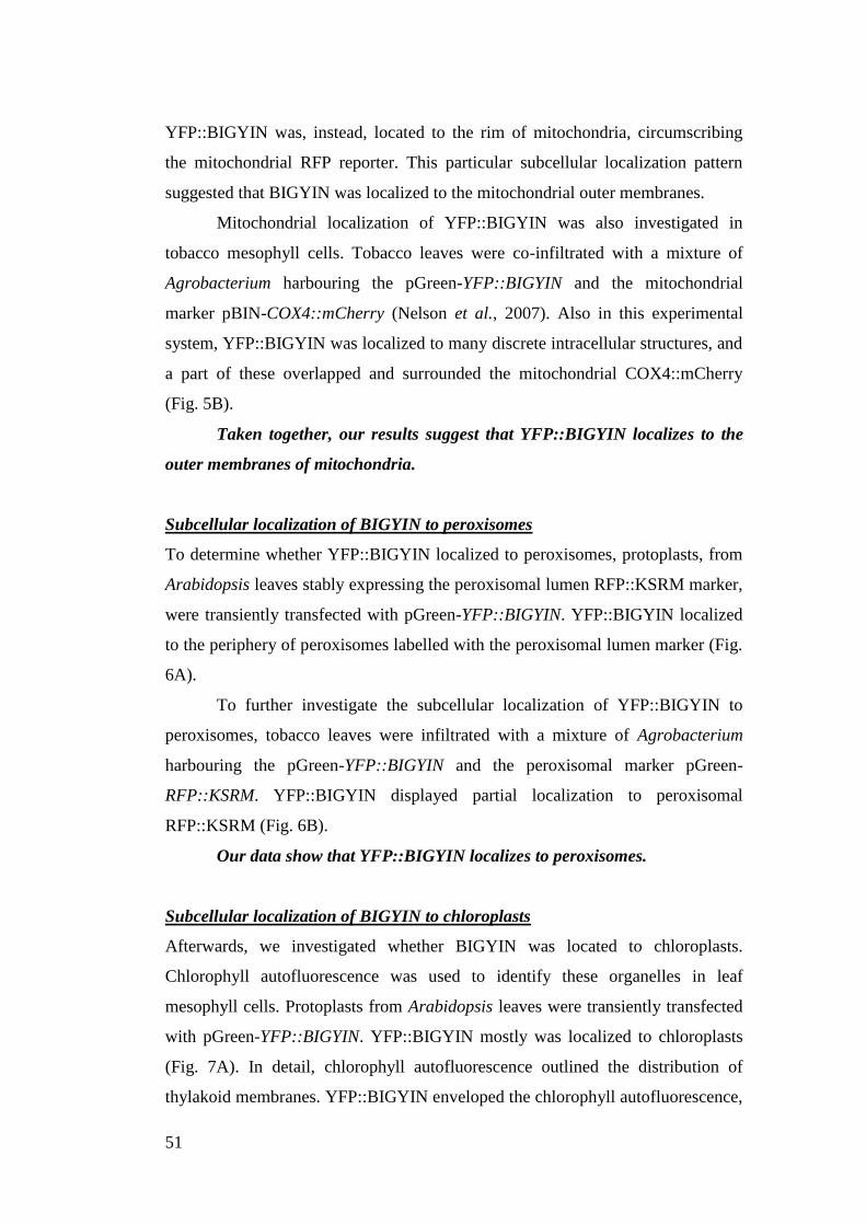
51
YFP::BIGYIN was, instead, located to the rim of mitochondria, circumscribing
the mitochondrial RFP reporter. This particular subcellular localization pattern
suggested that BIGYIN was localized to the mitochondrial outer membranes.
Mitochondrial localization of YFP::BIGYIN was also investigated in
tobacco mesophyll cells. Tobacco leaves were co-infiltrated with a mixture of
Agrobacterium harbouring the pGreen-YFP::BIGYIN and the mitochondrial
marker pBIN-COX4::mCherry (Nelson et al., 2007). Also in this experimental
system, YFP::BIGYIN was localized to many discrete intracellular structures, and
a part of these overlapped and surrounded the mitochondrial COX4::mCherry
(Fig. 5B).
Taken together, our results suggest that YFP::BIGYIN localizes to the
outer membranes of mitochondria.
Subcellular localization of BIGYIN to peroxisomes
To determine whether YFP::BIGYIN localized to peroxisomes, protoplasts, from
Arabidopsis leaves stably expressing the peroxisomal lumen RFP::KSRM marker,
were transiently transfected with pGreen-YFP::BIGYIN. YFP::BIGYIN localized
to the periphery of peroxisomes labelled with the peroxisomal lumen marker (Fig.
6A).
To further investigate the subcellular localization of YFP::BIGYIN to
peroxisomes, tobacco leaves were infiltrated with a mixture of Agrobacterium
harbouring the pGreen-YFP::BIGYIN and the peroxisomal marker pGreen-
RFP::KSRM. YFP::BIGYIN displayed partial localization to peroxisomal
RFP::KSRM (Fig. 6B).
Our data show that YFP::BIGYIN localizes to peroxisomes.
Subcellular localization of BIGYIN to chloroplasts
Afterwards, we investigated whether BIGYIN was located to chloroplasts.
Chlorophyll autofluorescence was used to identify these organelles in leaf
mesophyll cells. Protoplasts from Arabidopsis leaves were transiently transfected
with pGreen-YFP::BIGYIN. YFP::BIGYIN mostly was localized to chloroplasts
(Fig. 7A). In detail, chlorophyll autofluorescence outlined the distribution of
thylakoid membranes. YFP::BIGYIN enveloped the chlorophyll autofluorescence,
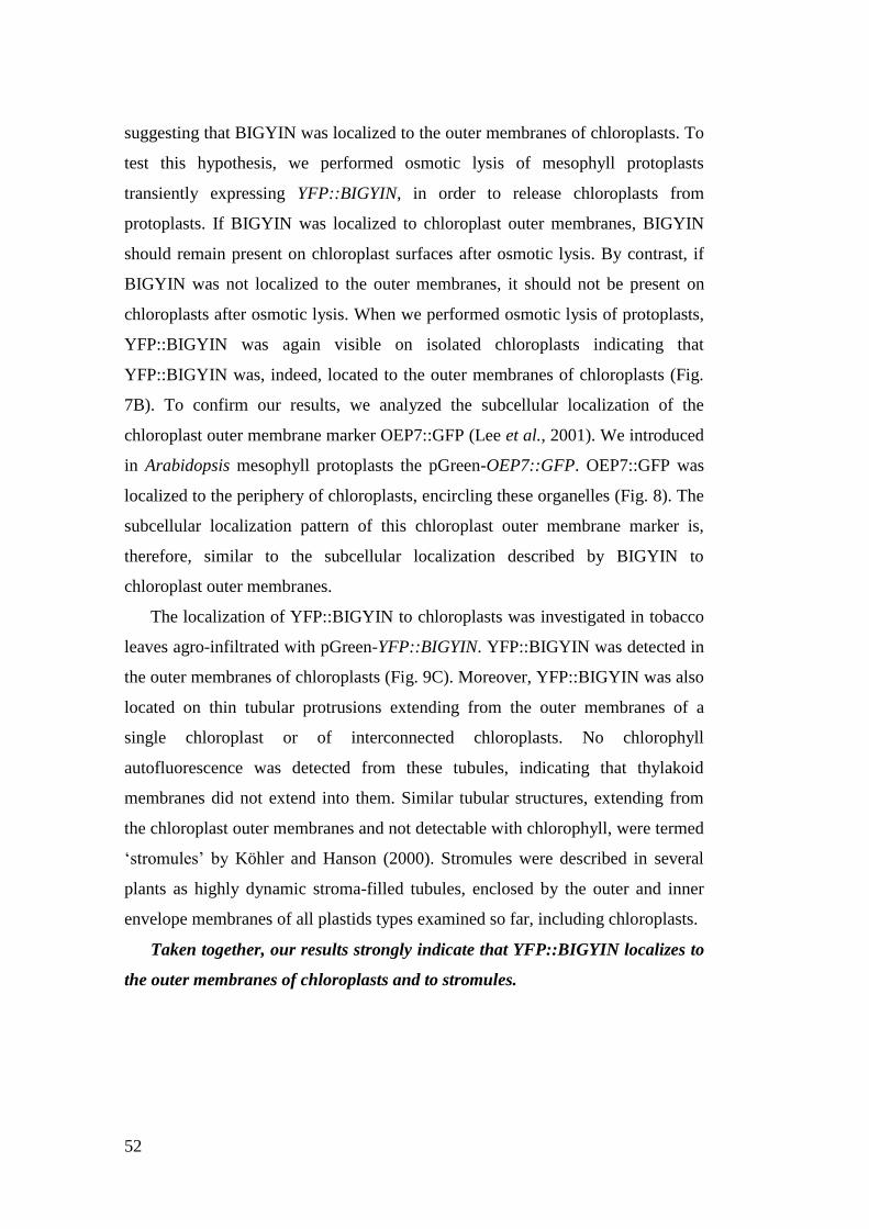
52
suggesting that BIGYIN was localized to the outer membranes of chloroplasts. To
test this hypothesis, we performed osmotic lysis of mesophyll protoplasts
transiently expressing YFP::BIGYIN, in order to release chloroplasts from
protoplasts. If BIGYIN was localized to chloroplast outer membranes, BIGYIN
should remain present on chloroplast surfaces after osmotic lysis. By contrast, if
BIGYIN was not localized to the outer membranes, it should not be present on
chloroplasts after osmotic lysis. When we performed osmotic lysis of protoplasts,
YFP::BIGYIN was again visible on isolated chloroplasts indicating that
YFP::BIGYIN was, indeed, located to the outer membranes of chloroplasts (Fig.
7B). To confirm our results, we analyzed the subcellular localization of the
chloroplast outer membrane marker OEP7::GFP (Lee et al., 2001). We introduced
in Arabidopsis mesophyll protoplasts the pGreen-OEP7::GFP. OEP7::GFP was
localized to the periphery of chloroplasts, encircling these organelles (Fig. 8). The
subcellular localization pattern of this chloroplast outer membrane marker is,
therefore, similar to the subcellular localization described by BIGYIN to
chloroplast outer membranes.
The localization of YFP::BIGYIN to chloroplasts was investigated in tobacco
leaves agro-infiltrated with pGreen-YFP::BIGYIN. YFP::BIGYIN was detected in
the outer membranes of chloroplasts (Fig. 9C). Moreover, YFP::BIGYIN was also
located on thin tubular protrusions extending from the outer membranes of a
single chloroplast or of interconnected chloroplasts. No chlorophyll
autofluorescence was detected from these tubules, indicating that thylakoid
membranes did not extend into them. Similar tubular structures, extending from
the chloroplast outer membranes and not detectable with chlorophyll, were termed
„stromules‟ by Köhler and Hanson (2000). Stromules were described in several
plants as highly dynamic stroma-filled tubules, enclosed by the outer and inner
envelope membranes of all plastids types examined so far, including chloroplasts.
Taken together, our results strongly indicate that YFP::BIGYIN localizes to
the outer membranes of chloroplasts and to stromules.
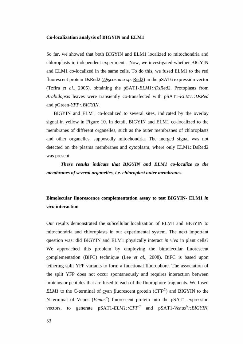
53
Co-localization analysis of BIGYIN and ELM1
So far, we showed that both BIGYIN and ELM1 localized to mitochondria and
chloroplasts in independent experiments. Now, we investigated whether BIGYIN
and ELM1 co-localized in the same cells. To do this, we fused ELM1 to the red
fluorescent protein DsRed2 (Discosoma sp. Red2) in the pSAT6 expression vector
(Tzfira et al., 2005), obtaining the pSAT1-ELM1::DsRed2. Protoplasts from
Arabidopsis leaves were transiently co-transfected with pSAT1-ELM1::DsRed
and pGreen-YFP::BIGYIN.
BIGYIN and ELM1 co-localized to several sites, indicated by the overlay
signal in yellow in Figure 10. In detail, BIGYIN and ELM1 co-localized to the
membranes of different organelles, such as the outer membranes of chloroplasts
and other organelles, supposedly mitochondria. The merged signal was not
detected on the plasma membranes and cytoplasm, where only ELM1::DsRed2
was present.
These results indicate that BIGYIN and ELM1 co-localize to the
membranes of several organelles, i.e. chloroplast outer membranes.
Bimolecular fluorescence complementation assay to test BIGYIN- ELM1 in
vivo interaction
Our results demonstrated the subcellular localization of ELM1 and BIGYIN to
mitochondria and chloroplasts in our experimental system. The next important
question was: did BIGYIN and ELM1 physically interact in vivo in plant cells?
We approached this problem by employing the bimolecular fluorescent
complementation (BiFC) technique (Lee et al., 2008). BiFC is based upon
tethering split YFP variants to form a functional fluorophore. The association of
the split YFP does not occur spontaneously and requires interaction between
proteins or peptides that are fused to each of the fluorophore fragments. We fused
ELM1 to the C-terminal of cyan fluorescent protein (CFPC) and BIGYIN to the
N-terminal of Venus (VenusN) fluorescent protein into the pSAT1 expression
vectors, to generate pSAT1-ELM1::CFPC and pSAT1-Venus
N::BIGYIN,
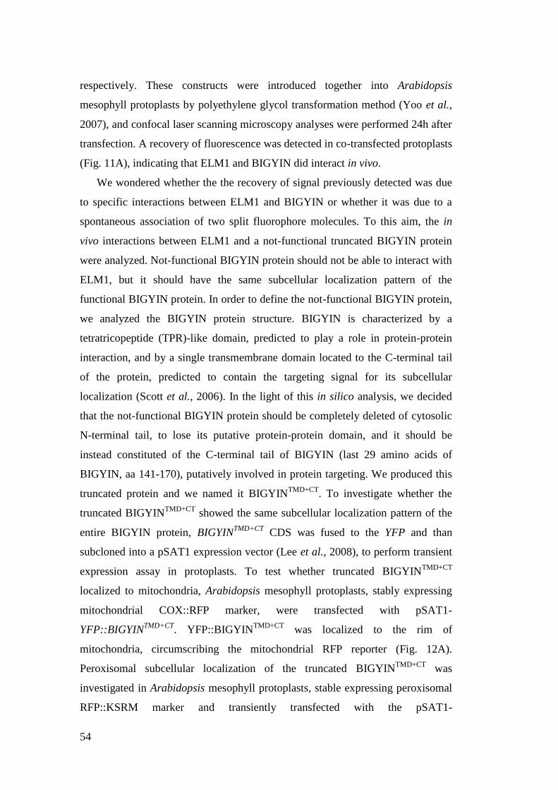
54
respectively. These constructs were introduced together into Arabidopsis
mesophyll protoplasts by polyethylene glycol transformation method (Yoo et al.,
2007), and confocal laser scanning microscopy analyses were performed 24h after
transfection. A recovery of fluorescence was detected in co-transfected protoplasts
(Fig. 11A), indicating that ELM1 and BIGYIN did interact in vivo.
We wondered whether the the recovery of signal previously detected was due
to specific interactions between ELM1 and BIGYIN or whether it was due to a
spontaneous association of two split fluorophore molecules. To this aim, the in
vivo interactions between ELM1 and a not-functional truncated BIGYIN protein
were analyzed. Not-functional BIGYIN protein should not be able to interact with
ELM1, but it should have the same subcellular localization pattern of the
functional BIGYIN protein. In order to define the not-functional BIGYIN protein,
we analyzed the BIGYIN protein structure. BIGYIN is characterized by a
tetratricopeptide (TPR)-like domain, predicted to play a role in protein-protein
interaction, and by a single transmembrane domain located to the C-terminal tail
of the protein, predicted to contain the targeting signal for its subcellular
localization (Scott et al., 2006). In the light of this in silico analysis, we decided
that the not-functional BIGYIN protein should be completely deleted of cytosolic
N-terminal tail, to lose its putative protein-protein domain, and it should be
instead constituted of the C-terminal tail of BIGYIN (last 29 amino acids of
BIGYIN, aa 141-170), putatively involved in protein targeting. We produced this
truncated protein and we named it BIGYINTMD+CT
. To investigate whether the
truncated BIGYINTMD+CT
showed the same subcellular localization pattern of the
entire BIGYIN protein, BIGYINTMD+CT
CDS was fused to the YFP and than
subcloned into a pSAT1 expression vector (Lee et al., 2008), to perform transient
expression assay in protoplasts. To test whether truncated BIGYINTMD+CT
localized to mitochondria, Arabidopsis mesophyll protoplasts, stably expressing
mitochondrial COX::RFP marker, were transfected with pSAT1-
YFP::BIGYINTMD+CT
. YFP::BIGYINTMD+CT
was localized to the rim of
mitochondria, circumscribing the mitochondrial RFP reporter (Fig. 12A).
Peroxisomal subcellular localization of the truncated BIGYINTMD+CT
was
investigated in Arabidopsis mesophyll protoplasts, stable expressing peroxisomal
RFP::KSRM marker and transiently transfected with the pSAT1-
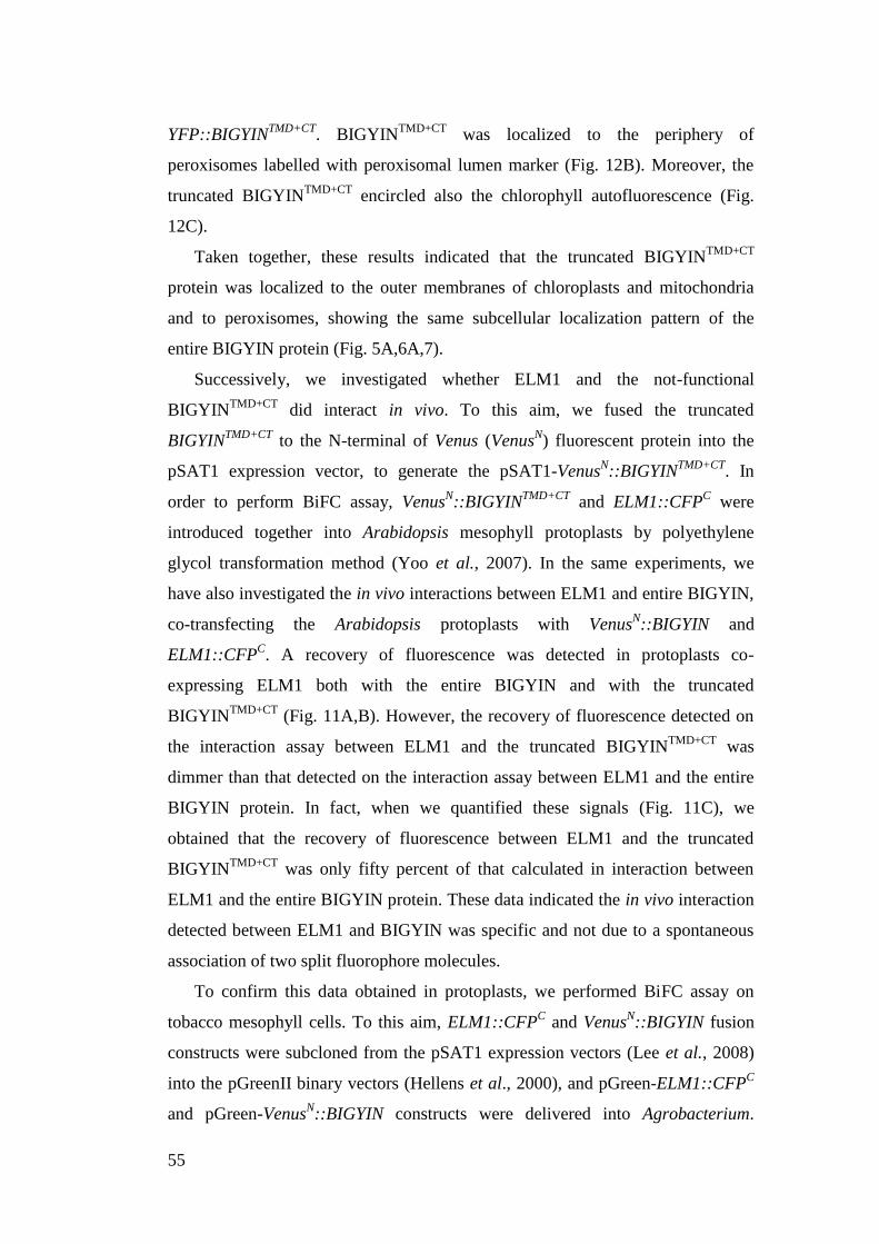
55
YFP::BIGYINTMD+CT
. BIGYINTMD+CT
was localized to the periphery of
peroxisomes labelled with peroxisomal lumen marker (Fig. 12B). Moreover, the
truncated BIGYINTMD+CT
encircled also the chlorophyll autofluorescence (Fig.
12C).
Taken together, these results indicated that the truncated BIGYINTMD+CT
protein was localized to the outer membranes of chloroplasts and mitochondria
and to peroxisomes, showing the same subcellular localization pattern of the
entire BIGYIN protein (Fig. 5A,6A,7).
Successively, we investigated whether ELM1 and the not-functional
BIGYINTMD+CT
did interact in vivo. To this aim, we fused the truncated
BIGYINTMD+CT
to the N-terminal of Venus (VenusN) fluorescent protein into the
pSAT1 expression vector, to generate the pSAT1-VenusN::BIGYIN
TMD+CT. In
order to perform BiFC assay, VenusN::BIGYIN
TMD+CT and ELM1::CFP
C were
introduced together into Arabidopsis mesophyll protoplasts by polyethylene
glycol transformation method (Yoo et al., 2007). In the same experiments, we
have also investigated the in vivo interactions between ELM1 and entire BIGYIN,
co-transfecting the Arabidopsis protoplasts with VenusN::BIGYIN and
ELM1::CFPC. A recovery of fluorescence was detected in protoplasts co-
expressing ELM1 both with the entire BIGYIN and with the truncated
BIGYINTMD+CT
(Fig. 11A,B). However, the recovery of fluorescence detected on
the interaction assay between ELM1 and the truncated BIGYINTMD+CT
was
dimmer than that detected on the interaction assay between ELM1 and the entire
BIGYIN protein. In fact, when we quantified these signals (Fig. 11C), we
obtained that the recovery of fluorescence between ELM1 and the truncated
BIGYINTMD+CT
was only fifty percent of that calculated in interaction between
ELM1 and the entire BIGYIN protein. These data indicated the in vivo interaction
detected between ELM1 and BIGYIN was specific and not due to a spontaneous
association of two split fluorophore molecules.
To confirm this data obtained in protoplasts, we performed BiFC assay on
tobacco mesophyll cells. To this aim, ELM1::CFPC and Venus
N::BIGYIN fusion
constructs were subcloned from the pSAT1 expression vectors (Lee et al., 2008)
into the pGreenII binary vectors (Hellens et al., 2000), and pGreen-ELM1::CFPC
and pGreen-VenusN::BIGYIN constructs were delivered into Agrobacterium.

56
Then, these constructs were co-expressed in tobacco leaves by agro-infiltration. In
co-infiltrated leaf mesophyll cells, there was a recovery of BiFC fluorescence
(Figure 12D), suggesting that BIGYIN and ELM1 interacted in vivo. As control,
the interactions between ELM1 and the truncated BIGYINTMD+CT
protein were
investigated. Tobacco mesophyll cells were agro-infiltrated with a mixture of
Agrobacterium harbouring the pGreen-ELM1::CFPC and the pGreen-
VenusN::BIGYIN
TMD+CT. Agro-infiltrated leaf cells did not show fluorescence or
only background fluorescence (Fig. 12E).
All together, these data strongly demonstrate that BIGYIN and ELM1 do
interact in vivo.
Subcellular localization of BIGYIN and ELM1-interaction sites
Successively, we investigated the subcellular compartments where BIGYIN and
ELM1 physically interacted in vivo. Arabidopsis mesophyll protoplasts were co-
transfected with BiFC constructs (ELM1::CFPC and Venus
N::BIGYIN), and they
were analysed by means of confocal microscopy 24h later. BiFC fluorescence,
chlorophyll autofluorescence and merged images were captured (Fig.13A). In co-
transformed protoplasts, the recovery of fluorescence was clearly detected on the
chloroplast outer membranes. To test whether BIGYIN and ELM1 interacted also
on mitochondria and peroxisomes, the ELM1::CFPC and Venus
N::BIGYIN were
introduced in Arabidopsis mesophyll protoplasts stably expressing the RFP
targeted to mitochondria (COX::RFP) and to peroxisomes (RFP::KSRM). BiFC
signal was enriched around mitochondria (Fig. 13B), while it was not localized to
peroxisomes (Figure 13C).
These data indicate that BIGYIN and ELM1 interact in vivo on outer
membranes of chloroplasts and on mitochondria, but not on peroxisomes.
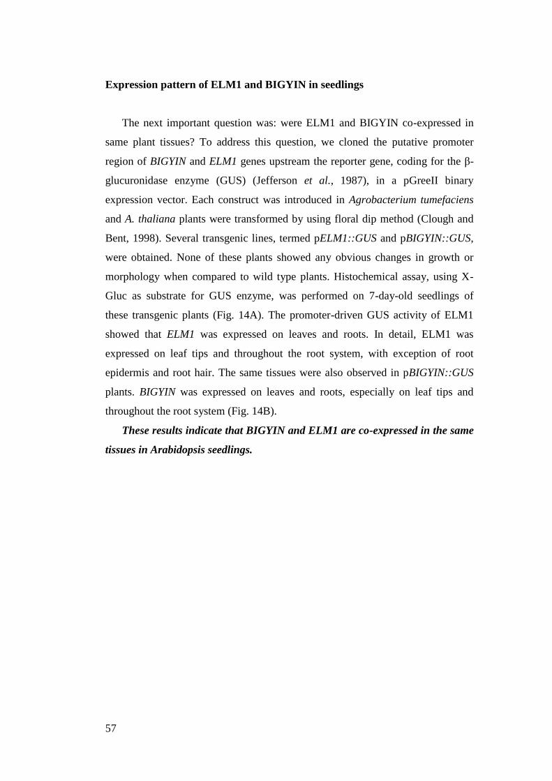
57
Expression pattern of ELM1 and BIGYIN in seedlings
The next important question was: were ELM1 and BIGYIN co-expressed in
same plant tissues? To address this question, we cloned the putative promoter
region of BIGYIN and ELM1 genes upstream the reporter gene, coding for the β-
glucuronidase enzyme (GUS) (Jefferson et al., 1987), in a pGreeII binary
expression vector. Each construct was introduced in Agrobacterium tumefaciens
and A. thaliana plants were transformed by using floral dip method (Clough and
Bent, 1998). Several transgenic lines, termed pELM1::GUS and pBIGYIN::GUS,
were obtained. None of these plants showed any obvious changes in growth or
morphology when compared to wild type plants. Histochemical assay, using X-
Gluc as substrate for GUS enzyme, was performed on 7-day-old seedlings of
these transgenic plants (Fig. 14A). The promoter-driven GUS activity of ELM1
showed that ELM1 was expressed on leaves and roots. In detail, ELM1 was
expressed on leaf tips and throughout the root system, with exception of root
epidermis and root hair. The same tissues were also observed in pBIGYIN::GUS
plants. BIGYIN was expressed on leaves and roots, especially on leaf tips and
throughout the root system (Fig. 14B).
These results indicate that BIGYIN and ELM1 are co-expressed in the same
tissues in Arabidopsis seedlings.

58
59
DISCUSSION
ELM1::YFP localizes to mitochondria chloroplasts, cytoplasm and plasma
membranes
In this study we have reported a detailed subcellular localization of ELM1, a
specific plant protein involved in mitochondrial fission (Arimura et al., 2008). To
this aim, we have cloned ELM1::YFP fusion construct downstream a constitutive
promoter into a plant expression vector, to perform transient expression
experiments in Arabidopsis leaves protoplasts and tobacco mesophyll cells. To
precisely localize ELM1::YFP to mitochondria and peroxisomes, several
organelle markers have been used.
We have shown that ELM1 localizes to multiple subcellular sites. In detail,
ELM1 localizes mainly to the outer membranes of mitochondria and chloroplasts,
while it does not located to peroxisomes. We have shown that ELM1 can be also
localizes to plasma membranes and cytoplasm. Mitochondrial localization of
ELM1 is in agreement with data reported by Arimura et al. (2008) and it is also
consistent with the involvement of ELM1 in mitochondrial fission machinery
(Arimura et al., 2008). By contrast, the subcellular localization of ELM1 to
chloroplasts has never been investigated before. So far, subcellular localization
analyses of BIGYIN have been performed only on epidermal leaves (Arimura et
al., 2008), where chloroplasts are absent. Subcellular localization of ELM1 to the
cytoplasm and to the plasma membrane was not previously reported. However, a
cytosolic localization of ELM1 could been expected, because in silico analysis of
ELM1 protein sequence does not show predicted transmembrane domains or
membrane-anchoring domains (Arimura et al., 2008).
Our data show that the over-expression of ELM1 leads to abnormal
mitochondrial morphology and distribution inside the cells: mitochondria, in fact,
aggregate in clusters. The observed pattern (Fig.1A) suggests that mitochondrial
fission occurs, but mitochondria seem not to be able to physically separate from
each other and to spread throughout the cytoplasm. There are no published data
concerning the consequences of over-expression of ELM1, because ELM1 over-
expression has not been investigated so far. Instead, it has been described that the
over-expression of DRP3A or DRP3B, two Arabidopsis proteins involved in
60
mitochondrial fission events, caused, in leaf epidermal cells, fragmentation and
not aggregation of mitochondria (Fujimoto et al., 2009). ELM1, DRP3A and
DRP3B seem therefore to have distinct functions during mitochondrial fission
events. Hence, we can speculate that the role of ELM1 could be not sufficient for
physical scission of mitochondrial membranes. By contrast, DRP3A and DRP3B
could act downstream ELM1 during mitochondrial fission events. DRP3A and
DRP3B seem to be actively involved in physical fission of mitochondrial
membranes, as suggested also by the homology of DRP3A and DPR3B with the
dynamin protein, a class of GTPase mechano-enzymes involved in membrane
scission.
YFP::BIGYIN localizes to mitochondria, peroxisomes and chloroplasts
We have shown that ELM1 localizes to different subcellular compartments. Since
transmembrane domains or other predicted membrane-anchoring domains are not
present in ELM1 structure, we could hypothesize that multiple subcellular
localization pattern of ELM1 depends on the interaction with different proteins
localized to different organelle compartments. BIGYIN is one of the proteins that
have been proposed to interact with ELM1 on mitochondria, because BIGYIN is a
transmembrane protein located to mitochondrial outer membranes and it is
involved in mitochondrial fission event. However, there are no published data to
support this hypothesis. Yet, our results put also another question: how can ELM1
reach the chloroplast outer membranes? To address this question, we have
reported a detailed subcellular localization of BIGYIN in the same experimental
systems previously adopted for the localizations of ELM1 (transient expression
assays in Arabidopsis leaves protoplasts and agro-infiltrated tobacco mesophyll
cells). To this aim, the YFP::BIGYIN was cloned downstream a constitutive
promoter into a plant expression vector and co-expressed with several organelle
markers. We have shown that BIGYIN localizes to the outer membranes of
mitochondria and to the membranes of peroxisomes. These data are in agreement
with the subcellular localization of BIGYIN already reported by Zhang and Hu
(2008a). These localization patterns are also consistent with the role of BIGYIN
as a fission protein involved in mitochondrial and peroxisomal fission events
(Zhang and Hu, 2008b).
61
If we compare the mitochondrial distribution in cells over-expressing
BIGYIN and in cells over-expressing ELM1, we observe that mitochondria are
not aggregated in clusters in BIGYIN-transformed cells, as we have instead
previously reported in ELM1-transformed cells. These findings indicate that
BIGYIN and ELM1 play distinct role(s) during mitochondrial fission events.
Interestingly, we show that BIGYIN localizes to chloroplasts outer
membrane both in Arabidopsis leaf protoplasts and in tobacco mesophyll cells.
Moreover, in tobacco mesophyll cells, unlike to Arabidopsis protoplasts, we have
shown that BIGYIN localizes also to thin protrusions (i.e. stromules), extending
from the outer membranes of single chloroplast or of interconnected chloroplasts.
Subcellular localization of BIGYIN to chloroplasts has not been previously
reported, probably because mesophyll cells, where chloroplasts are present, have
not been yet investigated. However, our subcellular localization of BIGYIN to
chloroplasts is in agreement with the identification of BIGYIN protein in
chloroplast proteome experiments (Zybailov et al., 2008).
Based on these results, BIGYIN is the first transmembrane protein, in
detail a tail-anchored (Nout-Cin) protein, localized to mitochondrial, chloroplast
and peroxisomal membranes.
BIGYIN and ELM1 co-localize to mitochondria and chloroplasts
So far, we have analyzed the subcellular localization of BIGYIN and ELM1
separately in independent experiments. In order to investigate their localization
pattern in the same cells, we have performed co-expression experiments in
Arabidopsis protoplasts. We have shown several sites where co-localization
between ELM1 and BIGYIN occurs, i.e. on the outer membranes of chloroplasts
and on other organelles, presumably mitochondria. These data are consistent with
our previous results of localization of BIGYIN and ELM1. The co-localization of
ELM1 and BIGYIN to the chloroplast membranes prompt a new question: do
ELM1 and BIGYIN play also a role on chloroplasts?
62
BIGYIN and ELM1 interact in vivo
Having demonstrated that ELM1 and BIGYIN localize in the same organelles, we
successively investigated whether they also physically interacted in vivo. To this
aim, we have employed the bimolecular fluorescent complementation (BiFC)
technique for in vivo protein-protein interactions in plant cells (Lee et al., 2008).
BiFC is based upon tethering split YFP variant to form a functional fluorophore.
BiFC assay was performed in Arabidopsis mesophyll protoplasts and in tobacco
mesophyll cells. We have shown that in both experimental systems there is a
recovery of fluorescence indicating that ELM1 and BIGYIN do physically interact
in vivo.
We have also verified that the recovery of fluorescence between ELM1 and
BIGYIN was specific and not due to a spontaneous interactions of two YFP split
fluorophore molecules. To this aim, we have analyzed the in vivo interactions
between ELM1 and a not-functional truncated BIGYIN (i.e. BIGYINTMD+CT
).
Truncated BIGYINTMD+CT
is constituted by the C-terminal end of BIGYIN, that is
predicted to contain the targeting signal to the subcellular localization of BIGYIN
(Scott et al., 2006). We have demonstrated that it shows the same subcellular
localization pattern of the entire BIGYIN protein in transiently transformed
Arabidopsis protoplasts. These data demonstrate that the C-terminal end of
BIGYIN protein contains the targeting signal sufficient to the subcellular
localization of BIGYIN to mitochondria, peroxisomes and chloroplasts. These
data are in agreement with the key role played by the C-terminal hydrophobic
anchor in targeting of tail anchored-proteins in other system (Borghese et al.,,
2007). Moreover, we have demonstrated that the N-terminal end of BIGYIN
protein is necessary for the protein-protein interactions, because when it has been
deleted, the interaction between ELM1 and BIGYIN is not detected.
Successively, we have performed BiFC assay between ELM1 and the
truncated BIGYINTMD+CT
protein in Arabidopsis leaf protoplasts. Our results
clearly demonstrate that interaction between ELM1 and BIGYIN is specific both
in vivo in Arabidopsis leaf protoplasts and in tobacco mesophyll cells.
So far, the interaction between BIGYIN and ELM1 was only hypothesized
due to the involvement of these two proteins in the mitochondrial fission
machinery, but clear evidences of their physical interaction were still missing.

63
Yeast-two hybrid experiments were performed to investigate an interaction
between ELM1 and BIGYIN, but these experiments didn‟t detect it (our data not
shown, Arimura et al., 2008), probably because BIGYIN expressed in yeast was
improperly folded or unstable. Our data, obtained through BiFC technique, have
instead demonstrated that BIGYIN and ELM1 do interact in vivo.
BIGYIN and ELM1 interact in vivo on mitochondria and chloroplasts
In order to identify the subcellular compartments where ELM1 and BIGYIN do
interact, BiFC assay have been performed in Arabidopsis leaf protoplasts,
expressing different organelle markers. We have shown that ELM1 and BIGYIN
interact on the outer membranes of mitochondria and on the outer membranes of
chloroplasts. The in vivo interaction between ELM1 and BIGYIN on
mitochondrial outer membranes is in agreement with their involvement in
mitochondrial fission machinery. In addition, we have demonstrated that BIGYIN
and ELM1 interact also on chloroplast outer membranes. The subcellular
localization of both proteins to chloroplasts has not been yet investigated. Their
specific interaction on chloroplast membrane is the first indication that ELM1 and
BIGYIN could play a role on chloroplasts. Therefore, ELM1 seems to be a protein
shared by mitochondrial and chloroplasts, and BIGYIN a tail-anchored protein
shared by mitochondria, peroxisomes and chloroplasts.
BIGYIN and ELM1 are expressed in the same plant tissues in Arabidopsis
seedlings
Afterwards we have analyzed whether the expression pattern of ELM1 and
BIGYIN in plant tissues. Our results, obtained through histochemical GUS assay
(Jefferson et al., 1987) on 7-day-old seedlings of pBIGYIN::GUS and
pELM1::GUS transgenic plants, have shown that BIGYIN and ELM1 are
expressed in the same organs (leaf and root) and in the same tissues (leaf tips and
throughout the root system) in seedlings.
Our subcellular localization analyses and BiFC assays have been performed
expressing BIGYIN and ELM1 proteins under the control of a constitutive
promoter, and adopting transient expression methods. Expression of BIGYIN and
ELM1, under its own promoter, in the same plant tissues at the same

64
developmental stage is a necessary confirm for our results of specific interaction
between ELM1 and BIGYIN on mitochondrial and chloroplast outer membranes.
65
MATERIAL AND METHODS
Plant materials and growth condition
All the Arabidopsis thaliana plants for this study were in the Columbia
background. Different Arabidopsis transgenic lines were used: plants
overexpressing the red fluorescent protein (RFP) targeted to mitochondria by the
cytocrome c oxidase-releated (COX) pre-sequence (35S::COX::RFP), and plants
overexpressing the RFP targeted to peroxisomes by the sequence tag KSRM fused
downstream the RFP CDS (35S::RFP::KSMR). These transgenic plants were
generated in our laboratory.
The Arabidopsis thaliana, Columbia ecotype, and Nicotiana tabacum plants were
incubated in an environmentally-controlled growth chamber with a long
photoperiod (16 hr light and 8 hr dark) at 25 ± 1°C, and a photosynthetic photon
flux of 35 mol m-2
s-1
Osram cool-white 18 W fluorescent lamps.
Genetic materials
The Arabidopsis BIGYIN and ELM1 coding sequence fragments were first
amplified by PCR from Arabidopsis cDNA with high-fidelity PCR enzymes
(Phusion High Fidelity DNA polymerase [Finnzymes]) and then cloned into the
vector of interest. To obtain the BIGYINTMD+CT
, only the C-terminal tail (421-510
bp, aa 141-170) of BIGYIN coding sequence was amplified, corresponding to the
transmembrane domain and the adjacent C-terminal end. All the cloned plasmids
were confirmed by sequencing.
Plasmids for the subcellular localization
For the expression of BIGYIN, BIGYINTMD+CT
and ELM1 in plants, the
pGreen0179 and pGreen0029 (Hellens et al., 2000) binary vectors were used. In
the pGreen0179 (HygromycinR) the p35S::YFP::BIGYIN and the
66
p35S::YFP::BIGYINTMD+CT
constructs were introduced, while in the pGreen0029
(KanamycinR) the p35S::ELM1:YFP was introduced.
To obtain the p35S::YFP::BIGYIN and the p35S::YFP::BIGYINTMD+CT
constructs,
the YFP coding sequence were subcloned from the pAVA554-35S::YFP plasmid
provided by Prof. Albrecht von Arnim (Von Arnim et al., 1998) to the pSAT1-
35S::VenusN vector (stock number E3228, Lee et al., 2008) by replacing into the
NcoI/BglII sites the VenusN cDNA sequence digesting with NCoI/BglII enzymes.
The BIGYIN and the BIGYINTTMD+CT
PCR products were amplified using
primers where the SacI/KpnI sites were introduced (For: 5‟-
CATGGAGCTCAAGGTGTTATAGGGATAGGGATCACG-3‟;
Rev: 5‟-CATGGGTACCTCATTTCTTGCGAGACATCG-3‟). The amplicons
were digested with SacI/KpnI and they were cloned into the pSAT1-p35S::YFP
vector. The pSAT1-p35S::YFP::BIGYIN and the pSAT1-
p35S::YFP::BIGYINTMD+CT
fusion constructs were subcloned with EcoRV/NotI
restriction sites into the pGreen0179 vector digested with SmaI/NotI to obtain the
pGreen0179-p35S::YFP::BIGYIN and pGreen0179-p35S::YFP::BIGYINTMD+CT
binary vectors.
The pGreen0029-p35S::ELM1::YFP binary vector was constructed introducing
first the p35S::YFP from the pAVA554-35S::YFP plasmid (von Arnim et al.,
1998) into the pGreen0029 vector between the KpnI/SacI sites and then cloning
the ELM1 PCR product into the NCoI site (Primer For: 5‟-
CATGCCATGGCCATGAGGCCAATCCT-3‟; Rev: 5‟-
CATGCCATGGCTGCAGACCGTAAACTCCATCC-3‟).
For the transiently co-expression of BIGYIN and ELM1 in Arabidopsis
protoplasts, the pSAT1-p35S::BIGYIN previously described and the pSAT6
p35S::ELM1::DsRed2 were used. The pSAT6-p35S::ELM1::DsRed2 was
obtained cloning with the KpnI restriction enzyme the ELM1 PCR product
(For:5'-CATGGGTACCGGATGAGGCCAATCCTATTGCCGG-3‟;Rev: 5‟-
CATGGGTACCCTGCAGACCGTAAACTCCATCCACGTGC-3‟) into the
pSAT6-DsRed2 vector provided by Citovsky (Tzifira et al., 2005).
67
Plasmids for the BiFC assay
The pSAT1-p35S::VenusN (stock number E3228) and the pSAT1-p35S::CFP
C
(stock number E3449) vectors were provided by Prof. Gelvin (Lee et al., 2008).
The BIGYINTMD+CT
cDNA were amplified by PCR (5‟-
CATGGAGCTCAAATGGATGCTAAGATCGGAC-3‟; Rev: 5‟-
CATGGGTACCTCATTTCTTGCGAGACATCG-3‟; ). The products were
digested with SacI/KpnI and cloned into the pSAT1-p35S::VenusN, to obtain the
pSAT1-p35S::VenusN::BIGYIN:: and p35S::Venus
N::BIGYIN
TMD+CT. The ELM1
coding sequence was amplified by PCR using specific 5‟ and 3‟ primers where the
KpnI/KpnI sites were introduced (5‟-
CATGGGTACCGGATGAGGCCAATCCTATTGCCGG-3‟; Rev: 5‟-
CATGGGTACCCTGCAGACCGTAAACTCCATCCACGTGC-3‟). The
amplicon was digested with KpnI enzyme and cloned into the pSAT1-
p35S::CFPC, to obtain the pSAT1-p35S::35S:ELM1::CFP
C.
pBIGYIN:GUS and pELM1::GUS constructs and generation of transgenic lines
Transgenic Arabidopsis lines used for histochemical studies carried the following
promoter-reporter gene fusions: pBIGYIN::GUS and pELM1::GUS.
The GUS (-glucuronidase)-coding sequence was fused to the BIGYIN promoter
(base pair -862 to -1). The 862 bp promoter fragment was amplified by PCR using
genomic DNA extracted from Arabidopsis leaves as template. The pair of
primers, both carrying an EcoRI restriction site, were as follows: forward primers
5‟-CATGGAATTCCTTTCGAGGCTCACCTCAAC-3 and reverse primer 5‟-
CATGGAATTCTGAAGGCGATTTTGAGCTTTGA-3‟). After digestion, the
promoter was cloned upstream of the GUS coding region, into a modified
pGreen0029 binary vector (KamaycinR; Hellens et al., 2000), where the GUS
coding sequence, fused with the nos terminator, was previously inserted in the
polylinker between KpnI-SacI restriction sites.
To obtain the pELM1::GUS fusion construct, a similar working strategy was
adopted. The ELM1 promoter consist of a 960 bp genomic fragment (base pair -
960 to -1) and it was amplified by PCR with specific primer containing the EcoRI
restriction site (Primer For: 5‟-
68
CATGCTCGAGCCTAACTGTTTACAACCTGCACA -3‟; Rev: 5‟-
CATGCTCGAGGCCGGTTAGATTATCGATTCC-3‟).
The pGreen-pBIGYIN::GUS and pGreen-pELM1::GUS constructs were
transferred into GV3101-pSoup Agrobacterium strain (Hellens et al., 2000) and
Arabidopsis plants were transformed by floral dip method (Clough and Bent,
1998) and screened on half-strength MS agar medium containing 50 mgL-1
kanamycin. The GUS-staining analyses were performed on T2 plants.
Organelle marker used in transient expression experiments
For the localization of the mitochondria in transient expression transformation, the
red fluorescent protein (RFP) targeted was fused to the Arabidopsis cytocrome c
oxidase-releated (COX) pre-sequence (p35S::COX::RFP; obtained in our
laboratory ). For the localization of the peroxisomes, the RFP was fused to
sequence tag KSRM (p35S::RFP::KSRM; obtained in our laboratory). To detect
the chloroplast outer membrane, we used the OEP7::GFP (Lee et al., 2001).
Accession Numbers
Sequence data from this article can be found in the GenBank/EMBL data libraries
under accession numbers At5g22350 for ELM1, named also NETWORK, and
At3g57090 for BIGYIN, previously termed FIS1A.
Protoplasts isolation
Protoplasts were isolated following the protocol of Yoo et al. (2007) with some
modifications. Leaves of 3-4 weeks old Arabidopsis plants were cut into
approximately 0.5-1mm strips, placed in a Petri dish containing the enzymatic
solution (1.25% cellulase R10 [Yakult Pharmaceutical, Japan], 0.3%
macerozyme R10 [Yakult Pharmaceutical, Japan], 0.4M mannitol, 20mM KCl,
20mM MES pH 5.7, 10mM CaCl2, 0.1% BSA; filter sterilized) and vacuum-
infiltrated for 30min. The digestion was continued for 3h without shaking in the
dark at 22°C. After incubation the solution containing protoplasts was filtered
with 50 m nylon mesh sieve and centrifuged in 10ml polystyrene tubes at 100xg
69
for 5min to pellet the protoplasts. The protoplasts were washed twice with W5
solution (154mM NaCl, 125mM CaCl2, 5mM KCl, 2mM MES, pH 5.8; filter
sterilized) and kept on ice for 30min. Then protoplasts were collected by
centrifuging at 100xg for 1min and the pellet was resuspended in an appropriate
volume of MMg solution (0.4M mannitol, 15mM MgCl2) in order to obtain
approximately 2x10⁴ protoplasts in 0.1 mL of MMg.
Protoplasts transfection assay
Protoplasts were transfected according to the procedure of Yoo et al. (2007) with
some modifications.
In a 2ml eppendorf tube 10g of plasmid DNA was added to 2x10⁴ protoplasts
and mixed with an equal volume of a freshly prepared polyethylene-glycol (PEG)
solution (40% w/v PEG4000 [Fluka], 0.1M CaCl2, 0.2M mannitol). The solution
was gently mixed and incubated for 20min in the dark at room temperature. After
incubation, 2 volumes of W5 solution (154mM NaCl, 125mM CaCl2, 5mM KCl,
2mM MES, pH 5.8; filter sterilized) were added to the tube to dilute and washed
out the PEG. The protoplasts were collected by 1min centrifugation at 100xg and
then resuspended in 1 ml of W5 solution. The protoplasts were incubated at 20°C
in the dark for at least 16 hr before the microscopy analysis.
Agrobacterium tumefaciens strain
For the use of pGreenII–derived binary vectors, the A. tumefaciens GV3101 strain
was co-transformed with the pSoup vector (Hellens et al., 2000). Competent cells
of A. tumefaciens GV3101 strain were prepared according to Main et al. (1995)
and the binary vectors were introduced by „freeze-thaw‟ method. 1g of plasmid
DNA was added to the competent cells, frozen in liquid nitrogen for 5min and
heated at 37°C for 5min. The bacterial culture was incubated at 28°C for 3hr with
gentle shaking in 1ml YEP medium (10g/L bacto-trypton, 10g/L yeast extract,
5g/L NaCl; pH 7.0) and then spread on a YEP agar plate containing the
appropriate antibiotic selection (gentamycin 50 mgL-1
, rifampicin 50 mgL-1
,
kanamycin 50 mgL-1
and tetracyclin 5 mgL-1
).
70
Tobacco leaf agroinfiltration
Agrobacterium-mediated transient expression was performed essentially as
described in Zottini et al. (2008). Single colonies of A. tumefaciens growing on
agar plate were inoculated in 3mL of YEP liquid medium supplemented with
specific antibiotics. The bacteria were incubated for 2 days at 28°C at 200rpm on
an orbital shaker. 25L of confluent bacterial culture was re-inoculated in 5mL
(1/200 ratio, v/v) of fresh YEP medium (10g/L bacto-trypton, 10g/L yeast extract,
5g/L NaCl; pH 7.0) containing the appropriate antibiotics, and this new culture
was grown under the same condition for an additional day. 2mL of bacterial
suspension was pellet by centrifugation at 1.500xg for 4min at room temperature.
The pellet was washed twice with 2mL of infiltration buffer [50mM MES pH 5.6,
2mM Na3PO4, 0.5% w/v glucose, and 100M acetosyringone (Aldrich, Italy)] and
then diluted with infiltration buffer to a final OD600 of 0.2. Approximately 300L
of this Agrobacterium mixture was infiltrated into a young leaf of N. tabacum
through the stomata of the lower epidermis by using 1-ml syringe without a
needle. For experiments requiring co-infection of more than one construct,
bacteria strains containing the constructs were mixed before performing the leaf
infection, with the inoculum of each construct adjusted to a final OD600 of 0.2.
After infiltration the plants were maintained in the environmentally-controlled
chamber under standard growth condition. The transient expression was assayed
four days after infection.
Confocal analyses
Confocal microscopies were performed by using a Nikon PCM2000 (Bio-Rad,
Germany) and an inverted SP/2 (Leica, http://www.leica.com). laser scanning
confocal imaging systems. For GFP/YFP and RFP detection, excitation was at
488nm and 543nm respectively, and emission between 515/530nm for YFP and
550/650nm for RFP, respectively. For the mCherry detection, excitation was at
543 nm and detection 550/650nm. For the chlorophyll detection, excitation was at
488nm and detection over 600nm. The images acquired from the confocal
microscope were processed using the software ImageJ bundle software
(http://rsb.info.nih.gov/ik/).
71
BiFC fluorescence quantification
The confocal acquisitions were made in the same experimental condition and 10
protoplasts were analyzed in three replicates. BiFC signal fluorescence
quantification was obtained normalizing the signal intensity of the interaction to
chlorophyll autofluorescence one. The data were plotted comparing the mean of
the BiFC signal intensity between ELM1 and BIGYINTMD+CT
with the mean of the
BiFC signal between ELM1 and BIGYIN. The error standard was calculated.
-glucuronidase (GUS) histochemical analyses
GUS histochemical staining was performed at four developmental stages:
germinating seeds (32 h after imbibition), 5-day-old seedlings (cotyledons open),
7-day-old seedlings (first leaves developing), and flowering mature plants.
Samples were analysed for GUS activity following the protocol described by
Jefferson et al. (1987). The samples were vacuum infiltrated for 30 min in the
following solutions: 2 mM X-gluc, 0.5% Triton X-100, 0.1% Tween 20, 0.5 mM
K3Fe(CN)6, 0.5 mM K4Fe(CN)6_3H2O, 10 mM Na2EDTA and 50 mM sodium
phosphate buffer, pH 7.0, and then incubated at 37°C for 16 h. After staining,
samples were cleared by several washes with methanol/acetic acid (3:1 v/v)
solution and kept at 4°C in 70% ethanol.
Statistic
All experiments were conducted al least in triplicate, and pictures represented
typical example.
72
REFERENCE
- Arimura SI, Fujimoto M, Doniwa Y, Kadoya N, Nakazono M, Sakamoto W,
Tsutsumi N (2008). Arabidopsis ELONGATED MITOCHONDRIA1 is
required for the localization of DYNAMIN-RELATED PROTEIN3A to
mitochondrial fission sites. The Plant Cell, 20(6):1555-1566
- Bauwe H, Hagemann M, Ferine AR (2010). Photorespiration: players,
partners and origin. Trends in Plant Science, 15 (6):330-336
- Borghese N, Brambilasca S, Colombo S (2007). How tails guide tail-anchored
proteins to their destinations. Current Opinion in Cell Biology, 19:368–375
- Clough SJ, Bent AF (1998). Floral dip: a simplified method for
Agrobacterium-mediated transformation of Arabidopsis thaliana. The Plant
Journal, 16 (6):735–743
- Fujimoto M, Arimura S, Mano S, Kondo M, Saito C, Ueda T, Nakazono M,
Nakano A, Nishimura M, Tsutsumi N (2009). Arabidopsis dynamin-related
proteins DRP3A and DRP3B are functionally redundant in mitochondrial
fission, but have distinct roles in peroxisomal fission. Plant Journal, 58
(3):388-400
- Hellens RP, Edwards EA, Leyland NR, Bean S, Mullineaux PM (2000).
pGreen: a versatile and flexible binary Ti vector for Agrobacterium-
mediated plant transformation. Plant Molecular Biology, 42:819–832
- Köhler RH, Hanson MR (2000). Plastid tubules of higher plants are tissue-
specific and developmentally regulated. Journal of Cell Science, 113 (1):81-89
- Jefferson RA, Kavanagh TA, Bevan MW (1987). GUS fusions: beta-
glucuronidase as a sensitive and versatile gene fusion marker in higher plants.
EMBO Journal, 6(13):3901-3907.
- Lee LY, Fang MJ, Kuang LY, Gelvin SB (2008). Vectors for multi-color
bimolecular fluorescence complementation to investigate protein-protein
interactions in living plant cells. Plant Methods, 4:24
- Lee YJ, Kim DH, Kim YW, Hwang I (2001). Identification of a signal that
distinguishes between the chloroplasts outer envelope membrane and the
endomembrane system in vivo. The Plant Cell, 13:2175-2190
73
- Logan DC (2010). The dynamic plant chondriome. Seminars in Cell &
Developmental Biology, 21 (6):550-557
- Logan DC (2003). Mitochondrial dynamics. New Phytologist, 160 (3): 463–
478
- Mai S, Klinkenberg M, Auburger G, Bereiter-Hahn J, Jendrach M (2010).
Decreased expression of Drp1 and Fis1 mediates mitochondrial elongation in
senescent cells and enhances resistance to oxidative stress through PINK1.
Journal of Cell Science, 123: 917-926
- Main GD, Reynolds S, Gartland JS (1995) Electroporation protocols in
Agrobacterium. In: Gartland KMA, Davey MR (eds) Methods in molecular
biology, vol 44: Agrobacterium protocols. Humana Press, Totowa, pp 405–
412
- Nelson BK, Cai X, Nebenführ A (2007). A multicolored set of in vivo
organelle markers for co-localization studies in Arabidopsis and other plants.
The Plant Journal, 51 (6):1126-36
- Noctor G, De Paepe R, Foyer CH (2007). Mitochondrial redox biology and
homeostasis in plants. TRENDS in Plant Science, 12 (3):125-134
- Scott I, Tobin AK, Logan DC (2006). BIGYIN, an orthologue of human and
yeast FIS1 genes functions in the control of mitochondrial size and number in
Arabidopsis thaliana. Journal of Experimental Botany, 57 (6):1275-1280
- Suen DF, Norris KL, Youle RJ (2008). Mitochondrial dynamics and
apoptosis, Genes and Development, 22: 1577-1590
- Suzuki M, Jeong SY, Karbowski M, Youle RJ, Tjandra N (2003). The
solution structure of human mitochondria fission protein Fis1 reveals a novel
TPR-like helix bundle. Journal of Molecular Biology, 334 (3):445-58
- Sweetlove L, Fait A, Nunes-Nesi A, Williams T, Fernie AR (2007). The
mitochondrion: an integration point of cellular metabolism and signalling.
Critical Reviews in Plant Sciences, 26:17-43
- Tzfira T, Tian GW, Lacroix B, Vyas S, Li J, Leitner-Dagan Y, Krichevsky A,
Taylor T, Vainstein A, Citovsky V (2005). pSAT vectors: a modular series of
plasmids for autofluorescent tagging and expression of multiple genes in
plants. Plant Molecular Biology, 57:503-516
74
- Vianello A, Zancani M, Peresson C, Petrussa E, Casolo V, Krajňáková J,
Patui S, Braidot E, Macrì F (2007). Plant mitochondrial pathway leading to
programmed cell death. Physiologia Plantarum, 129 (1):242-252
- Von Arnim AG, Deng XW, Stacey MG (1998). Cloning vectors for the
expression of green fluorescent protein fusion proteins in transgenic plants.
Gene, 221:35–43
- Yoo SD, Cho YH, Sheen J (2007). Arabidopsis mesophyll protoplasts: a
versatile cell system for transient gene expression analysis. Nature Protocols,
2(7):1565-1572
- Zhang XC, Hu JP (2008a).Two small protein families, DYNAMIN-
RELATED PROTEIN3 and FISSION1, are required for peroxisomal fission
in Arabidopsis. The Plant Journal, 57(1):146-59
- Zhang XC, Hu JP (2008b). FISSION1A and FISSION1B proteins mediate the
fission of peroxisomes and mitochondria in Arabidopsis. Molecular Plant,
1(6):1036-1047
- Zottini M, Barizza E, Costa A, Formentin E, Ruberti C, Carimi F, Lo Schiavo
F (2008). Agroinfiltration of grapevine leaves for fast transient assays of gene
expression and for long-term production of stable transformed cells. Plant Cell
Reports, 27(5):845-853
- Zybailov B, Rutschow H, Friso G, Rudella A, Emanuelsson O, Sun Q, Van
Wijk KJ (2008). Sorting signals, N-terminal modification and abundance of
the chloroplast proteome. PLoS One, 3(4):e1994
75
Chapter 3
The subcellular localization of BIGYIN, an
Arabidopsis protein involved in mitochondrial and
peroxisomal division, unveils a dynamic network of
tubules and organelles

76
77
INTRODUCTION
Mitochondria, peroxisomes and plastids are essential and ubiquitous subcellular
organelles in plants. Each of these organelles plays specific roles in the
metabolism of plant cells, for example mitochondria in oxidative phosphorylation,
peroxisomes in -oxidation of fatty acids, and chloroplasts in photosynthesis.
However, in higher plants, extensive metabolic interactions between
mitochondria, peroxisomes and chloroplasts have been described during several
processes, such as photorespiration, where individual reactions are distributed
over chloroplast, peroxisome, mitochondrion and cytosol (Bauwe et al., 2010).
This metabolically link requires an intimate physical contact among these
organelles. However, this inter-organellar physical continuity has not yet been
demonstrated. Although several ultrastructural studies of plant cells described
chloroplasts, mitochondria and peroxisomes often very close located each other,
only very few electron micrographs documented interactions among them. These
few electron micrographs showed that inter-organellar interactions could occur
both trough direct continuity between membranes of different organelles, or
trough membranous tubular protrusions extending from organellar membranes
(Crotty and Ledbetter, 1973). Nowadays, new tools are available in addition of
electron microscopy opening the possibility to further investigate the inter-
organellar interactions in plant cells. In detail, confocal microscopy studies have
documented the close vicinity between organelles and the presence of tubular
structures extending from organellar membranes in living cells. These tubular
structures were termed „stromules‟, when extending from plastidial outer
membranes (Natesan et al., 2005); „matrixules‟ if extending from mitochondrial
outer membranes (Scott et al., 2007); and „peroxules‟ if extending from
peroxisomal membranes (Jedd and Chua, 2002). The precise role(s) of these
tubular structures is so far unknown. It has been reported that stromules allow the
exchange of molecules between interconnected plastids (Kwok and Hanson,
2004a), and in a more general context, it has been proposed that peroxules might
exchange molecules between peroxisomes (Mano et al., 2002), and matrixules
between mitochondria (Scott et al., 2007). Yet, in the light of metabolic
interactions that occurred among organelles, it has been proposed that these
78
tubular structures might increase the transfer efficiency of inter-organelle
metabolites, through transient physical interactions between the tubules of
different organelles (Scott et al., 2007). However no data have been reported so
far to confirm this hypothesis.
Interestingly, mitochondria and peroxisomes share certain morphological
similarity. Both organelles are highly dynamic, capable of changing their shape,
of moving rapidly throughout the cell and of dividing (from one to at least two
peroxisomes or two mitochondria) as shown in yeasts, mammals and higher
plants. Yet, mitochondria and peroxisomes share also several components of their
division machinery, like the dynamin-related proteins (DRPs) and the Fission1-
like (FIS1) protein. These proteins have been extensively studied in yeast
Saccharomyces cerevisiae and mammals. These studies have indicated that DRPs
are members of the dynamin superfamily of membrane-remodeling GTPases.
DRPs are cytosolic proteins that are direct involved in mitochondrial and
peroxisomal membrane scission (Thomas and Erdmann, 2005). FIS1 protein is,
instead, integral membrane protein targeted to both peroxisomes and
mitochondria. This transmembrane protein is considered a member of tail-
anchored (Nout-Cin) family of membrane proteins, because it possesses a single
membrane-spanning domain located near the C-terminal tail and an N-terminal
region predicted to be exposed to cytosol (Borghese et al., 2007). During division
of mitochondria and peroxisomes in yeasts and mammals, FIS1 protein act as
adaptor for DRP proteins, recruiting cytosolic DRPs to organelles in order to
perform membrane fission (Kobayashi et al., 2007). The Arabidopsis genome has
two closely related DRPs, functional orthologs of yeast and mammalian DRPs
(Fujimoto et al., 2009), and two FIS1 orthologs termed BIGYIN (previously
named FIS1A, At3g57090) and FIS1B (At5g12390), both involved in
peroxisomal and mitochondrial fission (Zhang and Hu, 2008b). It has been
reported that, when BIGYIN is ectopically overexpressed by the cauliflower
mosaic virus 35S (CaMV 35S) promoter, it localizes to mitochondrial outer
membranes and peroxisomal membranes (Zhang and Hu, 2008a). We have shown
(Chapter 2), that BIGYIN displays also a subcellular localization to chloroplasts
in leaf mesophyll cells, when BIGYIN is overexpressed by CaMV 35S promoter
in Arabidopsis leaf mesophyll protoplasts transiently transfected or in tobacco
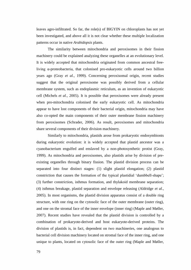
79
leaves agro-infiltrated. So far, the role(s) of BIGYIN on chloroplasts has not yet
been investigated, and above all it is not clear whether these multiple localization
patterns occur in native Arabidopsis plants.
The similarity between mitochondria and peroxisomes in their fission
machinery could be explained analyzing these organelles at an evolutionary level.
It is widely accepted that mitochondria originated from common ancestral free-
living α-proteobacteria, that colonised pro-eukaryotic cells around two billion
years ago (Gray et al., 1999). Concerning peroxisomal origin, recent studies
suggest that the original peroxisome was possibly derived from a cellular
membrane system, such as endoplasmic reticulum, as an invention of eukaryotic
cell (Michels et al., 2005). It is possible that peroxisomes were already present
when pro-mitochondria colonised the early eukaryotic cell. As mitochondria
appear to have lost components of their bacterial origin, mitochondria may have
also co-opted the main components of their outer membrane fission machinery
from peroxisomes (Schrader, 2006). As result, peroxisomes and mitochondria
share several components of their division machinery.
Similarly to mitochondria, plastids arose from prokaryotic endosymbionts
during eukaryotic evolution: it is widely accepted that plastid ancestor was a
cyanobacterium engulfed and enslaved by a non-photosynthetic protist (Gray,
1999). As mitochondria and peroxisomes, also plastids arise by division of pre-
existing organelles through binary fission. The plastid division process can be
separated into four distinct stages: (1) slight plastid elongation; (2) plastid
constriction that causes the formation of the typical plastidial „dumbbell-shape‟;
(3) further constriction, isthmus formation, and thylakoid membrane separation;
(4) isthmus breakage, plastid separation and envelope releasing (Aldridge et al.,
2005). In most organisms, the plastid division apparatus consist of a double ring
structure, with one ring on the cytosolic face of the outer membrane (outer ring),
and one on the stromal face of the inner envelope (inner ring) (Maple and Møller,
2007). Recent studies have revealed that the plastid division is controlled by a
combination of prokaryote-derived and host eukaryote-derived proteins. The
division of plastids is, in fact, dependent on two machineries, one analogous to
bacterial cell division machinery located on stromal face of the inner ring, and one
unique to plants, located on cytosolic face of the outer ring (Maple and Møller,

80
2007). Similarly to mitochondria and peroxisomes, also in plastids orthologs of
dynamin superfamily GTPases are involved in plastid division. In particular in
Arabidopsis, one of these orthologs is DRPB5 (previously namely ARC5,
At3g19720) and very recent data have demonstrated that this chloroplast division
protein plays a role also in peroxisomal division (Zhang and Hu, 2010).
All together, these findings indicate that, in Arabidopsis, peroxisomes and
chloroplasts share at least one component of their division machinery, and that
peroxisomes and mitochondria share several components of their division
machinery. The use of shared components could be a mechanism to promote
coordinated division among these organelles that are at least metabolically linked
(Schrader, 2006).
Here, we focus on the study of BIGYIN, an Arabidopsis Fission1-like
protein involved in mitochondrial and peroxisomal fission. We report, in
Arabidopsis plants, a detailed subcellular localization of BIGYIN, expressed
under control of its own promoter. We are in fact initially interested in elucidating
whether the multiple subcellular localization patterns of BIGYIN to mitochondria,
peroxisomes and chloroplasts, observed in our previously analyses (Chapter 2)
using the constitutive CaMV promoter, are maintained when BIGYIN expression
is driven by the native promoter. Then, we analyze the subcellular localization of
mutated BIGYIN protein showing on chloroplasts surfaces a very interesting
pattern that provides the first indication that this protein could be involved in
chloroplast fission. Yet, we dissect BIGYIN to define domains crucial for its
multiple targeting. In addition, we report the localization of BIGYIN to the
tubular protrusions extending from mitochondrial, peroxisomal and chloroplast
membranes, and we investigated whether inter-organellar interactions occurred
through these tubular structures in plants.
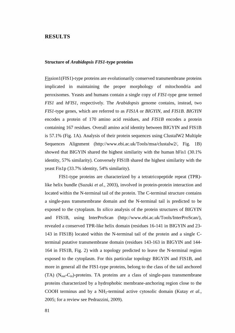
81
RESULTS
Structure of Arabidopsis FIS1-type proteins
Fission1(FIS1)-type proteins are evolutionarily conserved transmembrane proteins
implicated in maintaining the proper morphology of mitochondria and
peroxisomes. Yeasts and humans contain a single copy of FIS1-type gene termed
FIS1 and hFIS1, respectively. The Arabidopsis genome contains, instead, two
FIS1-type genes, which are referred to as FIS1A or BIGYIN, and FIS1B. BIGYIN
encodes a protein of 170 amino acid residues, and FIS1B encodes a protein
containing 167 residues. Overall amino acid identity between BIGYIN and FIS1B
is 57.1% (Fig. 1A). Analysis of their protein sequences using ClustalW2 Multiple
Sequences Alignment (http://www.ebi.ac.uk/Tools/msa/clustalw2/, Fig. 1B)
showed that BIGYIN shared the highest similarity with the human hFis1 (30.1%
identity, 57% similarity). Conversely FIS1B shared the highest similarity with the
yeast Fis1p (33.7% identity, 54% similarity).
FIS1-type proteins are characterized by a tetratricopeptide repeat (TPR)-
like helix bundle (Suzuki et al., 2003), involved in protein-protein interaction and
located within the N-terminal tail of the protein. The C-terminal structure contains
a single-pass transmembrane domain and the N-terminal tail is predicted to be
exposed to the cytoplasm. In silico analysis of the protein structures of BIGYIN
and FIS1B, using InterProScan (http://www.ebi.ac.uk/Tools/InterProScan/),
revealed a conserved TPR-like helix domain (residues 16-141 in BIGYIN and 23-
143 in FIS1B) located within the N-terminal tail of the protein and a single C-
terminal putative transmembrane domain (residues 143-163 in BIGYIN and 144-
164 in FIS1B, Fig. 2) with a topology predicted to leave the N-terminal region
exposed to the cytoplasm. For this particular topology BIGYIN and FIS1B, and
more in general all the FIS1-type proteins, belong to the class of the tail anchored
(TA) (Nout-Cin)-proteins. TA proteins are a class of single-pass transmembrane
proteins characterized by a hydrophobic membrane-anchoring region close to the
COOH terminus and by a NH2-terminal active cytosolic domain (Kutay et al.,
2005; for a review see Pedrazzini, 2009).
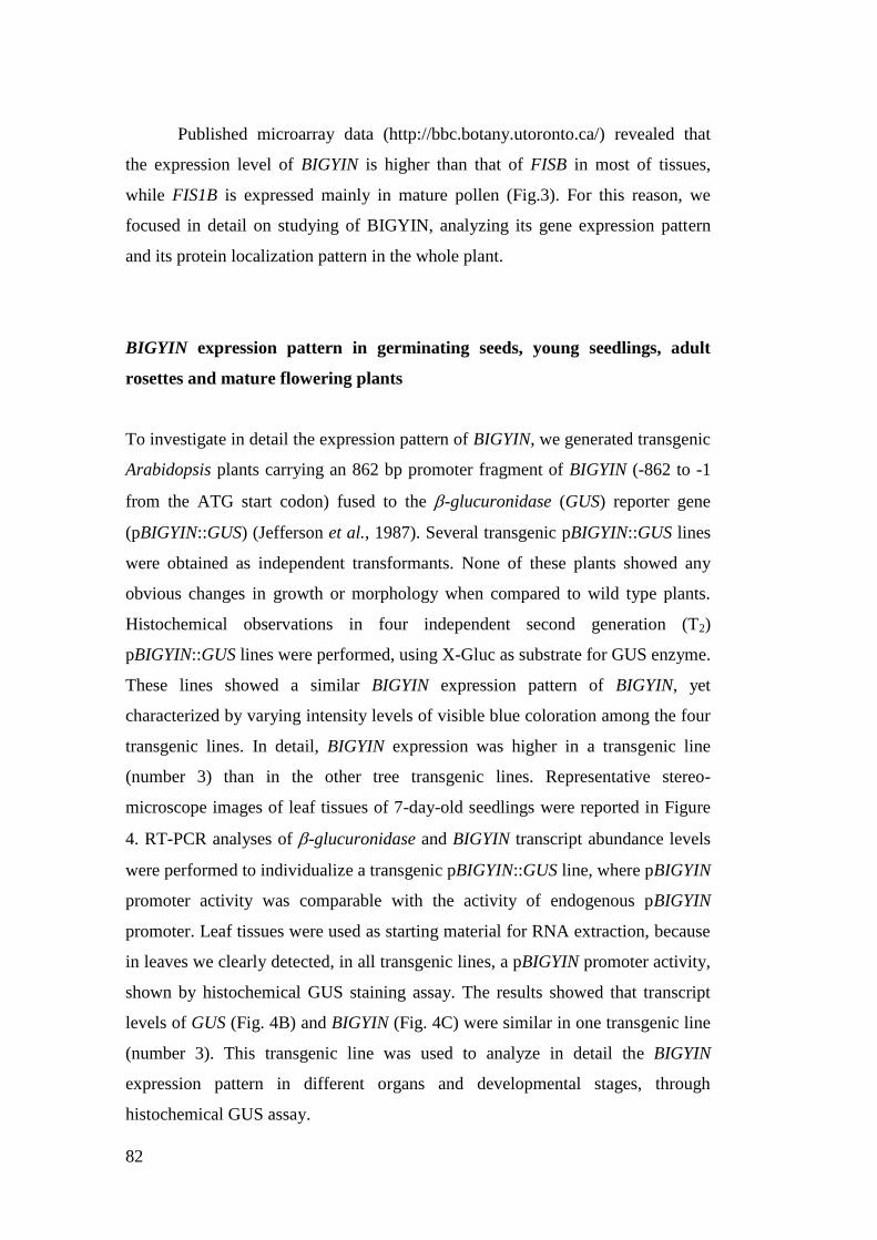
82
Published microarray data (http://bbc.botany.utoronto.ca/) revealed that
the expression level of BIGYIN is higher than that of FISB in most of tissues,
while FIS1B is expressed mainly in mature pollen (Fig.3). For this reason, we
focused in detail on studying of BIGYIN, analyzing its gene expression pattern
and its protein localization pattern in the whole plant.
BIGYIN expression pattern in germinating seeds, young seedlings, adult
rosettes and mature flowering plants
To investigate in detail the expression pattern of BIGYIN, we generated transgenic
Arabidopsis plants carrying an 862 bp promoter fragment of BIGYIN (-862 to -1
from the ATG start codon) fused to the -glucuronidase (GUS) reporter gene
(pBIGYIN::GUS) (Jefferson et al., 1987). Several transgenic pBIGYIN::GUS lines
were obtained as independent transformants. None of these plants showed any
obvious changes in growth or morphology when compared to wild type plants.
Histochemical observations in four independent second generation (T2)
pBIGYIN::GUS lines were performed, using X-Gluc as substrate for GUS enzyme.
These lines showed a similar BIGYIN expression pattern of BIGYIN, yet
characterized by varying intensity levels of visible blue coloration among the four
transgenic lines. In detail, BIGYIN expression was higher in a transgenic line
(number 3) than in the other tree transgenic lines. Representative stereo-
microscope images of leaf tissues of 7-day-old seedlings were reported in Figure
4. RT-PCR analyses of -glucuronidase and BIGYIN transcript abundance levels
were performed to individualize a transgenic pBIGYIN::GUS line, where pBIGYIN
promoter activity was comparable with the activity of endogenous pBIGYIN
promoter. Leaf tissues were used as starting material for RNA extraction, because
in leaves we clearly detected, in all transgenic lines, a pBIGYIN promoter activity,
shown by histochemical GUS staining assay. The results showed that transcript
levels of GUS (Fig. 4B) and BIGYIN (Fig. 4C) were similar in one transgenic line
(number 3). This transgenic line was used to analyze in detail the BIGYIN
expression pattern in different organs and developmental stages, through
histochemical GUS assay.
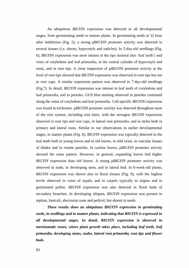
83
An ubiquitous BIGYIN expression was detected in all developmental
stages, from germinating seeds to mature plants. In germinating seeds at 32 hour
after imbibition (Fig. 5), a strong pBIGYIN promoter activity was detected in
several tissues (i.e. shoots, hypocotyls and radicles). In 5-day-old seedlings (Fig.
6), BIGYIN expression was most intense at the tips (termed also „leaf teeth‟) and
veins of cotyledons and leaf primordia, in the central cylinder of hypocotyls and
roots, and in root tips. A close inspection of pBIGYIN promoter activity at the
level of root tips showed that BIGYIN expression was observed in root tips but not
in root caps. A similar expression pattern was observed in 7-day-old seedlings
(Fig.7). In detail, BIGYIN expression was intense in leaf teeth of cotyledons and
leaf primordia, and in petioles. GUS blue staining observed in petioles continued
along the veins of cotyledons and leaf primordia. Cell-specific BIGYIN expression
was found in trichomes. pBIGYIN promoter activity was detected throughout most
of the root system, including root hairs, with the strongest BIGYIN expression
observed in root tips and root caps, in lateral root primordia, and in steles both in
primary and lateral roots. Similar to our observations in earlier developmental
stages, in mature plants (Fig. 8), BIGYIN expression was typically detected in the
leaf teeth both in young leaves and in old leaves, in mid veins, in vascular tissues
of blades and in rosette petioles. In cauline leaves, pBIGYIN promoter activity
showed the same pattern. However, in general, expanding leaves had higher
BIGYIN expression than old leaves. A strong pBIGYIN promoter activity was
observed in node, in developing stem, and in lateral bud. In 6-week-old plants,
BIGYIN expression was shown also in floral tissues (Fig. 9), with the highest
levels observed in veins of sepals, and in carpels typically in stigma and in
germinated pollen. BIGYIN expression was also detected in floral buds of
secondary branches. In developing siliques, BIGYIN expression was present in
replum, funiculi, abscission zone and pedicel, but absent in seeds.
These results show an ubiquitous BIGYIN expression in germinating
seeds, in seedlings and in mature plants, indicating that BIGYIN is expressed in
all developmental stages. In detail, BIGYIN expression is observed in
meristematic zones, where plant growth takes place, including leaf teeth, leaf
primordia, developing stems, nodes, lateral root primordia, root tips and flower
buds.
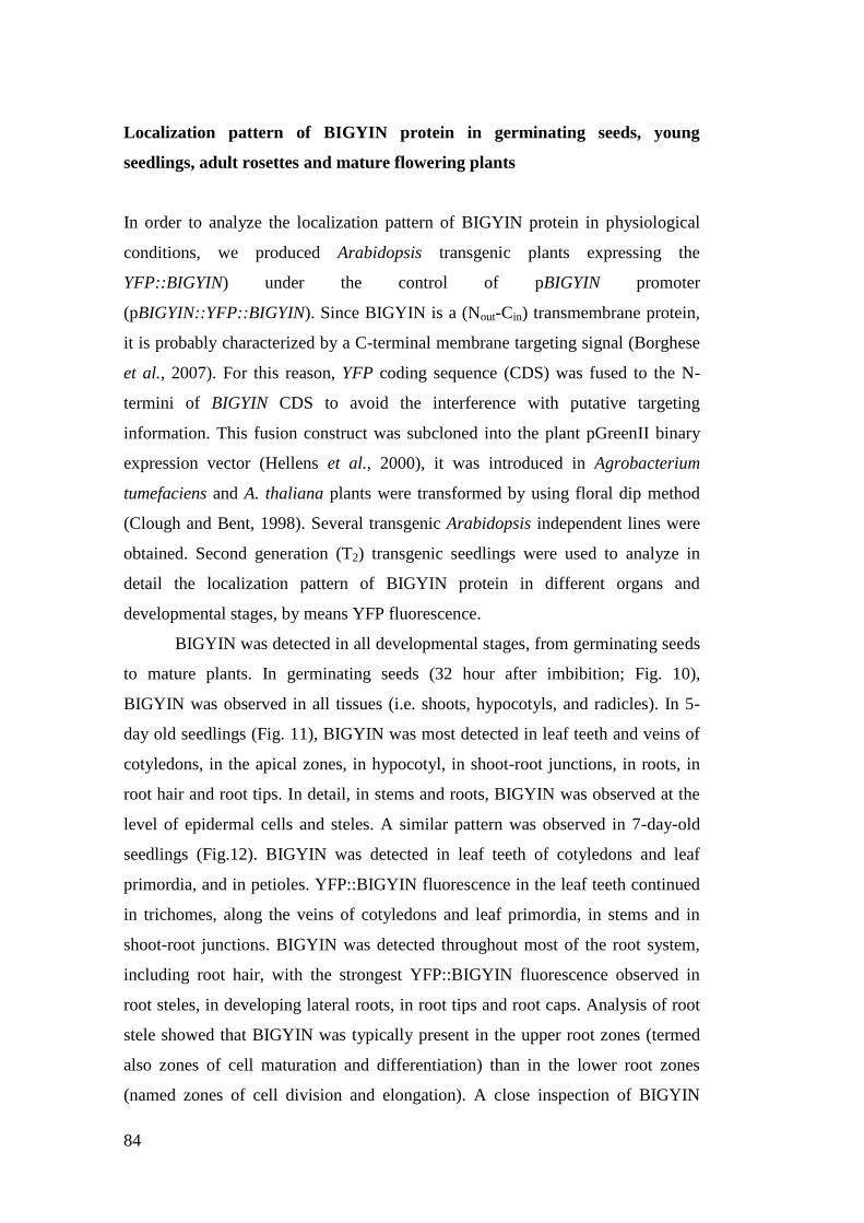
84
Localization pattern of BIGYIN protein in germinating seeds, young
seedlings, adult rosettes and mature flowering plants
In order to analyze the localization pattern of BIGYIN protein in physiological
conditions, we produced Arabidopsis transgenic plants expressing the
YFP::BIGYIN) under the control of pBIGYIN promoter
(pBIGYIN::YFP::BIGYIN). Since BIGYIN is a (Nout-Cin) transmembrane protein,
it is probably characterized by a C-terminal membrane targeting signal (Borghese
et al., 2007). For this reason, YFP coding sequence (CDS) was fused to the N-
termini of BIGYIN CDS to avoid the interference with putative targeting
information. This fusion construct was subcloned into the plant pGreenII binary
expression vector (Hellens et al., 2000), it was introduced in Agrobacterium
tumefaciens and A. thaliana plants were transformed by using floral dip method
(Clough and Bent, 1998). Several transgenic Arabidopsis independent lines were
obtained. Second generation (T2) transgenic seedlings were used to analyze in
detail the localization pattern of BIGYIN protein in different organs and
developmental stages, by means YFP fluorescence.
BIGYIN was detected in all developmental stages, from germinating seeds
to mature plants. In germinating seeds (32 hour after imbibition; Fig. 10),
BIGYIN was observed in all tissues (i.e. shoots, hypocotyls, and radicles). In 5-
day old seedlings (Fig. 11), BIGYIN was most detected in leaf teeth and veins of
cotyledons, in the apical zones, in hypocotyl, in shoot-root junctions, in roots, in
root hair and root tips. In detail, in stems and roots, BIGYIN was observed at the
level of epidermal cells and steles. A similar pattern was observed in 7-day-old
seedlings (Fig.12). BIGYIN was detected in leaf teeth of cotyledons and leaf
primordia, and in petioles. YFP::BIGYIN fluorescence in the leaf teeth continued
in trichomes, along the veins of cotyledons and leaf primordia, in stems and in
shoot-root junctions. BIGYIN was detected throughout most of the root system,
including root hair, with the strongest YFP::BIGYIN fluorescence observed in
root steles, in developing lateral roots, in root tips and root caps. Analysis of root
stele showed that BIGYIN was typically present in the upper root zones (termed
also zones of cell maturation and differentiation) than in the lower root zones
(named zones of cell division and elongation). A close inspection of BIGYIN
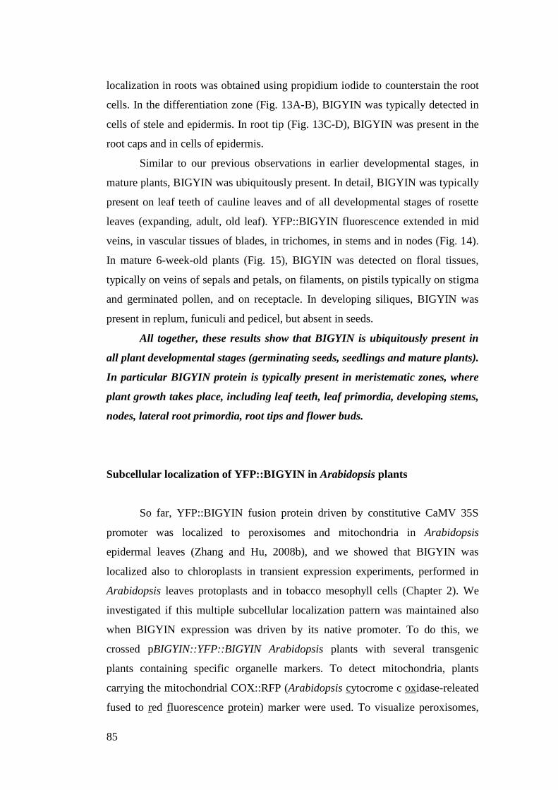
85
localization in roots was obtained using propidium iodide to counterstain the root
cells. In the differentiation zone (Fig. 13A-B), BIGYIN was typically detected in
cells of stele and epidermis. In root tip (Fig. 13C-D), BIGYIN was present in the
root caps and in cells of epidermis.
Similar to our previous observations in earlier developmental stages, in
mature plants, BIGYIN was ubiquitously present. In detail, BIGYIN was typically
present on leaf teeth of cauline leaves and of all developmental stages of rosette
leaves (expanding, adult, old leaf). YFP::BIGYIN fluorescence extended in mid
veins, in vascular tissues of blades, in trichomes, in stems and in nodes (Fig. 14).
In mature 6-week-old plants (Fig. 15), BIGYIN was detected on floral tissues,
typically on veins of sepals and petals, on filaments, on pistils typically on stigma
and germinated pollen, and on receptacle. In developing siliques, BIGYIN was
present in replum, funiculi and pedicel, but absent in seeds.
All together, these results show that BIGYIN is ubiquitously present in
all plant developmental stages (germinating seeds, seedlings and mature plants).
In particular BIGYIN protein is typically present in meristematic zones, where
plant growth takes place, including leaf teeth, leaf primordia, developing stems,
nodes, lateral root primordia, root tips and flower buds.
Subcellular localization of YFP::BIGYIN in Arabidopsis plants
So far, YFP::BIGYIN fusion protein driven by constitutive CaMV 35S
promoter was localized to peroxisomes and mitochondria in Arabidopsis
epidermal leaves (Zhang and Hu, 2008b), and we showed that BIGYIN was
localized also to chloroplasts in transient expression experiments, performed in
Arabidopsis leaves protoplasts and in tobacco mesophyll cells (Chapter 2). We
investigated if this multiple subcellular localization pattern was maintained also
when BIGYIN expression was driven by its native promoter. To do this, we
crossed pBIGYIN::YFP::BIGYIN Arabidopsis plants with several transgenic
plants containing specific organelle markers. To detect mitochondria, plants
carrying the mitochondrial COX::RFP (Arabidopsis cytocrome c oxidase-releated
fused to red fluorescence protein) marker were used. To visualize peroxisomes,
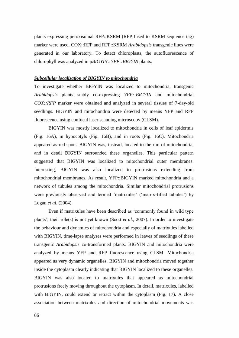
86
plants expressing peroxisomal RFP::KSRM (RFP fused to KSRM sequence tag)
marker were used. COX::RFP and RFP::KSRM Arabidopsis transgenic lines were
generated in our laboratory. To detect chloroplasts, the autofluorescence of
chlorophyll was analyzed in pBIGYIN::YFP::BIGYIN plants.
Subcellular localization of BIGYIN to mitochondria
To investigate whether BIGYIN was localized to mitochondria, transgenic
Arabidopsis plants stably co-expressing YFP::BIGYIN and mitochondrial
COX::RFP marker were obtained and analyzed in several tissues of 7-day-old
seedlings. BIGYIN and mitochondria were detected by means YFP and RFP
fluorescence using confocal laser scanning microscopy (CLSM).
BIGYIN was mostly localized to mitochondria in cells of leaf epidermis
(Fig. 16A), in hypocotyls (Fig. 16B), and in roots (Fig. 16C). Mitochondria
appeared as red spots. BIGYIN was, instead, located to the rim of mitochondria,
and in detail BIGYIN surrounded these organelles. This particular pattern
suggested that BIGYIN was localized to mitochondrial outer membranes.
Interesting, BIGYIN was also localized to protrusions extending from
mitochondrial membranes. As result, YFP::BIGYIN marked mitochondria and a
network of tubules among the mitochondria. Similar mitochondrial protrusions
were previously observed and termed „matrixules‟ („matrix-filled tubules‟) by
Logan et al. (2004).
Even if matrixules have been described as „commonly found in wild type
plants‟, their role(s) is not yet known (Scott et al., 2007). In order to investigate
the behaviour and dynamics of mitochondria and especially of matrixules labelled
with BIGYIN, time-lapse analyses were performed in leaves of seedlings of these
transgenic Arabidopsis co-transformed plants. BIGYIN and mitochondria were
analyzed by means YFP and RFP fluorescence using CLSM. Mitochondria
appeared as very dynamic organelles. BIGYIN and mitochondria moved together
inside the cytoplasm clearly indicating that BIGYIN localized to these organelles.
BIGYIN was also located to matrixules that appeared as mitochondrial
protrusions freely moving throughout the cytoplasm. In detail, matrixules, labelled
with BIGYIN, could extend or retract within the cytoplasm (Fig. 17). A close
association between matrixules and direction of mitochondrial movements was
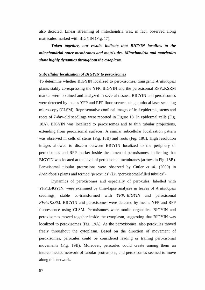
87
also detected. Linear streaming of mitochondria was, in fact, observed along
matrixules marked with BIGYIN (Fig. 17).
Taken together, our results indicate that BIGYIN localizes to the
mitochondrial outer membranes and matrixules. Mitochondria and matrixules
show highly dynamics throughout the cytoplasm.
Subcellular localization of BIGYIN to peroxisomes
To determine whether BIGYIN localized to peroxisomes, transgenic Arabidopsis
plants stably co-expressing the YFP::BIGYIN and the peroxisomal RFP::KSRM
marker were obtained and analyzed in several tissues. BIGYIN and peroxisomes
were detected by means YFP and RFP fluorescence using confocal laser scanning
microscopy (CLSM). Representative confocal images of leaf epidermis, stems and
roots of 7-day-old seedlings were reported in Figure 18. In epidermal cells (Fig.
18A), BIGYIN was localized to peroxisomes and to thin tubular projections,
extending from peroxisomal surfaces. A similar subcellular localization pattern
was observed in cells of stems (Fig. 18B) and roots (Fig. 18C). High resolution
images allowed to discern between BIGYIN localized to the periphery of
peroxisomes and RFP marker inside the lumen of peroxisomes, indicating that
BIGYIN was located at the level of peroxisomal membranes (arrows in Fig. 18B).
Peroxisomal tubular protrusions were observed by Cutler et al. (2000) in
Arabidopsis plants and termed „peroxules‟ (i.e. „peroxisomal-filled tubules‟).
Dynamics of peroxisomes and especially of peroxules, labelled with
YFP::BIGYIN, were examined by time-lapse analyses in leaves of Arabidopsis
seedlings, stable co-transformed with YFP::BIGYIN and peroxisomal
RFP::KSRM. BIGYIN and peroxisomes were detected by means YFP and RFP
fluorescence using CLSM. Peroxisomes were motile organelles. BIGYIN and
peroxisomes moved together inside the cytoplasm, suggesting that BIGYIN was
localized to peroxisomes (Fig. 19A). As the peroxisomes, also peroxules moved
freely throughout the cytoplasm. Based on the direction of movement of
peroxisomes, peroxules could be considered leading or trailing peroxisomal
movements (Fig. 19B). Moreover, peroxules could create among them an
interconnected network of tubular protrusions, and peroxisomes seemed to move
along this network.
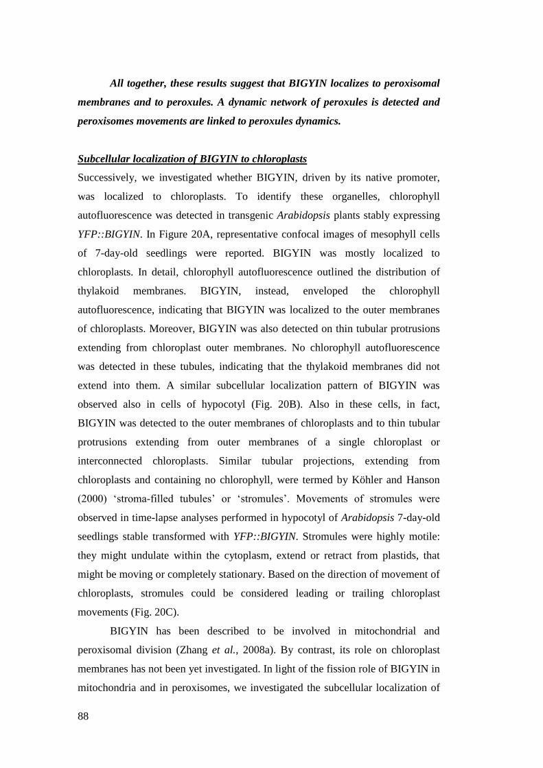
88
All together, these results suggest that BIGYIN localizes to peroxisomal
membranes and to peroxules. A dynamic network of peroxules is detected and
peroxisomes movements are linked to peroxules dynamics.
Subcellular localization of BIGYIN to chloroplasts
Successively, we investigated whether BIGYIN, driven by its native promoter,
was localized to chloroplasts. To identify these organelles, chlorophyll
autofluorescence was detected in transgenic Arabidopsis plants stably expressing
YFP::BIGYIN. In Figure 20A, representative confocal images of mesophyll cells
of 7-day-old seedlings were reported. BIGYIN was mostly localized to
chloroplasts. In detail, chlorophyll autofluorescence outlined the distribution of
thylakoid membranes. BIGYIN, instead, enveloped the chlorophyll
autofluorescence, indicating that BIGYIN was localized to the outer membranes
of chloroplasts. Moreover, BIGYIN was also detected on thin tubular protrusions
extending from chloroplast outer membranes. No chlorophyll autofluorescence
was detected in these tubules, indicating that the thylakoid membranes did not
extend into them. A similar subcellular localization pattern of BIGYIN was
observed also in cells of hypocotyl (Fig. 20B). Also in these cells, in fact,
BIGYIN was detected to the outer membranes of chloroplasts and to thin tubular
protrusions extending from outer membranes of a single chloroplast or
interconnected chloroplasts. Similar tubular projections, extending from
chloroplasts and containing no chlorophyll, were termed by Köhler and Hanson
(2000) „stroma-filled tubules‟ or „stromules‟. Movements of stromules were
observed in time-lapse analyses performed in hypocotyl of Arabidopsis 7-day-old
seedlings stable transformed with YFP::BIGYIN. Stromules were highly motile:
they might undulate within the cytoplasm, extend or retract from plastids, that
might be moving or completely stationary. Based on the direction of movement of
chloroplasts, stromules could be considered leading or trailing chloroplast
movements (Fig. 20C).
BIGYIN has been described to be involved in mitochondrial and
peroxisomal division (Zhang et al., 2008a). By contrast, its role on chloroplast
membranes has not been yet investigated. In light of the fission role of BIGYIN in
mitochondria and in peroxisomes, we investigated the subcellular localization of
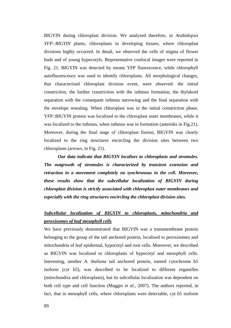
89
BIGYIN during chloroplast division. We analyzed therefore, in Arabidopsis
YFP::BIGYIN plants, chloroplasts in developing tissues, where chloroplast
divisions highly occurred. In detail, we observed the cells of stigma of flower
buds and of young hypocotyls. Representative confocal images were reported in
Fig. 21. BIGYIN was detected by means YFP fluorescence, while chlorophyll
autofluorescence was used to identify chloroplasts. All morphological changes,
that characterized chloroplast division event, were observed: the initial
constriction, the further constriction with the isthmus formation, the thylakoid
separation with the consequent isthmus narrowing and the final separation with
the envelope resealing. When chloroplast was in the initial constriction phase,
YFP::BIGYIN protein was localized to the chloroplast outer membranes, while it
was localized to the isthmus, when isthmus was in formation (asterisks in Fig.21).
Moreover, during the final stage of chloroplast fission, BIGYIN was clearly
localized to the ring structures encircling the division sites between two
chloroplasts (arrows, in Fig. 21).
Our data indicate that BIGYIN localizes to chloroplasts and stromules.
The outgrowth of stromules is characterized by transient extension and
retraction in a movement completely no synchronous in the cell. Moreover,
these results show that the subcellular localization of BIGYIN during
chloroplast division is strictly associated with chloroplast outer membranes and
especially with the ring structures encircling the chloroplast division sites.
Subcellular localization of BIGYIN to chloroplasts, mitochondria and
peroxisomes of leaf mesophyll cells
We have previously demonstrated that BIGYIN was a transmembrane protein
belonging to the group of the tail anchored protein, localized to peroxisomes and
mitochondria of leaf epidermal, hypocotyl and root cells. Moreover, we described
as BIGYIN was localized to chloroplasts of hypocotyl and mesophyll cells.
Interesting, another A. thaliana tail anchored protein, named cytochrome b5
isoform (cyt b5), was described to be localized to different organelles
(mitochondria and chloroplasts), but its subcellular localization was dependent on
both cell type and cell function (Maggio et al., 2007). The authors reported, in
fact, that in mesophyll cells, where chloroplasts were detectable, cyt b5 isoform
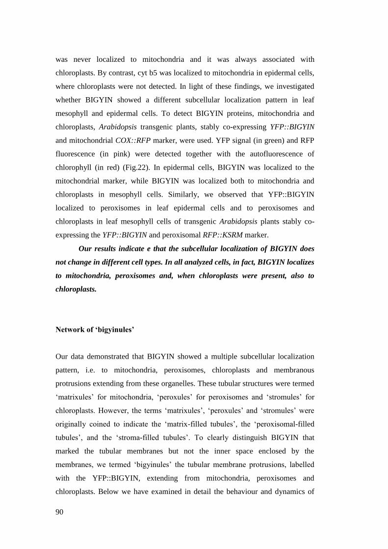
90
was never localized to mitochondria and it was always associated with
chloroplasts. By contrast, cyt b5 was localized to mitochondria in epidermal cells,
where chloroplasts were not detected. In light of these findings, we investigated
whether BIGYIN showed a different subcellular localization pattern in leaf
mesophyll and epidermal cells. To detect BIGYIN proteins, mitochondria and
chloroplasts, Arabidopsis transgenic plants, stably co-expressing YFP::BIGYIN
and mitochondrial COX::RFP marker, were used. YFP signal (in green) and RFP
fluorescence (in pink) were detected together with the autofluorescence of
chlorophyll (in red) (Fig.22). In epidermal cells, BIGYIN was localized to the
mitochondrial marker, while BIGYIN was localized both to mitochondria and
chloroplasts in mesophyll cells. Similarly, we observed that YFP::BIGYIN
localized to peroxisomes in leaf epidermal cells and to peroxisomes and
chloroplasts in leaf mesophyll cells of transgenic Arabidopsis plants stably co-
expressing the YFP::BIGYIN and peroxisomal RFP::KSRM marker.
Our results indicate e that the subcellular localization of BIGYIN does
not change in different cell types. In all analyzed cells, in fact, BIGYIN localizes
to mitochondria, peroxisomes and, when chloroplasts were present, also to
chloroplasts.
Network of ‘bigyinules’
Our data demonstrated that BIGYIN showed a multiple subcellular localization
pattern, i.e. to mitochondria, peroxisomes, chloroplasts and membranous
protrusions extending from these organelles. These tubular structures were termed
„matrixules‟ for mitochondria, „peroxules‟ for peroxisomes and „stromules‟ for
chloroplasts. However, the terms „matrixules‟, „peroxules‟ and „stromules‟ were
originally coined to indicate the „matrix-filled tubules‟, the „peroxisomal-filled
tubules‟, and the „stroma-filled tubules‟. To clearly distinguish BIGYIN that
marked the tubular membranes but not the inner space enclosed by the
membranes, we termed „bigyinules‟ the tubular membrane protrusions, labelled
with the YFP::BIGYIN, extending from mitochondria, peroxisomes and
chloroplasts. Below we have examined in detail the behaviour and dynamics of
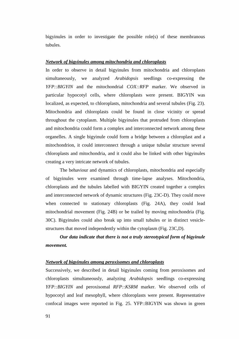
91
bigyinules in order to investigate the possible role(s) of these membranous
tubules.
Network of bigyinules among mitochondria and chloroplasts
In order to observe in detail bigyinules from mitochondria and chloroplasts
simultaneously, we analyzed Arabidopsis seedlings co-expressing the
YFP::BIGYIN and the mitochondrial COX::RFP marker. We observed in
particular hypocotyl cells, where chloroplasts were present. BIGYIN was
localized, as expected, to chloroplasts, mitochondria and several tubules (Fig. 23).
Mitochondria and chloroplasts could be found in close vicinity or spread
throughout the cytoplasm. Multiple bigyinules that protruded from chloroplasts
and mitochondria could form a complex and interconnected network among these
organelles. A single bigyinule could form a bridge between a chloroplast and a
mitochondrion, it could interconnect through a unique tubular structure several
chloroplasts and mitochondria, and it could also be linked with other bigyinules
creating a very intricate network of tubules.
The behaviour and dynamics of chloroplasts, mitochondria and especially
of bigyinules were examined through time-lapse analyses. Mitochondria,
chloroplasts and the tubules labelled with BIGYIN created together a complex
and interconnected network of dynamic structures (Fig. 23C-D). They could move
when connected to stationary chloroplasts (Fig. 24A), they could lead
mitochondrial movement (Fig. 24B) or be trailed by moving mitochondria (Fig.
30C). Bigyinules could also break up into small tubules or in distinct vesicle-
structures that moved independently within the cytoplasm (Fig. 23C,D).
Our data indicate that there is not a truly stereotypical form of bigyinule
movement.
Network of bigyinules among peroxisomes and chloroplasts
Successively, we described in detail bigyinules coming from peroxisomes and
chloroplasts simultaneously, analyzing Arabidopsis seedlings co-expressing
YFP::BIGYIN and peroxisomal RFP::KSRM marker. We observed cells of
hypocotyl and leaf mesophyll, where chloroplasts were present. Representative
confocal images were reported in Fig. 25. YFP::BIGYIN was shown in green
92
colour; peroxisomes in red; while chloroplasts were easily distinguish by their
dimension. BIGYIN was localized, as expected, to chloroplasts, peroxisomes and
several tubules. As previously observed in the case of mitochondria and
chloroplasts, also peroxisomes and chloroplasts could be close localized or
dispersed within the cytoplasm. Several bigyinules protruded from chloroplasts
and peroxisomes forming a complex network interconnecting these different
organelles.
The behaviour and dynamics of chloroplasts, peroxisomes and especially
of bigyinules were examined by time-lapse analyses (Fig. 26). Peroxisomes,
chloroplasts and tubules, labelled with BIGYIN, described together a complex and
interconnected network of dynamic structures. Bigyinules could extend or retract
freely from organelles. Bigyinules could also protrude from peroxisomes to
chloroplasts or vice versa. Based on the direction of movements of peroxisomes,
bigyinules could be considered leading or trailing the peroxisomal movements:
bigyinules could, in fact, be trailed by moving peroxisomes or, on the contrary,
bigyinules could lead peroxisomes. Bigyinules could break up into small tubules
or in distinct vesicle-structures that moved independently within the cytoplasm
(Fig. 26C).
These results confirmed that there are not truly stereotypical forms of
bigyinule movement.
Analysis of the anti-BIGYIN antibody specificity
A specific polyclonal antibody was generated against a peptide of 15aa located to
the N-terminal tail of BIGYIN (aa 33-47), in order to confirm the multiple
subcellular localization of BIGYIN by means biochemical techniques (i.e.
immunogold electron microscopy assay and immunoblot assay of protein extracts
enriched in specific subcellular compartments). So far, we tested the anti-
BIGYIN-antibody in western blot assay using crude extracts from transgenic
plants stably expressing YFP::BIGYIN and from wild type plants. As result, we
obtained that the anti-BIGYIN antibody was specific, given that it specifically
recognized in wild type plants the BIGYIN protein (18,7 kDa) (Fig. 27), and in
93
pBIGYIN::YFP::BIGYIN transgenic plants both the BIGYIN protein (18,7 kDa)
and the YFP::BIGYIN fusion protein (46,3 kDa). Moreover, we observed that the
YFP::BIGYIN fusion protein was more intense that BIGYIN in the crude extracts
obtained from the transgenic pBIGYIN::YFP::BIGYIN plants.
All together, these results indicate that the anti-BIGYIN-antibody is
specific for BIGYIN. Moreover, our data indicate that in
pBIGYIN::YFP::BIGYIN plants, although the YFP::BIGYIN is driven by the
pBIGYIN promoter, the YFP::BIGYIN fusion protein is more abundant than
the endogenous BIGYIN protein.
Characterization of pBIGYIN::YFP::BIGYIN transgenic plants
It was been reported that in BIGYIN overexpressing plants the number of
peroxisomes increased to about three fold compared with the wild type, and that
the total peroxisomal area in BIGYIN overexpressing plants increased of five to
six times from that of wild type plants (Zhang and Hu, 2008b). In order to
investigate if in our pBIGYIN::YFP::BIGYIN plants a similar aberrant
morphology of peroxisomes occurred, we used ImageJ software to measure the
peroxisomal area and the peroxisomal number in plant co-expressing the
pBIGYIN::YFP::BIGYIN and the peroxisomal (RFP::KSRM) marker and in plant
expressing the peroxisomal RFP::KSRM, used as control of peroxisomal
morphology (Fig. 28). We obtained that the number of peroxisomes was increased
to about two fold compared with the control, and that the area of peroxisomes per
cell was decreased to about two fold compared with the control. The total
peroxisomal area did not show any difference between the transgenic and wild
type plants.
All together, our results demonstrate that in pBIGYIN::YFP::BIGYIN
plants the number of peroxisomes increase to about two fold compared with the
control, while the total peroxisomal area per cell remain invariable.
94
Effects of an actin inhibitor on movement of organelles labelled with
YFP::BIGYIN and bigyinules
In plants the intracellular movements of mitochondria, peroxisomes and
chloroplasts are modulated by a microfilament-based cytoskeleton. We wondered
whether the actin cytoskeleton played a role in the movements of organelles
marked with YFP::BIGYIN and of bigyinules. To this aim, we treated
YFP::BIGYIN seedlings with Lantruculin B (LatB), an inhibitor of actin
polymerization. The treatment with Lat B inhibited the movement of the
subcellular structures marked by YFP::BIGYIN. As result, the distribution of
organelles and of tubules labelled with YFP::BIGYIN in the hypocotyl cells did
not change over 40 seconds (Fig. 29B,C). To verify the inhibitor action,
transgenic reporter plants were included in the analyses. Seedlings expressing
mitochondrial COX::RFP or peroxisomal RFP::KSRM marker were treated with
Lat B and we observed that mitochondria and peroxisomes were almost stationary
(Fig. 30B,D). These data confirmed the treatment efficacy.
Taken together these results demonstrated that the actin-based
cytoskeleton plays a role in the movement of bigyinules and of organelles
marked with YFP::BIGYIN.
Effects of an actin inhibitor on movement of YFP::BIGYIN within organelle
membranes
We wondered whether the YFP::BIGYIN fusion protein could also move
within the organelle membranes that it marked. Fluorescence recovery after
photobleaching (FRAP) was used to monitor the movement of YFP::BIGYIN
between membranes of two connected chloroplasts in pBIGYIN::YFP::BIGYIN
seedlings (Fig. 31). FRAP assay was performed on chloroplasts because their big
dimension and their low dynamics guaranteed a perfect experimental condition.
When chloroplast membranes, marked with YFP::BIGYIN, were bleached,
fluorescence gradually recovered, at the expense of YFP fluorescence in the
connected plastid (Fig. 31B). Recovery was complete within 50 seconds. As
95
control, an unbleached and unconnected plastid in the same observation area did
not show loss fluorescence over time, indicating that no photobleaching was
occurred. These data indicated that there was a trafficking of YFP::BIGYIN
proteins among the outer membranes of connected chloroplasts.
Successively, we investigated whether the YFP::BIGYIN could move
among membranes of chloroplasts, mitochondria and peroxisomes. To this aim, a
single chloroplast not connected with other chloroplasts was bleached and then
monitored over 150 seconds (Fig. 32). Interestingly, during the observation period
many organelles labelled with YFP::BIGYIN were observed to go to the bleached
chloroplast. As result, the bleached chloroplast regained completely the
fluorescence within 94 seconds. As control, an unbleached and unconnected
plastid in the same observation area showed a minimal loss of fluorescence after
the 90 seconds as result of photobleaching. Since the bleached chloroplast was not
connected with other chloroplasts, and many YFP::BIGYIN-marked organelles
were observed approaching the bleached chloroplast during its fluorescence
recovery, it could be possible that trafficking of YFP::BIGYIN proteins occurred
among membranes of these different organelles.
To determine whether the movement of YFP::BIGYIN through the
organelle membranes was dependent on actin microfilaments, photobleaching
experiments were conducted after treatment with Lantruculin B (LatB) in
pBIGYIN::YFP::BIGYIN seedlings. To monitor YFP::BIGYIN trafficking, FRAP
experiment was performed on a chloroplast connected via bigyinules with another
chloroplast (Fig.33). When chloroplast membranes, marked with YFP::BIGYIN,
were bleached, no significant recovery of fluorescence was detected over time. In
addition, no organelles labelled with YFP::BIYIN were observed to go to the
bleached chloroplast. As control, an unbleached plastid in the same observation
area did not show a significant loss of fluorescence indicating that no
photobleaching was occurred during analysis. These results indicated that the
trafficking of the YFP::BIGYIN proteins among the organelle membranes is
dependent on actin-based cytoskeleton.
All together, our data demonstrate that a YFP::BIGYIN trafficking
actin-dependent occurs among membranes of different organelles labelled with
YFP::BIGYIN.
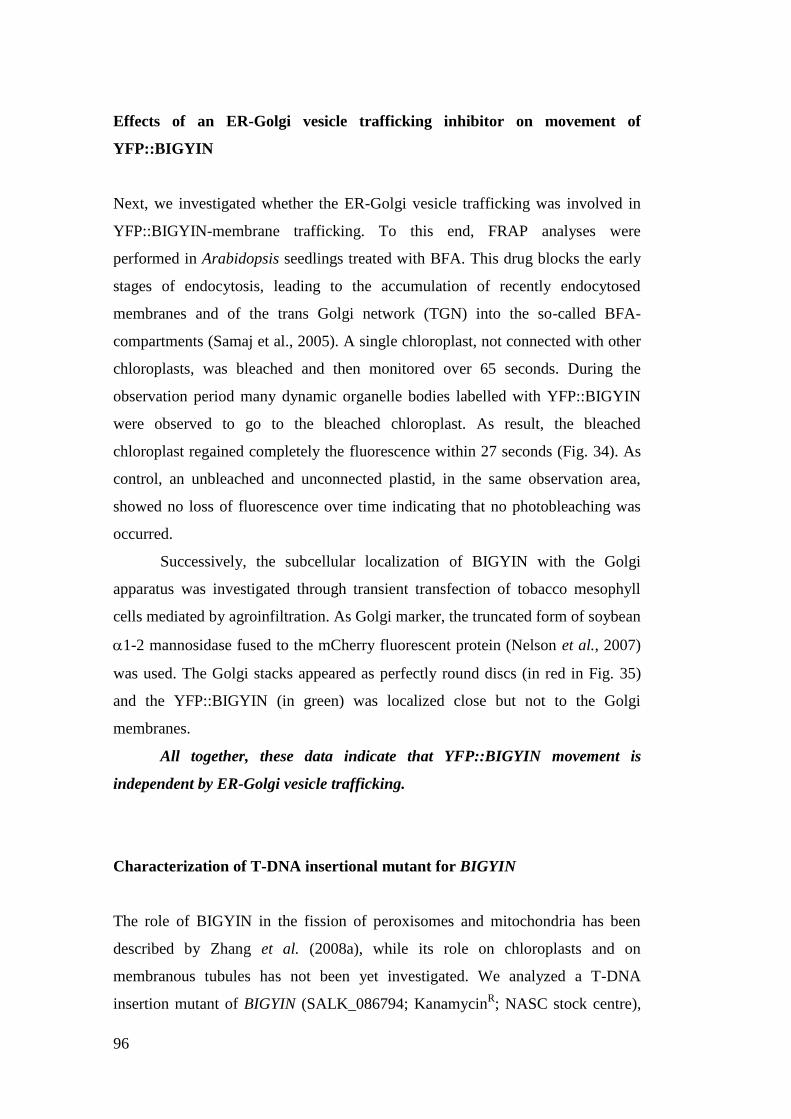
96
Effects of an ER-Golgi vesicle trafficking inhibitor on movement of
YFP::BIGYIN
Next, we investigated whether the ER-Golgi vesicle trafficking was involved in
YFP::BIGYIN-membrane trafficking. To this end, FRAP analyses were
performed in Arabidopsis seedlings treated with BFA. This drug blocks the early
stages of endocytosis, leading to the accumulation of recently endocytosed
membranes and of the trans Golgi network (TGN) into the so-called BFA-
compartments (Samaj et al., 2005). A single chloroplast, not connected with other
chloroplasts, was bleached and then monitored over 65 seconds. During the
observation period many dynamic organelle bodies labelled with YFP::BIGYIN
were observed to go to the bleached chloroplast. As result, the bleached
chloroplast regained completely the fluorescence within 27 seconds (Fig. 34). As
control, an unbleached and unconnected plastid, in the same observation area,
showed no loss of fluorescence over time indicating that no photobleaching was
occurred.
Successively, the subcellular localization of BIGYIN with the Golgi
apparatus was investigated through transient transfection of tobacco mesophyll
cells mediated by agroinfiltration. As Golgi marker, the truncated form of soybean
1-2 mannosidase fused to the mCherry fluorescent protein (Nelson et al., 2007)
was used. The Golgi stacks appeared as perfectly round discs (in red in Fig. 35)
and the YFP::BIGYIN (in green) was localized close but not to the Golgi
membranes.
All together, these data indicate that YFP::BIGYIN movement is
independent by ER-Golgi vesicle trafficking.
Characterization of T-DNA insertional mutant for BIGYIN
The role of BIGYIN in the fission of peroxisomes and mitochondria has been
described by Zhang et al. (2008a), while its role on chloroplasts and on
membranous tubules has not been yet investigated. We analyzed a T-DNA
insertion mutant of BIGYIN (SALK_086794; KanamycinR; NASC stock centre),
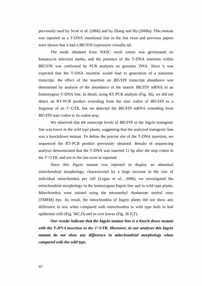
97
previously used by Scott et al. (2006) and by Zhang and Hu (2008a). This mutant
was reported as a T-DNA insertional line in the last exon and previous papers
were shown that it had a BIGYIN expression virtually nil.
The seeds obtained from NASC stock centre was germinated on
kanamycin selection media, and the presence of the T-DNA insertion within
BIGYIN was confirmed by PCR analyses on genomic DNA. Since it was
expected that the T-DNA insertion would lead to generation of a nonsense
transcript, the effect of the insertion on BIGYIN transcript abundance was
determined by analysis of the abundance of the mature BIGYIN mRNA in an
homozygous T-DNA line. In detail, using RT-PCR analysis (Fig. 36), we did not
detect an RT-PCR product extending from the start codon of BIGYIN to a
fragment of its 3‟-UTR, but we detected the BIGYIN mRNA extending from
BIGYIN start codon to its codon stop.
We observed that the transcript levels of BIGYIN in the bigyin transgenic
line was lower in the wild type plants, suggesting that the analyzed transgenic line
was a knockdown mutant. To define the precise site of the T-DNA insertion, we
sequenced the RT-PCR product previously obtained. Results of sequencing
analysis demonstrated that the T-DNA was inserted 11 bp after the stop codon in
the 3‟-UTR, and not in the last exon as reported.
Since this bigyin mutant was reported to display an abnormal
mitochondrial morphology, characterized by a large increase in the size of
individual mitochondria per cell (Logan et al., 2006), we investigated the
mitochondrial morphology in the homozygous bigyin line and in wild type plants.
Mitochondria were stained using the tetramethyl rhodamine methyl ester
(TMRM) dye. As result, the mitochondria of bigyin plants did not show any
difference in size when compared with mitochondria in wild type both in leaf
epidermal cells (Fig. 36C,D) and in root leaves (Fig. 36 E,F).
Our results indicate that the bigyin mutant line is a knock-down mutant
with the T-DNA insertion in the 3’-UTR. Moreover, in our analyses this bigyin
mutant do not show any difference in mitochondrial morphology when
compared with the wild type.
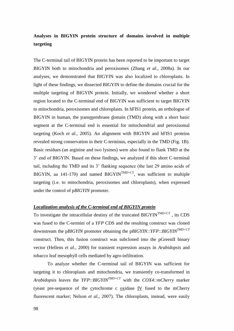
98
Analyses in BIGYIN protein structure of domains involved in multiple
targeting
The C-terminal tail of BIGYIN protein has been reported to be important to target
BIGYIN both to mitochondria and peroxisomes (Zhang et al., 2008a). In our
analyses, we demonstrated that BIGYIN was also localized to chloroplasts. In
light of these findings, we dissected BIGYIN to define the domains crucial for the
multiple targeting of BIGYIN protein. Initially, we wondered whether a short
region located to the C-terminal end of BIGYIN was sufficient to target BIGYIN
to mitochondria, peroxisomes and chloroplasts. In hFIS1 protein, an orthologue of
BIGYIN in human, the transmembrane domain (TMD) along with a short basic
segment at the C-terminal end is essential for mitochondrial and peroxisomal
targeting (Koch et al., 2005). An alignment with BIGYIN and hFIS1 proteins
revealed strong conservation in their C-terminus, especially in the TMD (Fig. 1B).
Basic residues (an arginine and two lysines) were also found to flank TMD at the
3‟ end of BIGYIN. Based on these findings, we analyzed if this short C-terminal
tail, including the TMD and its 3‟ flanking sequence (the last 29 amino acids of
BIGYIN, aa 141-170) and named BIGYINTMD+CT
, was sufficient to multiple
targeting (i.e. to mitochondria, peroxisomes and chloroplasts), when expressed
under the control of pBIGYIN promoter.
Localization analysis of the C-terminal end of BIGYIN protein
To investigate the intracellular destiny of the truncated BIGYINTMD+CT
, its CDS
was fused to the C-termini of a YFP CDS and the resulting construct was cloned
downstream the pBIGYIN promoter obtaining the pBIGYIN::YFP::BIGYINTMD+CT
construct. Then, this fusion construct was subcloned into the pGreenII binary
vector (Hellens et al., 2000) for transient expression assays in Arabidopsis and
tobacco leaf mesophyll cells mediated by agro-infiltration.
To analyze whether the C-terminal tail of BIGYIN was sufficient for
targeting it to chloroplasts and mitochondria, we transiently co-transformed in
Arabidopsis leaves the YFP::BIGYINTMD+CT
with the COX4::mCherry marker
(yeast pre-sequence of the cytochrome c oxidase IV fused to the mCherry
fluorescent marker; Nelson et al., 2007). The chloroplasts, instead, were easily
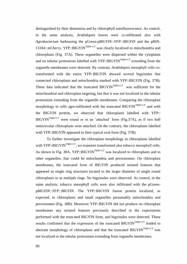
99
distinguished by their dimension and by chlorophyll autofluorescence. As control,
in the same analysis, Arabidopsis leaves were co-infiltrated also with
Agrobacterium harbouring the pGreen-pBIGYIN::YFP::BIGYIN and the pBIN-
COX4::mCherry. YFP::BIGYINTMD+CT
was clearly localized to mitochondria and
chloroplasts (Fig. 37A). These organelles were dispersed within the cytoplasm
and no tubular protrusions labelled with YFP::BIGYINTMD+CT
extending from the
organelle membranes were detected. By contrast, Arabidopsis mesophyll cells co-
transformed with the entire YFP::BIGYIN showed several bigyinules that
connected chloroplasts and mitochondria marked with YFP::BIGYIN (Fig. 37B).
These data indicated that the truncated BIGYINTMD+CT
was sufficient for the
mitochondrial and chloroplast targeting, but that it was not localized to the tubular
protrusions extending from the organelle membranes. Comparing the chloroplast
morphology in cells agro-infiltrated with the truncated BIGYINTMD+CT
and with
the BIGYIN protein, we observed that chloroplasts labelled with YFP::
BIGYINTMD+CT
were round or in an „attached‟ form (Fig.37A), as if two half
semicircular chloroplasts were attached. On the contrary, the chloroplasts labelled
with YFP::BIGYIN appeared in their typical oval form (Fig. 37B).
To further investigate the chloroplast morphology in chloroplasts labelled
with YFP::BIGYINTMD+CT
, we transient transformed also tobacco mesophyll cells.
As shown in Fig. 38A, YFP::BIGYINTMD+CT
was localized to chloroplasts and to
other organelles, that could be mitochondria and peroxisomes. On chloroplast
membranes, the truncated form of BIGYIN produced striated features that
appeared as single ring structures located to the larger diameter of single round
chloroplasts or as multiple rings. No bigyinules were observed. As control, in the
same analysis, tobacco mesophyll cells were also infiltrated with the pGreen-
pBIGYIN::YFP::BIGYIN. The YFP::BIGYIN fusion protein localized, as
expected, to chloroplasts and small organelles presumably mitochondria and
peroxisomes (Fig. 38B). Moreover YFP::BIGYIN did not produce on chloroplast
membranes any striated features previously described in the experiments
performed with the truncated BIGYIN form, and bigyinules were detected. These
results confirmed that the expression of the truncated BIGYINTMD+CT
leaded to
aberrant morphology of chloroplasts and that the truncated BIGYINTMD+CT
was
not localized to the tubular protrusions extending from organelle membranes.
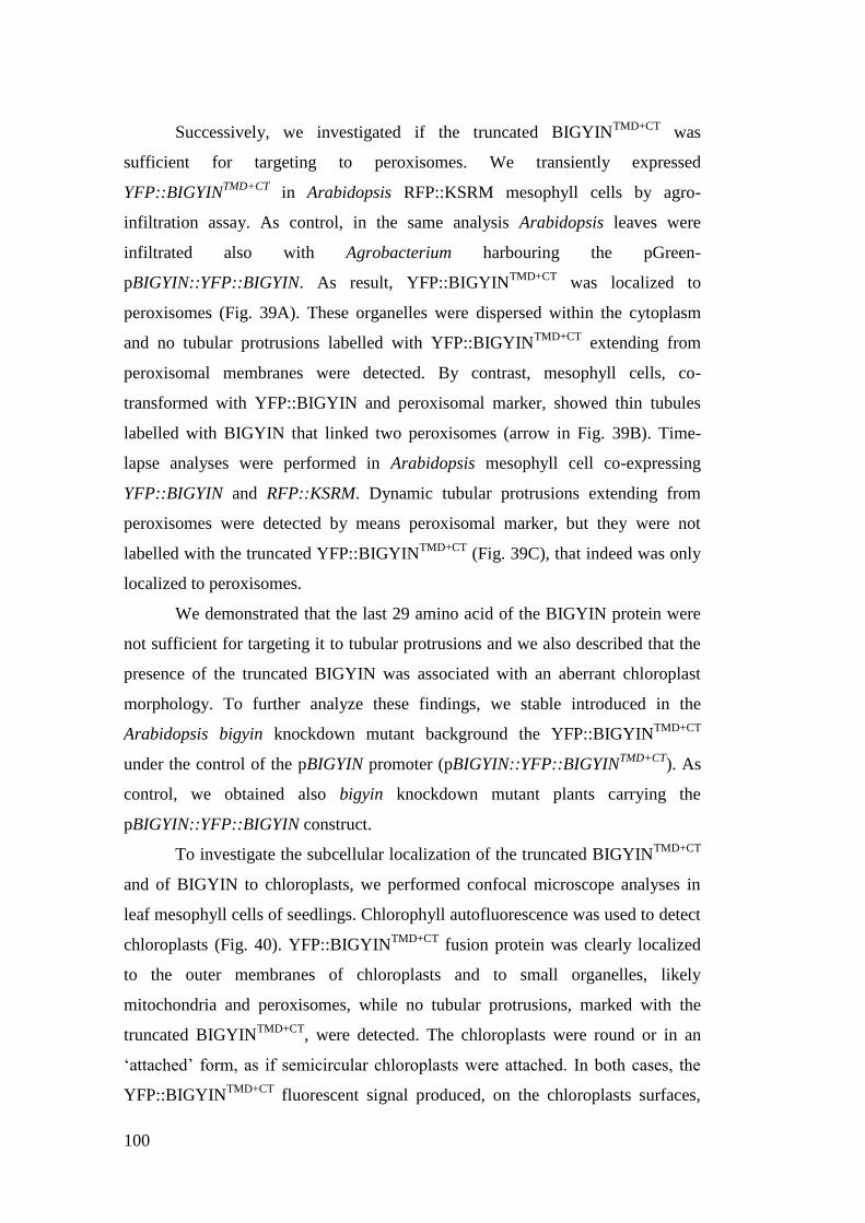
100
Successively, we investigated if the truncated BIGYINTMD+CT
was
sufficient for targeting to peroxisomes. We transiently expressed
YFP::BIGYINTMD+CT
in Arabidopsis RFP::KSRM mesophyll cells by agro-
infiltration assay. As control, in the same analysis Arabidopsis leaves were
infiltrated also with Agrobacterium harbouring the pGreen-
pBIGYIN::YFP::BIGYIN. As result, YFP::BIGYINTMD+CT
was localized to
peroxisomes (Fig. 39A). These organelles were dispersed within the cytoplasm
and no tubular protrusions labelled with YFP::BIGYINTMD+CT
extending from
peroxisomal membranes were detected. By contrast, mesophyll cells, co-
transformed with YFP::BIGYIN and peroxisomal marker, showed thin tubules
labelled with BIGYIN that linked two peroxisomes (arrow in Fig. 39B). Time-
lapse analyses were performed in Arabidopsis mesophyll cell co-expressing
YFP::BIGYIN and RFP::KSRM. Dynamic tubular protrusions extending from
peroxisomes were detected by means peroxisomal marker, but they were not
labelled with the truncated YFP::BIGYINTMD+CT
(Fig. 39C), that indeed was only
localized to peroxisomes.
We demonstrated that the last 29 amino acid of the BIGYIN protein were
not sufficient for targeting it to tubular protrusions and we also described that the
presence of the truncated BIGYIN was associated with an aberrant chloroplast
morphology. To further analyze these findings, we stable introduced in the
Arabidopsis bigyin knockdown mutant background the YFP::BIGYINTMD+CT
under the control of the pBIGYIN promoter (pBIGYIN::YFP::BIGYINTMD+CT
). As
control, we obtained also bigyin knockdown mutant plants carrying the
pBIGYIN::YFP::BIGYIN construct.
To investigate the subcellular localization of the truncated BIGYINTMD+CT
and of BIGYIN to chloroplasts, we performed confocal microscope analyses in
leaf mesophyll cells of seedlings. Chlorophyll autofluorescence was used to detect
chloroplasts (Fig. 40). YFP::BIGYINTMD+CT
fusion protein was clearly localized
to the outer membranes of chloroplasts and to small organelles, likely
mitochondria and peroxisomes, while no tubular protrusions, marked with the
truncated BIGYINTMD+CT
, were detected. The chloroplasts were round or in an
„attached‟ form, as if semicircular chloroplasts were attached. In both cases, the
YFP::BIGYINTMD+CT
fluorescent signal produced, on the chloroplasts surfaces,
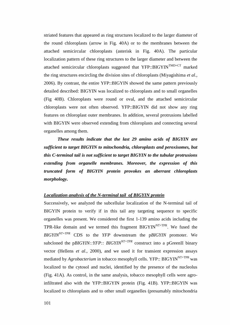
101
striated features that appeared as ring structures localized to the larger diameter of
the round chloroplasts (arrow in Fig. 40A) or to the membranes between the
attached semicircular chloroplasts (asterisk in Fig. 40A). The particular
localization pattern of these ring structures to the larger diameter and between the
attached semicircular chloroplasts suggested that YFP::BIGYINTMD+CT
marked
the ring structures encircling the division sites of chloroplasts (Miyagishima et al.,
2006). By contrast, the entire YFP::BIGYIN showed the same pattern previously
detailed described: BIGYIN was localized to chloroplasts and to small organelles
(Fig 40B). Chloroplasts were round or oval, and the attached semicircular
chloroplasts were not often observed. YFP::BIGYIN did not show any ring
features on chloroplast outer membranes. In addition, several protrusions labelled
with BIGYIN were observed extending from chloroplasts and connecting several
organelles among them.
These results indicate that the last 29 amino acids of BIGYIN are
sufficient to target BIGYIN to mitochondria, chloroplasts and peroxisomes, but
this C-terminal tail is not sufficient to target BIGYIN to the tubular protrusions
extending from organelle membranes. Moreover, the expression of this
truncated form of BIGYIN protein provokes an aberrant chloroplasts
morphology.
Localization analysis of the N-terminal tail of BIGYIN protein
Successively, we analyzed the subcellular localization of the N-terminal tail of
BIGYIN protein to verify if in this tail any targeting sequence to specific
organelles was present. We considered the first 1-139 amino acids including the
TPR-like domain and we termed this fragment BIGYINNT+TPR
. We fused the
BIGYINNT+TPR
CDS to the YFP downstream the pBIGYIN promoter. We
subcloned the pBIGYIN::YFP:: BIGYINNT+TPR
construct into a pGreenII binary
vector (Hellens et al., 2000), and we used it for transient expression assays
mediated by Agrobacterium in tobacco mesophyll cells. YFP:: BIGYINNT+TPR
was
localized to the cytosol and nuclei, identified by the presence of the nucleolus
(Fig. 41A). As control, in the same analysis, tobacco mesophyll cells were agro-
infiltrated also with the YFP::BIGYIN protein (Fig. 41B). YFP::BIGYIN was
localized to chloroplasts and to other small organelles (presumably mitochondria
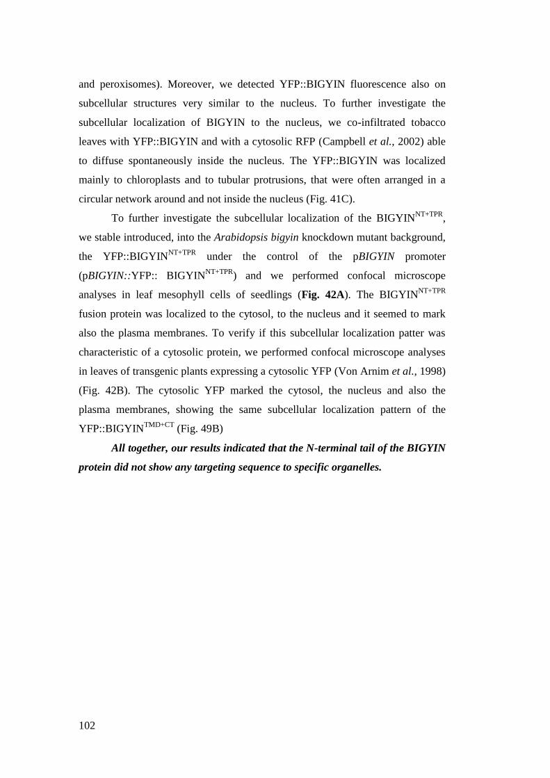
102
and peroxisomes). Moreover, we detected YFP::BIGYIN fluorescence also on
subcellular structures very similar to the nucleus. To further investigate the
subcellular localization of BIGYIN to the nucleus, we co-infiltrated tobacco
leaves with YFP::BIGYIN and with a cytosolic RFP (Campbell et al., 2002) able
to diffuse spontaneously inside the nucleus. The YFP::BIGYIN was localized
mainly to chloroplasts and to tubular protrusions, that were often arranged in a
circular network around and not inside the nucleus (Fig. 41C).
To further investigate the subcellular localization of the BIGYINNT+TPR
,
we stable introduced, into the Arabidopsis bigyin knockdown mutant background,
the YFP::BIGYINNT+TPR
under the control of the pBIGYIN promoter
(pBIGYIN::YFP:: BIGYINNT+TPR
) and we performed confocal microscope
analyses in leaf mesophyll cells of seedlings (Fig. 42A). The BIGYINNT+TPR
fusion protein was localized to the cytosol, to the nucleus and it seemed to mark
also the plasma membranes. To verify if this subcellular localization patter was
characteristic of a cytosolic protein, we performed confocal microscope analyses
in leaves of transgenic plants expressing a cytosolic YFP (Von Arnim et al., 1998)
(Fig. 42B). The cytosolic YFP marked the cytosol, the nucleus and also the
plasma membranes, showing the same subcellular localization pattern of the
YFP::BIGYINTMD+CT
(Fig. 49B)
All together, our results indicated that the N-terminal tail of the BIGYIN
protein did not show any targeting sequence to specific organelles.
103
DISCUSSION
BIGYIN is a tail anchored protein belonging to the FIS1-type proteins
The FISSION1 (FIS1)-type proteins are evolutionarily conserved integral
membrane proteins involved in maintaining the morphology of mitochondria and
peroxisomes in mammals (James et al., 2003), yeast (Zhang, 2007), and plants
(Zhang and Hu, 2008b). The Arabidopsis thaliana genome contains two closely
related FIS1-type proteins, orthologs of yeast FIS1 and human hFis1: BIGYIN
(previously named FIS1A, At3g57090) and FIS1B (At5g12390).
Analyzing published microarray data (http://bbc.botany.utoronto.ca/), we observe
that that both BIGYIN and FIS1B are constitutively expressed in Arabidopsis.
However, FIS1B is especially expressed only in mature pollen, while the
expression level of BIGYIN is higher than that of FISB in most of tissues.
In silico analyses show that BIGYIN and FIS1B, as all FIS1-type proteins,
are characterized by a tetratricopeptide (TPR)-like domain, predicted to be
exposed to cytosol, and by a single transmembrane domain located to the C-
terminal end of the protein. Based on this topology, we can considered these
proteins as members of tail-anchored (TA; Nout-Cin) family of membrane proteins
(Borghese et al., 2007).
Expression and localization pattern of BIGYIN in Arabidopsis
In this study, we have shown that pBIGYIN promoter is constitutively active in all
plant developmental stages from germinating seeds to mature plants and in all
tissues (leaves, stems and roots). Our data are in agreement with microarray data
already published (http://bbc.botany.utoronto.ca/). Yet, we have reported that
BIGYIN protein is constitutively present in all developmental stages and in all
tissues. In addition, we have shown that tissues where plant growth takes place
(i.e. leaf teeth, leaf primordia, developing stems, nodes, flower buds, lateral root
primordia and root tips) are characterized by the highest pBIGYIN promoter
activity and by the highest presence of BIGYIN protein. By contrast, pBIGYIN
promoter activity and BIGYIN protein are low detected in fully expanded leaves
or in mature stems.
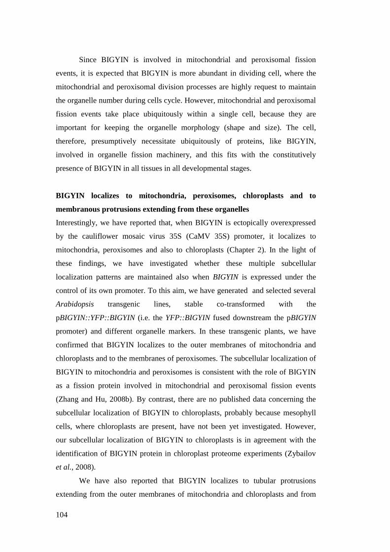
104
Since BIGYIN is involved in mitochondrial and peroxisomal fission
events, it is expected that BIGYIN is more abundant in dividing cell, where the
mitochondrial and peroxisomal division processes are highly request to maintain
the organelle number during cells cycle. However, mitochondrial and peroxisomal
fission events take place ubiquitously within a single cell, because they are
important for keeping the organelle morphology (shape and size). The cell,
therefore, presumptively necessitate ubiquitously of proteins, like BIGYIN,
involved in organelle fission machinery, and this fits with the constitutively
presence of BIGYIN in all tissues in all developmental stages.
BIGYIN localizes to mitochondria, peroxisomes, chloroplasts and to
membranous protrusions extending from these organelles
Interestingly, we have reported that, when BIGYIN is ectopically overexpressed
by the cauliflower mosaic virus 35S (CaMV 35S) promoter, it localizes to
mitochondria, peroxisomes and also to chloroplasts (Chapter 2). In the light of
these findings, we have investigated whether these multiple subcellular
localization patterns are maintained also when BIGYIN is expressed under the
control of its own promoter. To this aim, we have generated and selected several
Arabidopsis transgenic lines, stable co-transformed with the
pBIGYIN::YFP::BIGYIN (i.e. the YFP::BIGYIN fused downstream the pBIGYIN
promoter) and different organelle markers. In these transgenic plants, we have
confirmed that BIGYIN localizes to the outer membranes of mitochondria and
chloroplasts and to the membranes of peroxisomes. The subcellular localization of
BIGYIN to mitochondria and peroxisomes is consistent with the role of BIGYIN
as a fission protein involved in mitochondrial and peroxisomal fission events
(Zhang and Hu, 2008b). By contrast, there are no published data concerning the
subcellular localization of BIGYIN to chloroplasts, probably because mesophyll
cells, where chloroplasts are present, have not been yet investigated. However,
our subcellular localization of BIGYIN to chloroplasts is in agreement with the
identification of BIGYIN protein in chloroplast proteome experiments (Zybailov
et al., 2008).
We have also reported that BIGYIN localizes to tubular protrusions
extending from the outer membranes of mitochondria and chloroplasts and from
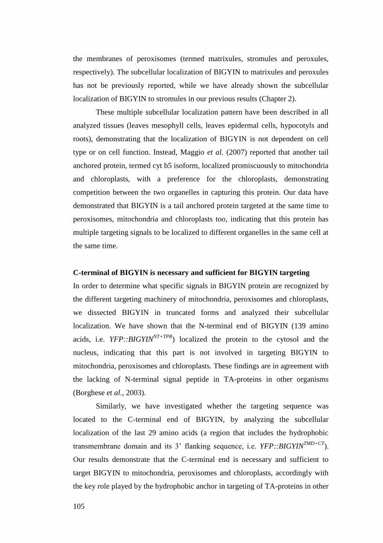
105
the membranes of peroxisomes (termed matrixules, stromules and peroxules,
respectively). The subcellular localization of BIGYIN to matrixules and peroxules
has not be previously reported, while we have already shown the subcellular
localization of BIGYIN to stromules in our previous results (Chapter 2).
These multiple subcellular localization pattern have been described in all
analyzed tissues (leaves mesophyll cells, leaves epidermal cells, hypocotyls and
roots), demonstrating that the localization of BIGYIN is not dependent on cell
type or on cell function. Instead, Maggio et al. (2007) reported that another tail
anchored protein, termed cyt b5 isoform, localized promiscuously to mitochondria
and chloroplasts, with a preference for the chloroplasts, demonstrating
competition between the two organelles in capturing this protein. Our data have
demonstrated that BIGYIN is a tail anchored protein targeted at the same time to
peroxisomes, mitochondria and chloroplasts too, indicating that this protein has
multiple targeting signals to be localized to different organelles in the same cell at
the same time.
C-terminal of BIGYIN is necessary and sufficient for BIGYIN targeting
In order to determine what specific signals in BIGYIN protein are recognized by
the different targeting machinery of mitochondria, peroxisomes and chloroplasts,
we dissected BIGYIN in truncated forms and analyzed their subcellular
localization. We have shown that the N-terminal end of BIGYIN (139 amino
acids, i.e. YFP::BIGYINNT+TPR
) localized the protein to the cytosol and the
nucleus, indicating that this part is not involved in targeting BIGYIN to
mitochondria, peroxisomes and chloroplasts. These findings are in agreement with
the lacking of N-terminal signal peptide in TA-proteins in other organisms
(Borghese et al., 2003).
Similarly, we have investigated whether the targeting sequence was
located to the C-terminal end of BIGYIN, by analyzing the subcellular
localization of the last 29 amino acids (a region that includes the hydrophobic
transmembrane domain and its 3‟ flanking sequence, i.e. YFP::BIGYINTMD+CT
).
Our results demonstrate that the C-terminal end is necessary and sufficient to
target BIGYIN to mitochondria, peroxisomes and chloroplasts, accordingly with
the key role played by the hydrophobic anchor in targeting of TA-proteins in other
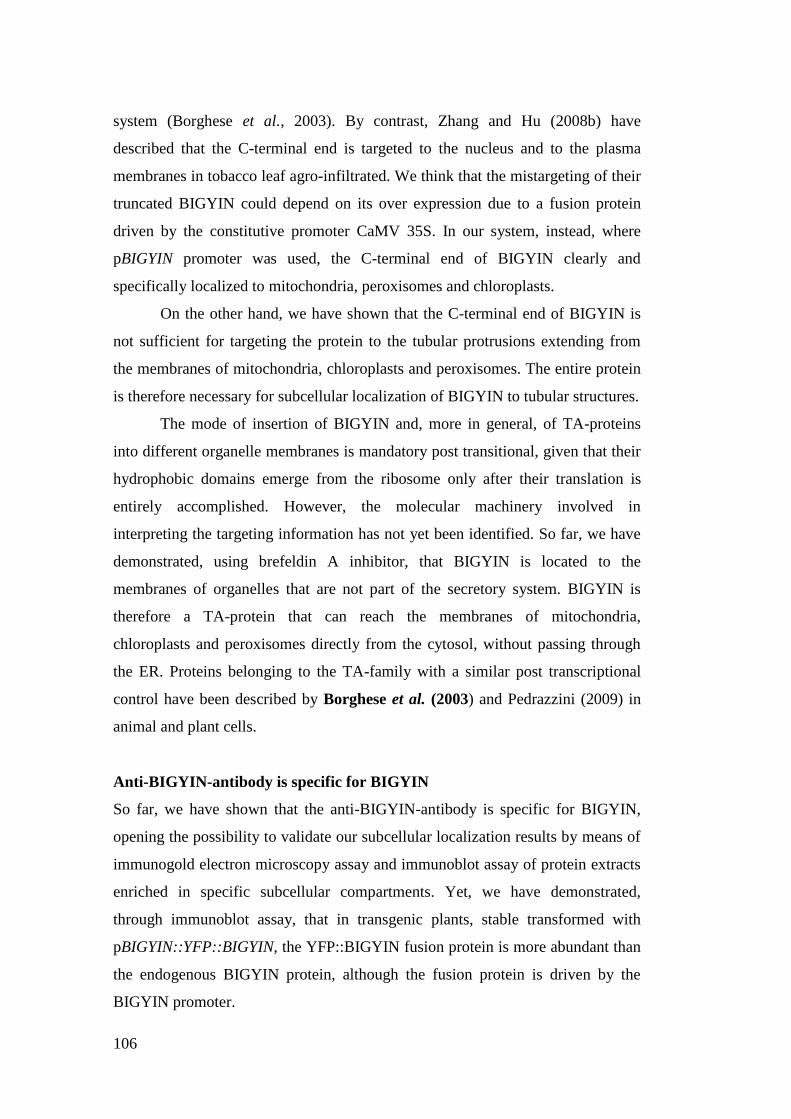
106
system (Borghese et al., 2003). By contrast, Zhang and Hu (2008b) have
described that the C-terminal end is targeted to the nucleus and to the plasma
membranes in tobacco leaf agro-infiltrated. We think that the mistargeting of their
truncated BIGYIN could depend on its over expression due to a fusion protein
driven by the constitutive promoter CaMV 35S. In our system, instead, where
pBIGYIN promoter was used, the C-terminal end of BIGYIN clearly and
specifically localized to mitochondria, peroxisomes and chloroplasts.
On the other hand, we have shown that the C-terminal end of BIGYIN is
not sufficient for targeting the protein to the tubular protrusions extending from
the membranes of mitochondria, chloroplasts and peroxisomes. The entire protein
is therefore necessary for subcellular localization of BIGYIN to tubular structures.
The mode of insertion of BIGYIN and, more in general, of TA-proteins
into different organelle membranes is mandatory post transitional, given that their
hydrophobic domains emerge from the ribosome only after their translation is
entirely accomplished. However, the molecular machinery involved in
interpreting the targeting information has not yet been identified. So far, we have
demonstrated, using brefeldin A inhibitor, that BIGYIN is located to the
membranes of organelles that are not part of the secretory system. BIGYIN is
therefore a TA-protein that can reach the membranes of mitochondria,
chloroplasts and peroxisomes directly from the cytosol, without passing through
the ER. Proteins belonging to the TA-family with a similar post transcriptional
control have been described by Borghese et al. (2003) and Pedrazzini (2009) in
animal and plant cells.
Anti-BIGYIN-antibody is specific for BIGYIN
So far, we have shown that the anti-BIGYIN-antibody is specific for BIGYIN,
opening the possibility to validate our subcellular localization results by means of
immunogold electron microscopy assay and immunoblot assay of protein extracts
enriched in specific subcellular compartments. Yet, we have demonstrated,
through immunoblot assay, that in transgenic plants, stable transformed with
pBIGYIN::YFP::BIGYIN, the YFP::BIGYIN fusion protein is more abundant than
the endogenous BIGYIN protein, although the fusion protein is driven by the
BIGYIN promoter.
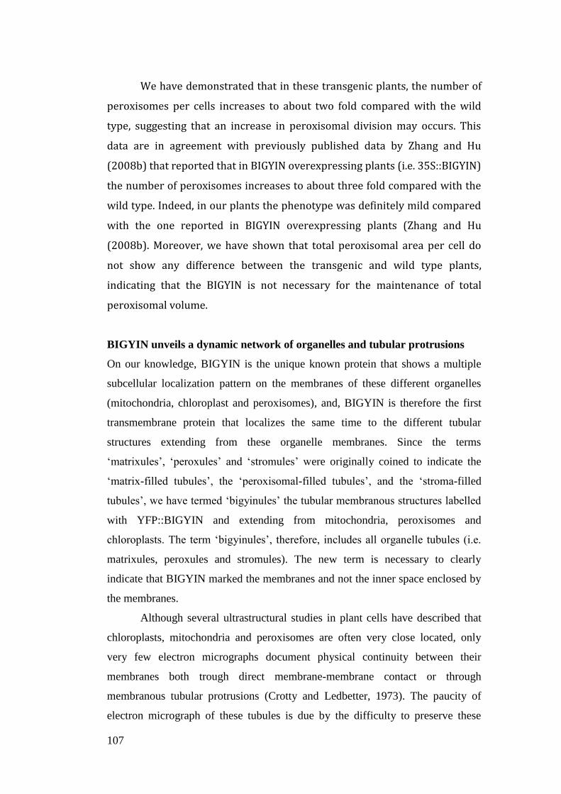
107
We have demonstrated that in these transgenic plants, the number of
peroxisomes per cells increases to about two fold compared with the wild
type, suggesting that an increase in peroxisomal division may occurs. This
data are in agreement with previously published data by Zhang and Hu
(2008b) that reported that in BIGYIN overexpressing plants (i.e. 35S::BIGYIN)
the number of peroxisomes increases to about three fold compared with the
wild type. Indeed, in our plants the phenotype was definitely mild compared
with the one reported in BIGYIN overexpressing plants (Zhang and Hu
(2008b). Moreover, we have shown that total peroxisomal area per cell do
not show any difference between the transgenic and wild type plants,
indicating that the BIGYIN is not necessary for the maintenance of total
peroxisomal volume.
BIGYIN unveils a dynamic network of organelles and tubular protrusions
On our knowledge, BIGYIN is the unique known protein that shows a multiple
subcellular localization pattern on the membranes of these different organelles
(mitochondria, chloroplast and peroxisomes), and, BIGYIN is therefore the first
transmembrane protein that localizes the same time to the different tubular
structures extending from these organelle membranes. Since the terms
„matrixules‟, „peroxules‟ and „stromules‟ were originally coined to indicate the
„matrix-filled tubules‟, the „peroxisomal-filled tubules‟, and the „stroma-filled
tubules‟, we have termed „bigyinules‟ the tubular membranous structures labelled
with YFP::BIGYIN and extending from mitochondria, peroxisomes and
chloroplasts. The term „bigyinules‟, therefore, includes all organelle tubules (i.e.
matrixules, peroxules and stromules). The new term is necessary to clearly
indicate that BIGYIN marked the membranes and not the inner space enclosed by
the membranes.
Although several ultrastructural studies in plant cells have described that
chloroplasts, mitochondria and peroxisomes are often very close located, only
very few electron micrographs document physical continuity between their
membranes both trough direct membrane-membrane contact or through
membranous tubular protrusions (Crotty and Ledbetter, 1973). The paucity of
electron micrograph of these tubules is due by the difficulty to preserve these
108
protrusions during the standard fixation required by electron microscopy (Köhler
and Hanson, 2000). Our transgenic plants expressing BIGYIN are significant
because allow to analyze at the same time different organelles and especially the
tubular protrusions that extend from different organelle membranes, observing the
dynamics and behaviour of these interesting interconnected tubules.
Mitochondria, chloroplasts and peroxisomes could be close located or
spread randomly throughout the cytoplasm. Interestingly, even when
mitochondria, chloroplasts or peroxisomes are spread within the cell, the presence
of bigyinules could allow interconnections among them: several bigyinules could
protrude from chloroplasts, mitochondria and peroxisomes forming a complex and
highly dynamic network of tubules. A similar motility have been described for
stromules (Gunning, 2005) and peroxules (Sincalir et al., 2009), but not for
matrixules. How is it this motility achieved? We have demonstrated that the actin-
based cytoskeleton plays a role in movement of bigyinules and of mitochondria,
chloroplasts and peroxisomes marked with YFP::BIGYIN. This is in agreement
with previous studies on effect of actin-polymerization inhibitors on movements
of mitochondria, of peroxisomes, chloroplasts and stromules (Gestel et al., 2002;
Jedd and Chua, 2002; Kandesamy and Meagher, 1999).
Moreover, we have also shown a trafficking of BIGYIN through the
membranes of connected organelles (for example two chloroplasts, Fig. 31) or
through the membranes un-connected organelles (Fig. 32). These data support the
existence of physical inter-organellar connections and of a trafficking of proteins
among physical un-connected organelles. There are few published data
concerning physical inter-organellar interactions, although their existence has
been often proposed in the light of several metabolic and functional interactions
among organelles (i.e. chloroplast, mitochondria and peroxisomes, for example
during respiration). The existence of a trafficking of molecules for example
between mitochondria and chloroplasts has been proposed in the literature by
Kwok and Hanson (2004a-b) and supported by Jouhet et al. (2004), when they
demonstrated the transfer of digalactosyldiacylglycerol glycolipids between
plastids and mitochondria. Moreover, the authors suggested that this transfer
depended upon contacts between plastids and mitochondria or upon vesicles
without the involvement of the endomembrane system. Similarly, we have
109
demonstrated that endomembrane system is not involved in the trafficking of
structures labelled with BIGYIN, and we have also shown that free vesicle-like
structures labelled with BIGYIN can separate from bigyinules or organelles (Fig.
22, 26C ). Vesicle-structures originated from stromules and detached from by
stromules have been also described by Pyke and Howells (2002). Authors
suggested that these vesicle-structures could be an export mechanism for the
secretion of plastid products. In the light of these findings, we can suggest that the
vesicle-structures labelled with BIGYIN could be an export mechanism for the
secretion of organelle product.
Moreover, we have shown that chloroplasts and bigyinules could be close
located to the surface of nuclei suggesting that this close vicinity may facilitate
the exchange of molecules and signal between chloroplasts and nuclei. This data
are in agreement with Kwok and Hanson (2004b), that proposed a similar role for
stromules located near nuclei.
BIGYIN may play a role also in chloroplast division
BIGYIN has been demonstrated to be involved in mitochondrial and peroxisomal
division (Zhang and Hu, 2008a). By contrast its role(s) on chloroplasts and on
membranous tubules has not yet been analyzed. In order to further investigate the
function(s) played by BIGYIN on chloroplasts and on tubular protrusions, we
have analyzed an Arabidopsis T-DNA insertional line for BIGYIN, reported to be
a knock-out mutant and having a T-DNA insertion in the last exon (Scott et al.,
2006; Zhang et al., 2008a). We have demonstrated this line as a knock down
mutant with the T-DNA insertion in the 3‟-UTR. In addition, we have not
observed any difference in mitochondrial morphology between bigyin mutant and
wild type plants, as instead reported by previous paper (Scott and Logan, 2006;
Zhang and Hu, 2008a). Based on these findings, we could not use this mutant for
our analyses.
In the light of the fission role of BIGYIN in mitochondria and in
peroxisomes, we have investigated the subcellular localization of BIGYIN during
chloroplast division in developing tissues (flower buds and young hypocotyls),
where chloroplast fission highly occurs (Miyagishima et al., 2006). We have
shown that BIGYIN localizes to the outer membranes of chloroplasts and around

110
the isthmus in formation. Interestingly, BIGYIN localizes also to the constriction
sites between two chloroplasts in division, when organelles are in the typical
„dumbbell‟-form, step preceding the last phase of chloroplast fission (Aldridge et
al., 2005). Moreover, we have reported that transgenic plants stably expressing a
truncated form of BIGYIN (i.e. YFP::BIGYINTMD+CT
, completely deleted in the
cytosolic tail involved in protein-protein interactions, as demonstrated in Chapter
2), shows a particular chloroplast morphology and a very interesting subcellular
localization pattern on chloroplast membranes. In these transgenic plants, we have
shown that chloroplasts could be round or constricted in the „dumbbell‟-form,
usually observed when chloroplast fission takes place (Aldridge et al., 2005),
although we performed these analyses on fully expanded leaves, where
chloroplast division does not usually occur. Moreover, the truncated BIGYIN
creates, on the chloroplast surfaces, striated features that appear to mark the ring
structures encircling the division sites of chloroplasts. By contrast, transgenic
plants expressing the entire BIGYIN shows a normal chloroplast morphology
(typically oval or round in fully expanded leaves), and BIGYIN is located to the
chloroplast surfaces without create any striated features. These findings suggested
that the particular chloroplast morphology, observed in plants expressing the
truncated BIGYIN, might be the result of a dominant-negative effect of the deleted
and un-functional protein. Similar constricted „dumbbell‟-form chloroplasts have
been reported in Arabidopsis mutant with defects in chloroplast division
(Miyagishima et al., 2006). In the light of these findings, we suggest a possible
role of BIGYIN in chloroplast division. BIGYIN proteins, therefore, is an
Arabidopsis Fission1-type protein that is involved in mitochondrial and
peroxisomal fission, as reported by Zhang and Hu (2008b), and that could also
play a role in chloroplasts division.
111
MATERIAL AND METHODS
Plant materials and growth condition
All the Arabidopsis thaliana plants for this study were in Columbia (Col-
0) background. Different Arabidopsis transgenic lines were used: plants
overexpressing red fluorescent protein (RFP) targeted to mitochondria by
Arabidopsis cytocrome c oxidase-releated (COX) pre-sequence
(35S::COX::RFP), plants overexpressing RFP targeted to peroxisomes by
sequence tag KSRM fused downstream RFP coding sequence
(35S::RFP::KSMR), and plants transformed with the cytosolic YFP (35S::YFP) or
cytosolic RFP (35S::RFP). These transgenic lines were generated in our
laboratory.
Arabidopsis thaliana, Columbia ecotype, and Nicotiana tabacum plants
were incubated in an environmentally-controlled growth chamber with a long
photoperiod (16 hr light and 8 hr dark) at 25 ± 1°C, and a photosynthetic photon
flux of 35 mol m-2
s-1
Osram cool-white 18 W fluorescent lamps. When in vitro
growth of Arabidopsis plants was required, Arabidopsis seeds were sterilized
using the vapour-phase surface-sterilization. Seeds and a beaker containing 100
ml bleach were placed in a dessicator jar. 3 ml concentrate HCl was added to the
bleach and then immediately the jar was sealed to allow sterilization by chlorine
fumes to proceed for 5-6 hours. Then the seed were sown on half-strength
Murashige and Skoog (MS) medium (Sigma).
DNA constructs
All the cloned plasmids were confirmed by sequencing.
pBIGYIN::GUS constructs and generation of transgenic lines
The GUS (-glucuronidase)-coding sequence was fused to the BIGYIN promoter
(base pair -862 to -1). The 862 bp promoter fragment was amplified by PCR using
genomic DNA extracted from Arabidopsis leaves as template. The pair of
primers, both carrying an EcoRI restriction site, were as follows: forward primer
112
5‟-CATGGAATTCCTTTCGAGGCTCACCTCAAC-3 and reverse primer 5‟-
CATGGAATTCTGAAGGCGATTTTGAGCTTTGA-3‟). After digestion, the
promoter was cloned upstream of the GUS coding region, into a modified
pGreen0029 binary vector (KamaycinR; Hellens et al., 2000), where the GUS
coding sequence, fused with the nos terminator, was previously inserted in the
polylinker between KpnI-SacI restriction sites. The pGreen-pBIGYIN::GUS
construct was transferred into GV3101-pSoup Agrobacterium strain (Hellens et
al., 2000) and Arabidopsis plants were transformed by floral dip method (Clough
and Bent, 1998) and screened on half-strength MS agar medium containing 50
mgL-1
kanamycin. The GUS-staining analyses were performed on T2 plants. No
one of the transgenic lines selected, with the different constructs, showed
phenotypic differences or abnormalities in our standard growth conditions.
pBIGYIN::YFP::BIGYIN, pBIGYIN::YFP::BIGYINTMD+CT
and
pBIGYIN::YFP::BIGYINNT+TPR
constructs and generation of the Arabidopsis
transgenic lines
The Arabidopsis BIGYIN coding sequence was first amplified by PCR
from Arabidopsis cDNA with proofreading PCR enzymes (Phusion High Fidelity
DNA polymerase [Finnzymes]) and then cloned into the vector of interest.
To obtain the BIGYINTMD+CT
, the C-terminal tail of BIGYIN coding sequence was
amplified (Primer For: 5‟-
CATGGAGCTCAAGGTGTTATAGGGATAGGGATCACG-3‟; Rev: 5‟-
CATGGGTACCTCATTTCTTGCGAGACATCG-3‟), corresponding to the
transmembrane domain and the adjacent C-terminal tail (421-510 bp,
corresponding to the last 29 amino acids of BIGYIN, aa 141-170).
To obtain the BIGYINNT+TPR
, the N-terminal tail of BIGYIN coding sequence was
amplified (Primer For: 5‟-catgGAGCTCAAATGGATGCTAAGATCGGAC-3‟;
Rev: 5‟-CATGggtaccTCACTTTGTGATTTTGTCTTCGATGGTC-3‟),
corresponding to the N-terminal end and of TPR-like domain(417 bp,
corresponding to the first 139 amino acids of BIGYIN, aa 1-139). For the
expression of BIGYIN, BIGYINTMD+CT
BIGYINNT+TPR
in plants, the pGreen0179
(hygromycinR; Hellens et al., 2000) binary vector was used.
113
pBIGYIN::YFP::BIGYIN construct contained the promoter region of BIGYIN (862
bp upstream region, base pair -862 to -1) fused to YFP::BIGYIN coding sequence,
and the cauliflower mosaic virus 35S terminator. The promoter region was
amplified by PCR using specific 5‟ and 3‟ primers (forward primers 5‟-
CATGGAATTCCTTTCGAGGCTCACCTCAAC-3 and reverse primer 5‟-
CATGGAATTCTGAAGGCGATTTTGAGCTTTGA-3‟) where the EcoRI/EcoRI
sites were introduced. The pBIGYIN amplicon was then digested with EcoRI and
cloned into the pGreen0179, obtaining the pGreen0179-pBIGYIN vector. At the
same time the YFP coding sequence was subcloned from the pAVA554-35S::YFP
plasmid provided by Prof. Albrecht von Arnim (von Arnim et al., 1998) to the
pSAT1-35S::nVenus vector (stock number E3228, Lee et al., 2008) by replacing
the nVenus cDNA sequence digesting with NCoI/BglII enzymes. The BIGYIN
PCR product was amplified using primers where the SacI/KpnI sites were
introduced (Primer For: 5‟-catgGAGCTCAAATGGATGCTAAGATCGGAC-3‟;
Rev: 5‟- CATGggtaccTCATTTCTTGCGAGACATCG-3‟). Amplicons were then
digested with SacI/KpnI and cloned into the pSAT1-35S::YFP vector. The
pSAT1-35S::YFP::BIGYIN fusion construct was then subcloned with
EcoRV/NotI restriction sites into the pGreen0179-pBIGYIN digested with
SmaI/NotI to obtain the pGreen0179-pBIGYIN::YFP::BIGYIN binary vector. A
similar strategy was used to obtain the Green0179-pBIGYIN:YFP::BIGYINTMD+CT
and Green0179-pBIGYIN:YFP::BIGYINNT+TPR
binary vectors. These binary
vectors were introduced into Agrobacterium. tumefaciens strain GV3101
(Labereke et al., 1974) and transferred to A. thaliana wild type and bigyin mutant
plants by floral dip method (Clough and Bent, 1998), in order to obtained plants
stable transformed with the pBIGYIN::YFP::BIGYIN,
pBIGYIN:YFP::BIGYINTMD+CT
or pBIGYIN:YFP::BIGYINNT+TPR
. Homozygous
T2-indipendent lines were then selected by segregation analysis on MS agar plates
containing 15 mgL-1
hygromycin. No one of the transgenic lines selected, with the
different constructs, showed phenotypic differences or abnormalities in our
standard growth conditions.
114
Generation of the Arabidopsis transgenic lines co-transformed with
pBIGYIN::YFP::BIGYIN and p35S::COX::RFP, or pBIGYIN::YFP::BIGYIN and
p35S::RFP::KSRM
The transgenic Arabidopsis plants stable co-transformed with
pBIGYIN::YFP::BIGYIN and p35S::COX::RFP, or pBIGYIN::YFP::BIGYIN and
p35S::RFP::KSRM were obtained crossing their mature flowers: a recipient
flower was prepared removing all the flower parts with exception of the ovary and
then this prepared ovary was brushed by the pollen of stamens of fully mature
flowers of the plant of interest. Siliques obtained were sown and seedlings were
selected by fluorescent signals.
Organelle marker used in tansient expression experiments
For the localization of the Golgi apparatus, the cytoplasmatic tail and
transmembrane domain (first 49 amino acids) of soybean -1,2-mannosidase-I
fused to the mCherry marker (Nelson et al., 2007) were used. For the localization
of mitochondria, the first 29 amino acids of yeast (Saccharomyces cerevisiae)
cytochrome c oxidase IV fused to the mCherry marker (Nelson et al., 2007) were
used.
Semi-quantitative RT-PCR analysis in pBIGYIN::GUS plants
Total RNA was extracted from leaves of 3-week-old plants as described by
Formentin et al. (2004.)
After DNAse I treatment (Ambion Ltd, UK), first strand synthesis and PCR were
carried out starting from 1 g of total RNA, according to the manufacturer‟s
instructions (M-MLV Reverse Transcriptase, Promega). After first strand cDNA
synthesis, samples were diluted 5 times and used as templates for semi-
quantitative RT-PCR.
RT-PCR analyses were performed to compare the transcript levels
between -glucuronidase and BIGYIN in each of the four transgenic
pBIGYIN::GUS lines. Specific primers were design to amplified a fragment of
575 bp into the GUS and of 511 bp into BIGYIN.
RT-PCR reactions were performed using GoTaq®
DNA Polymerase (Promega), in
a total reaction volume of 50µL according to manufacturer's recommendations
115
containing 5µL of cDNA. PCR amplification cycle was performed with an initial
denaturation step at 94°C for 2min, followed by 34 cycles (94°C for 30s; 61°C for
20s; 72°C for 40s), and finally with an elongation step at 72°C for 5min. The
number of cycles was determined as described in the QuantumRNA protocol.
PCR products were observed in 1% agarose gel electrophoresis, stained with
ethidium bromide. Ultra-pure water was used as the negative template control.
-glucuronidase (GUS) histochemical analysis
GUS histochemical staining was performed at four developmental stages:
germinating seeds (32 h after imbibition), 5-day-old seedlings (cotyledons open),
7-day-old seedlings (first leaves developing), and flowering mature plants.
Samples were analysed for GUS activity following the protocol described by
Jefferson et al. (1987). The samples were vacuum infiltrated for 30 min in the
following solutions: 2 mM X-gluc, 0.5% Triton X-100, 0.1% Tween 20, 0.5 mM
K3Fe(CN)6, 0.5 mM K4Fe(CN)6_3H2O, 10 mM Na2EDTA and 50 mM sodium
phosphate buffer, pH 7.0, and then incubated at 37°C for 16 h. After staining,
samples were cleared by several washes with methanol/acetic acid (3:1 v/v)
solution and kept at 4°C in 70% ethanol.
Inhibitor treatment
For Brefeldin A treatment, we used a 50 mM stock solution (made in dimethyl
sulfoxide) further diluted in distilled water to achieve an effective working
solution of 50 M before submerge the seedlings of 7-day old for 2 h (Geldner et
al., 2001). Lantruculin B was used at 500 nM for 0.5 h.
Antibody
Polyclonal antibodies against BIGYIN were designed in our laboratory and
produced by BioGenes GmbH (Berlin, Germany). In detail, polyclonal antibody
was raised in rabbits against an antigenic peptide corresponding to amino acids
33-47 of the BIGYIN protein.
116
Protein extraction and Western blot analysis
0,1 g of fresh leaves were grinded with a mortar and pestle and 2 ml Elution
Buffer (320 mM Sucrose, 50 mM Tris-HCl pH 7.4, 1 mM EDTA, 10 mM DTT,
1,17 M Leupeptin, 1,75M Pepstatin, 1 M PMSF), centrifugate for 15 min at
500 xg at 4 degrees Celsius. Supernatant was kept and centrifuged again for 20
min at xg at 4 degrees Celsius. The surnatant contained the total protein extracts.
Proteins (25 mg) were separated on 10% polyacrylamide gels (Laemmli, 1970)
and transferred to nitrocellulose membranes (Sartorius, Germany). Membranes
were blocked and incubated with the affinity-purified anti-BIGYIN antibody
diluted 1:500, followed by incubation with an anti-rabbit antibody conjugated to
alkaline phosphatase (SantaCruz, USA) diluted 1:10.000. After washing, blots
were visualised using the BCIP/NBT (Sigma-Aldrich, Italy) reagent system.
BIGYIN insertion mutant
Arabidopsis thaliana T-DNA insertion line (SALK_086794, KanR) of the
BIGYIN gene (At4g25000) was obtained from the Salk Institute Genomic
Analyses Laboratory. To identify individuals homozygous for the T-DNA
insertion, genomic DNA was obtained from kanamycin-resistant seedlings and
subjected to PCR-based genotyping using the specific primers designed according
to the related data from Signal (http://signal.salk.edu/isectprimers.html). PCR
reactions were performed using the GoTaq® DNA Polymerase (Promega), in a
total reaction volume of 50µL according to manufacturer's recommendations
containing 2µL of genomic DNA. PCR amplification cycle was performed with
an initial denaturation step at 94°C for 3min, followed by 35 cycles (94°C for 45s;
56°C for 45s; 72°C for 60s), and finally with an elongation step at 72°C for 5min.
PCR products were observed in 1% agarose gel electrophoresis, stained with
ethidium bromide. Ultra-pure water was used as the negative template control.
Other primers were designed to identify the precise site of the T-DNA insertion
and to sequence the PCR product obtained. The sequencing was performed by the
BMR genomics (Padova).
117
Analysis of mitochondria
The tetramethylrhodamine methyl ester dye (TMRM, Molecular Probes, Leiden,
the Netherlands), a mitochondrial membrane potential sensor, was used for
visualizing mitochondria in seedlings of bigyin mutants and wild type plants. 7-
day-old seedlings of were incubated in an eppendorf tube with 1 mL MSR2
medium containing 0.5 M TMRM for 15 minutes. Then the seedlings were
washed three times with MSR2 medium and visualized under a confocal
microscope (excitation 548 nm, emission 573 nm).
Transient expression experiments
Agrobacterium tumefaciens strain
For the use of pGreen–derived binary vectors, the A. tumefaciens GV3101 strain
was co-transformed with the pSoup vector (Hellens et al., 2000). Competent cells
of A. tumefaciens GV3101 strain were prepared according to Main et al. (1995)
and the binary vectors were introduced by „freeze-thaw‟ method. 1g of plasmid
DNA was added to the competent cells, frozen in liquid nitrogen for 5min and
heated at 37°C for 5min. The bacterial culture was incubated at 28°C for 3hr with
gentle shaking in 1ml YEP medium (10g/L bacto-trypton, 10g/L yeast extract,
5g/L NaCl; pH 7.0) and then spread on a YEP agar plate containing the
appropriate antibiotic selection (gentamycin 50 mgL-1
, rifampicin 50 mgL-1
,
kanamycin 50 mgL-1
and tetracyclin 5 mgL-1
).
Arabidopsis leaf agroinfiltration
Single colonies of A. tumefaciens growing on agar plate were inoculated in 5mL
of YEP liquid medium supplemented with specific antibiotics. The bacteria were
incubated overnight at 28°C at 150 rpm on an orbital shaker. 2mL of overnight
bacterial culture was collected in 15-mL sterile tubes by centrifugation (3000xg
for g minutes), and resuspended in 4 mL induction medium supplemented with
100 M Acetosyringone (4'-Hydroxy-3',5'-dimethoxyacetophenone; 3',5'-
Dimethoxy-4'-hydroxyacetophenone) and appropriate antibiotics. The agroacterial
suspension was incubated for 5 hours at 28°C and than collected by centrifugation
118
(3000xg for 5min). The bacterial cells were resuspended in the infiltration
medium (containing 200 M acetosyringone) to a final OD600 of 0.4.
Approximately 300L of Agrobacterium suspension was infiltrated into a young
fingernail-sized leaf of 4-week-old Arabidopsis plants through the stomata of the
lower epidermis by using 1-ml syringe without a needle. For experiments
requiring co-infection of more than one construct, bacteria strains containing the
constructs were mixed before performing the leaf infection, with the inoculum of
each construct adjusted to a final OD600 of 0.4. Competence for transient
transformation was enhanced by placing plants under complete darkness for 16 h
before infiltration. After infiltration the plants were maintained in the
environmentally-controlled chamber under standard growth condition. The
transient expression was assayed four days after infection.
Tobacco leaf agroinfiltration
Agrobacterium-mediated transient expression was performed essentially as
described in Zottini et al. (2008). Single colonies of A. tumefaciens growing on
agar plate were inoculated in 3mL of YEP liquid medium supplemented with
specific antibiotics. The bacteria were incubated for 2 days at 28°C at 200rpm on
-inoculated in 5mL
(1/200 ratio, v/v) of fresh YEP medium containing the appropriate antibiotics, and
this new culture was grown under the same condition for an additional day. 2mL
of bacterial suspension was pellet by centrifugation at 1.500xg for 4min at room
temperature. The pellet was washed twice with 2mL of infiltration buffer [50mM
MES pH 5.6, 2mM Na3PO4, 0.5% w/v glucose, and 100M acetosyringone
(Aldrich, Italy)] and then diluted with infiltration buffer to a final OD600 of 0.2.
Approximately 300L of this Agrobacterium mixture was infiltrated into a young
leaf of N. tabacum through the stomata of the lower epidermis by using 1-ml
syringae without a needle. For experiments requiring co-infection of more than
one construct, bacteria strains containing the constructs were mixed before
performing the leaf infection, with the inoculum of each construct adjusted to a
final OD600 of 0.2. After infiltration the plants were maintained in the
environmentally-controlled chamber under standard growth condition. The
transient expression was assayed four days after infection.
119
Confocal analyses
Four developmental stages were examined: germinating seeds (32 h after
imbibition), 5-day-old seedlings (cotyledons open), 7-day-old seedlings (first
leaves developing), and flowering mature plants.
Confocal microscopies were performed by using a Nikon PCM2000 (Bio-Rad,
Germany) and an inverted SP/2 (Leica, http://www.leica.com). laser scanning
confocal imaging systems. For YFP and RFP detection, excitation was at 488nm
and 543nm respectively, and emission between 515/530nm for YFP and
550/650nm for RFP, respectively. For the mCherry detection, excitation was at
543 nm and detection 550/650nm. For the chlorophyll detection, excitation was at
488nm and detection over 600nm. The images acquired from the confocal
microscope were processed using the software ImageJ bundle software
(http://rsb.info.nih.gov/ik/).
Fluorescence recovery after photobleaching
Fluorescence recovery after photobleaching (FRAP) experiments were performed
by using the Leica TCS-SP2 FRAP wizard application in the “FlyMode”
configuaration. For each experiment, a suitable plastid was selected and a pre-
bleached image was acquired. YFP photo-bleaching was accomplished with three
scans by using 100% 488 nm laser power. Recovery images were collected at 1.6
second intervals for the time indicated in each analysis. Images were analysed
using ImageJ software. Total fluorescence was measured in the regions of interest
(ROI) around the analysed plastids during the entire images acquisition and the
data were plotted on X(time)/Y(pixel intensity) graph. Total fluorescence was
expressed as relative fluorescence normalizing the fluorescence by each ROI with
the fluorescence of the same ROI in the in the pre-bleach image.
Accession Numbers
Sequence data from this article can be found in the GenBank/EMBL data libraries
under accession numbers At3g57090 for BIGYIN previously named FIS1A, and
At5g12390 for FIS1B.

120
Statistic
All experiments were conducted al least in triplicate, and pictures represented
typical example.
121
REFERENCE
- Aldridge C, Maple J, Møller SG (2005).The molecular biology and plastid
division in higher plant. Journal of Experimental Botany, 56 (414):1016-
1077
- Bauwe H, Hagemann M, Ferine AR (2010). Photorespiration: players,
partners and origin. Trends in Plant Science, 15 (6):330-336
- Borghese N, Colombo S, Pedrazzini e (2003). The tale anchored proteins:
coming from the cytosol and looking for a membrane. Journal of Cell
Biology, 161 (6): 1013-1019
- Campbell RE, Tour O, Palmer AE, Steinbach PA, Baird GS, Zacharias
DA, Tsien RT (2002). A monomeric red fluorescein protein. PNAS, 99:
7877-7882
- Clough SJ, Bent AF (1998). Floral dip: a simplified method for
Agrobacterium-mediated transformation of Arabidopsis thaliana. The
Plant Journal, 16 (6):735–743
- Crotty WD, Ledbetter MC (1973). Membrane continuities involving
chloroplasts and other organelles in plant cells. Science, 182: 839-841
- Cutler SR, Ehrhardt DW, Griffitts JS, Sommerville CR (2000). Random
GFP::cDNA fusions enable visualization of subcellular structures in cells
of Arabidopsis at high frequency. PNAS, 97 (7):3718-3723
- Formentin E, Varotto S, Costa A, Downey P, Bregante M, Naso A, Picco
C, Gambale F, Lo Schiavo F (2004). DKT1, a novel K+ channel from
carrot, forms functional heteromeric channel with KDC1. FEBS Letters,
573(1-3):61-7
- Fujimoto M, Arimura S, Mano S, Kondo M, Saito C, Ueda T, Nakazono
M, Nakano A, Nishimura M, Tsutsumi N (2009). Arabidopsis dynamin-
related proteins DRP3A and DRP3B are functionally redundant in
mitochondrial fission, but have distinct roles in peroxisomal fission. Plant
Journal, 58 (3):388-400
- Geldner N, Friml J, Stierhof YD, Jürgens G, Palme K (2001). Auxin
transport inhibitors block PIN1 cycling and vesicle trafficking. Nature,
413 (6854):425-8.
122
- Gestel KV, Kohler RH, Verbelen JP (2002). Plant mitochondria move on
F-actin, but their positioning in the cortical cytoplasm depends on both F-
actin and microtubules. Journal of Experimental Botany, 5 (369):659-667
- Gray MW, Burger G, Lang BF (1999). Mitochondrial evolution. Science,
283 (5407):1476-1481
- Gray MV (1999). Evolution of organellar genomes. Current Opinion in
Genetics and Development, 9 (6):678-687
- Gunning BES (2005). Plastid stromules: video microscopy of their
outgrowth, retraction, tensioning, anchoring, branching, bridging, and tip-
shedding. Protoplasma, 225: 33-42
- Hellens RP, Edwards EA, Leyland NR, Bean S, Mullineaux PM (2000).
pGreen: a versatile and flexible binary Ti vector for Agrobacterium-
mediated plant transformation. Plant Molecular Biology, 42:819–832
- James DJ, Parone PA, Mattenberger Y, Martinou JC (2003). hFIS1, a
novel component of the mammalian mitochondrial fission machinery. The
Journal of Biological Chemistry, 278 (38):36373-36379
- Jedd G, Chua NH (2002). Visualization of peroxisomes in living plant
cells reveals acto-myosin dependent cytoplasmic streaming and
peroxisome budding. Plant Cell Physiology, 43 (4):384-392
- Jefferson RA, Kavanagh TA, Bevan MW (1987). GUS fusions: beta-
glucuronidase as a sensitive and versatile gene fusion marker in higher
plants. EMBO Journal, 6(13):3901-3907.
- Jouhet J, Maréchal E, Baldan B, Bligny R, Joyard J, Block MA (2004).
Phosphate deprivation induces transfer of DGDG galactolipid from
chloroplast to mitochondria. The Journal of Cell Biology, 167 (5):863-874
- Kandasamy MK, Meagher RB (1999). Actin-organelle interaction:
association with chloroplast in arabidopsis leaf mesophyll cells. Cell
motily and the cytoskeleton, 44 (2):110-118
- Kobayashi S, Tanaka A, Fujiji Y (2007 o 2006). Fis1, DLP1, and Pex11p
coordinately regulate peroxisome morphogenesis. Experimental Cell
Research, 313 (8):1675-1686
123
- Koch A, Yoon Y, Bonekamp NA, McNiven MA, Schrader M (2005). A
role for Fis1 in both mitochondrial and peroxisomal fission in mammalian
cells. Molecular Biology of the Cell, 16 (11):5077–5086,
- Köhler RH, Hanson MR (2000). Plastid tubules of higher plants are
tissue-specific and developmentally regulated. Journal of Cell Science,
113 (1):81-89
- Kutay U, Ahnert-Hilgerl G, Hartmann E, Wiedenmann B, Rapoport TA
(1995). Transport route for synaptobrevin via a novel pathway of insertion
into the endoplasmatic reticulum membrane. The EMBO Journal, 14
(2):217-23
- Kwok EY, Hanson MR (2004a). GFP-labelled Rubisco and aspartate
aminotransferase are present in plastid stromules and traffic between
plastids. Journal of Experimental Botany, 55(397):595-604
- Kwok EY, Hanson MR (2004b). Stromules and dynamic nature of plastid
morphology. Journal of Microscopy, 214 (2):124-137
- Labereke NV, Engler G, Holster J, Elsacker SV, Zaenen J, Schilperoort
RA, Schell J (1974). Large plasmid in Agrobacterium tumefaciens
essential for crown gall-inducing ability. Nature, 252: 169–170
- Laemmli U K (1970). Cleavage of structural proteins during the assembly
of the head of bacteriophage T4. Nature 227, 680-685
- Logan DC, Scott I, Tobin AK (2004).AL2a, like ADL2b, is involved in
the control of higher plant mitochondrial morphology. Journal of
Experimental Botany, 55(397):783-785
- Maggio C, Barbante A, Ferro F, Frigerio L, Pedrazzini E (2007).
Intracellular sorting of the tail-anchored protein cytochrome b5 in plants: a
comparative study using different isoforms from rabbit and Arabidopsis.
Journal of Experimental Botany, 58 (6):1365-1379
- Main GD, Reynolds S, Gartland JS (1995) Electroporation protocols in
Agrobacterium. In: Gartland KMA, Davey MR (eds) Methods in
molecular biology, vol 44: Agrobacterium protocols. Humana Press,
Totowa, pp 405–412
- Mano S, Nakamori C, Hayashi M, Kato A, Kondo M, Nishimura M
(2002). Distribution and characterization of peroxisomes in Arabidopsis
124
by visualization with GFP: dynamic morphology and actin-dependent
movement. Plant Cell Physiology, 43 (3):331-341
- Maple J and Møller SG (2007). Plastid division coordination across a
double-membraned structure. FEBS Letter, 581 (11):2162-2167.
- Michels PA, Moyersoen J, Krazy H, Galland N, Herman M, Hannaert V
(2005). Peroxisomes, glyoxysomes and glycosomes. Molecular membrane
biology, 22(1-2):133-145
- Miyagishima SY, Froehlich JE, Osteryoung KW (2006). PDV1 and
PDV2 mediate recruitment of the dynamin-related protein ARC5 to the
plastid division site. The Plant Cell, 18 (10):2517-2530
- Natesan SK, Sullivan JA, Gray JC (2005). Stromules: a characteristic
cell-specific feature of plastid morphology. Journal of Experimental
Botany, 56 (413):787-797
- Nelson BK, Cai X, Nebenführ A (2007). A multicolored set of in vivo
organelle markers for co-localization studies in Arabidopsis and other
plants. The Plant Journal, 51 (6):1126-36
- Pedrazzini E (2009). Tail-anchored proteins in plants. Journal of Plant
Biology, 52 (2):88-101
- Pyke KA, Howells CA (2002). Plastid and stromule morphogenesis in
tomato. Annals of Botany, 90 (5):559-566
- Samaj J, Read ND, Volkmann D, Menzel D, Baluska F (2005). The
endocytic network in plants. Trends in Cell Biology ,15 (8):425-33.
- Scott I, Tobin AK, Logan DC (2006). BIGYIN, an orthologue of human
and yeast FIS1 genes functions in the control of mitochondrial size and
number in Arabidopsis thaliana. Journal of Experimental Botany, 57
(6):1275-1280
- Schrader M (2006). Shared components of mitochondrial and
peroxisomal division. Biochimica et Biophysica Acta, 1763(5-6):531-541
- Scott I, Sparkes IA, Logan DC (2007). The missing link: inter-organellar
connection in mitochondria and peroxisomes? TRENDS in Plant Science,
12 (9):380-381
125
- Sinclair AM, Trobacher CP, Mathur N, Greenwood JS, Mathur J (2009).
Peroxule extension over ER-defined paths constitutes a rapid subcellular
response to hydroxyl stress. The Plant Journal, 59:231-247
- Suzuki M, Jeong SY, Karbowski M, Youle RJ, Tjandra N (2003). The
solution structure of human mitochondria fission protein Fis1 reveals a
novel TPR-like helix bundle. Journal of Molecular Biology, 334 (3):445-
58
- Thomas S, Erdmann R (2005). Dynamin-related proteins and Pex11
proteins in peroxisome division and proliferation. FEBS Journal, 272:
(20)5169-5181
- Von Arnim AG, Deng XW, Stacey MG (1998). Cloning vectors for the
expression of green fluorescence protein fusion proteins in transgenic
plants. Gene, 221 (1):35:43
- Zhang Y, Chan DC (2007). Structural basis for recruitment of
mitochondrial fission complexes by Fis1. Proceedings of the National
Academy of Sciences of the United States of America, 104 (47):18526-30
- Zhang XC, Hu JP (2008a).Two small protein families, DYNAMIN-
RELATED PROTEIN3 and FISSION1, are required for peroxisome
fission in Arabidopsis. The Plant Journal, 57(1):146-59
- Zhang XC, Hu JP (2008b). FISSION1A and FISSION1B proteins
mediate the fission of peroxisomes and mitochondria in Arabidopsis.
Molecular Plant, 1 (6): 1036-1047
- Zhang XC, Hu JP (2010). The Arabidopsis chloroplast division protein
DYNAMIN-RELATED PROTEIN5B also mediates peroxisomes division.
The Plant Cell, 22 (2):431-42.
- Zottini M, Barizza E, Costa A, Formentin E, Ruberti C, Carimi F, Lo
Schiavo F (2008). Agroinfiltration of grapevine leaves for fast transient
assays of gene expression and for long-term production of stable
transformed cells. Plant Cell Reports, 27(5):845-853
- Zybailov B, Rutschow H, Friso G, Rudella A, Emanuelsson O, Sun Q,
Van Wijk KJ (2008). Sorting signals, N-terminal modification and
abundance of the chloroplast proteome. PLoS One, 3(4):e1994

126
127
CONCLUSIONS
Analyses of morphological mitochondrial changes during growth and senescence
was observed in grapevine cultured cells and leaves allowing us to show a link
between characteristic mitochondrial morphology and ageing cells. In particular,
in senescent cultured cells and leaf tissues, a decrease in mitochondria number
and an increase in their volume was detected.
These results clearly indicate that similar events take place in both
experimental systems, suggesting that analyses performed in a not complex
experimental system, as cultured cells, may contribute positively to understand
cellular mechanisms of important physiological processes, that occurred in
complex systems, such as plant organ and tissues.
The observed morphological variations prompt questions of why and how
different mitochondrial shapes are associated with different stages of cell ageing,
and how these mitochondrial changes are regulated.
To start to answer these open questions, the molecular mechanisms
responsible for mitochondrial fission in Arabidopsis plants, a model organism in
plant biology was analysed. Initially, the subcellular localization of two proteins
involved in mitochondrial fission events (ELM1 and BIGYIN) was analysed. Our
results have shown that ELM1 and BIGYIN localized and interacted in vivo on
mitochondrial outer membranes. These results allow us to elucidate and to better
define the interaction pattern of proteins involved in mitochondrial fission events.
In our analyses, the subcellular localization of BIGYIN on peroxisomes was
observed. In addition, we have shown that ELM1 and BIGYIN localized and
interacted in vivo on chloroplast outer membranes. This is the first indication that
these two proteins may play a role on chloroplasts.
Yet, our data suggest that ELM1 is a protein shared by mitochondria and
chloroplasts and that BIGYIN is a protein, properly a transmembrane protein,
belonging to the tail anchored (Nout-Cin) proteins, localized to mitochondria,
peroxisomes and chloroplasts. On my knowledge, BIGYIN is the first
transmembrane proteins characterized to have these multiple targeting. Our results
also demonstrated that BIGYIN reached the organelle membranes directly from
cytosol, without passing through the ER. The BIGYIN protein was also dissected,

128
and the C-terminal tail was identified to be sufficient to target BIGYIN to
mitochondria, peroxisomes and chloroplasts. Moreover, our data unveiled an
association between BIGYIN and chloroplast morphology. In fact,we observed
that BIGYIN localized to the chloroplast outer membranes and to the ring
structures encircling the chloroplast division sites during chloroplast division. But,
a mutated BIGYIN protein provoked chloroplast morphology changes. Our data
are the first indication of the involvement of BIGYIN with chloroplast division.
Recently, an anti-BIGYIN-antibody has been designed, and tested for its
specificity. This antibody will allow us in a near future to validate our subcellular
localization results, by means of immunogold electron microscopy assay and
immunoblot assay of protein extracts, enriched in subcellular compartments.
Last, the molecular aspects of physical organellar interactions among
mitochondria, peroxisomes and chloroplasts in plants were investigated by
imaging analyses of a protein (i.e. BIGYIN) localized to these different
organelles. BIGYIN unveiled a dynamic network of tubular protrusions extending
from the membranes of chloroplasts (i.e. stromules), mitochondria (i.e.
matrixules), and peroxisomes (peroxules). These tubules connected different
organelles among them, and we demonstrated that the actin-based cytoskeleton
played a role in the movement of tubules and organelles. Interestingly, we
demonstrated that a BIGYIN trafficking occurred among membranes of different
organelles, demonstrating, in this way, physical inter-organellar interactions.