RIANIMAZIONE CARDIOPOLMONARE Il ruolo dell’Ecografia · J. Soar et al. Resuscitation 95 (2015)...
21
RIANIMAZIONE CARDIOPOLMONARE Il ruolo dell’Ecografia A. ANILE RIANIM OVE-POLICL CT
Transcript of RIANIMAZIONE CARDIOPOLMONARE Il ruolo dell’Ecografia · J. Soar et al. Resuscitation 95 (2015)...
RIANIMAZIONE
CARDIOPOLMONARE Il ruolo dell’Ecografia
A. ANILE RIANIM OVE-POLICL CT
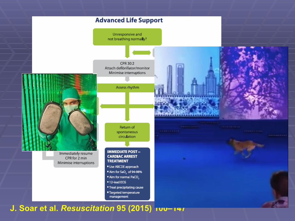
J. Soar et al. Resuscitation 95 (2015) 100–147
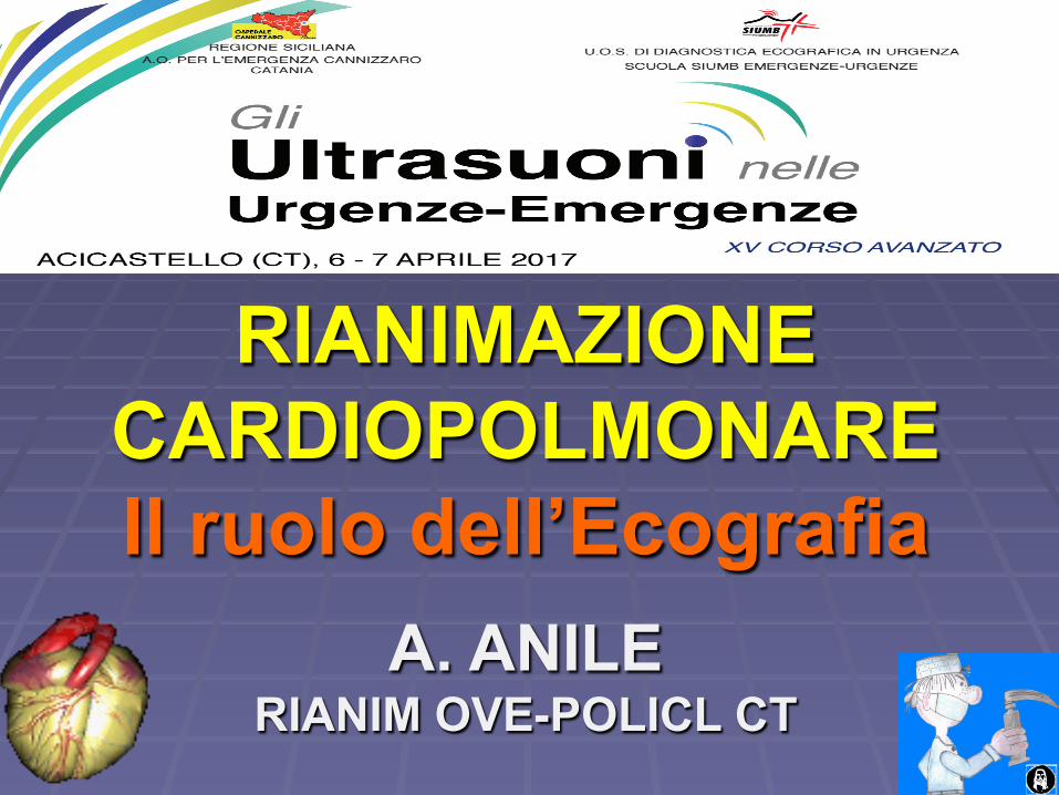
Nuove linee Guida (2015) !!!
J. Soar et al. Resuscitation 95 (2015) 100–147

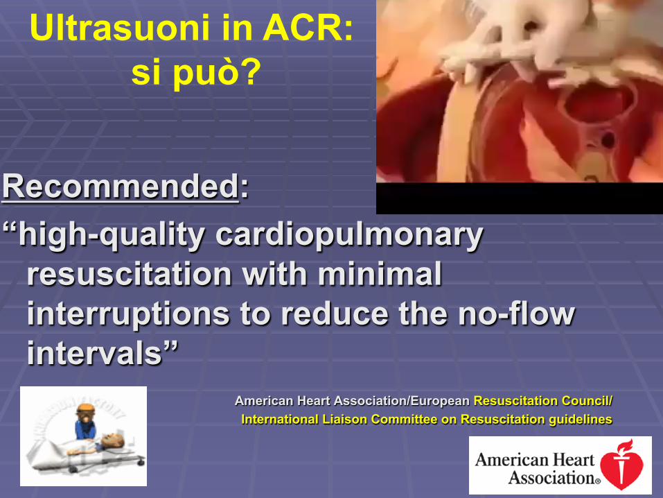
Recommended: “high-quality cardiopulmonary
resuscitation with minimal interruptions to reduce the no-flow intervals”
American Heart Association/European Resuscitation Council/ International Liaison Committee on Resuscitation guidelines
Ultrasuoni in ACR: si può?
• CPR e preparazione
• Esecuzione dell’ecografia
• Ripresa della CPR
• Interpretazione comunicazione e conseguenze
4 Fasi
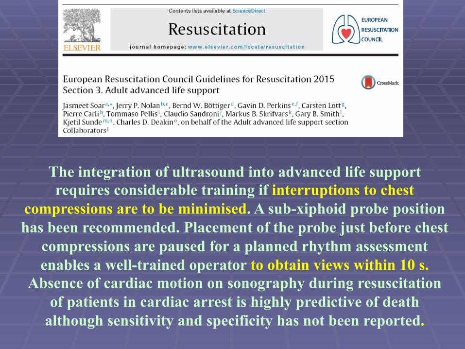
The integration of ultrasound into advanced life support requires considerable training if interruptions to chest
compressions are to be minimised. A sub-xiphoid probe position has been recommended. Placement of the probe just before chest
compressions are paused for a planned rhythm assessment enables a well-trained operator to obtain views within 10 s.
Absence of cardiac motion on sonography during resuscitation of patients in cardiac arrest is highly predictive of death
although sensitivity and specificity has not been reported.
Come integrarli Ultrasuoni in ACR
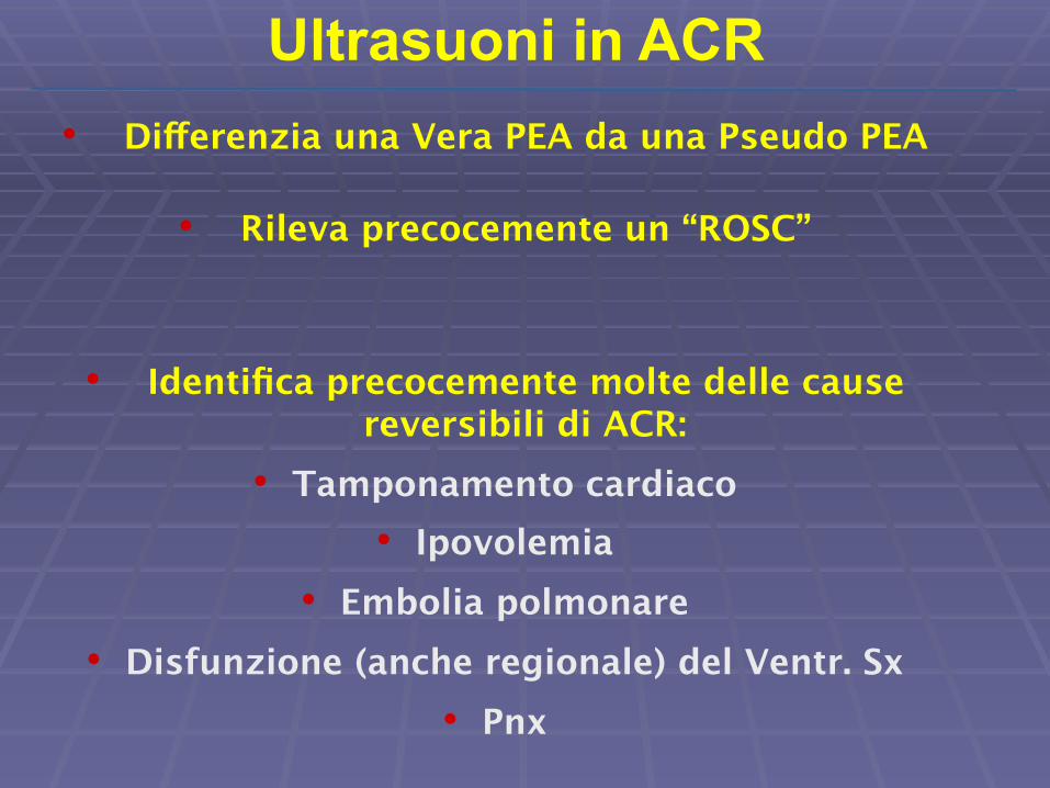
• Differenzia una Vera PEA da una Pseudo PEA
• Rileva precocemente un “ROSC”
• Identifica precocemente molte delle cause reversibili di ACR:
• Tamponamento cardiaco • Ipovolemia
• Embolia polmonare • Disfunzione (anche regionale) del Ventr. Sx
• Pnx
Ultrasuoni in ACR
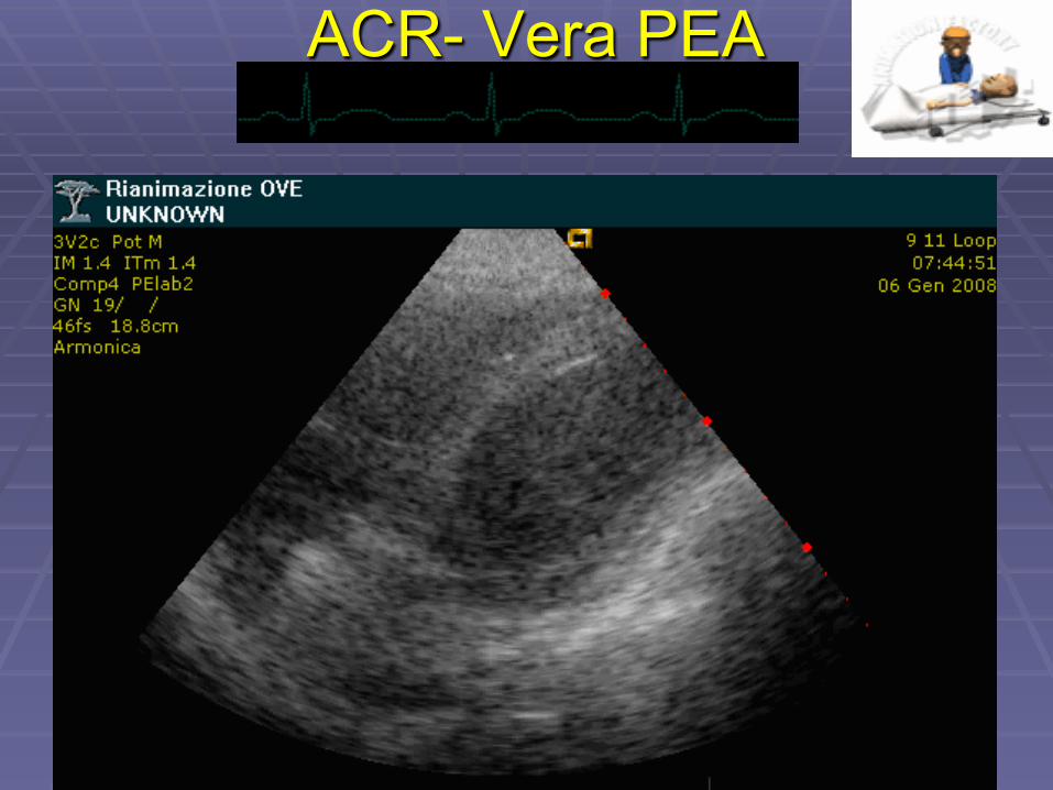
ACR- Vera PEA
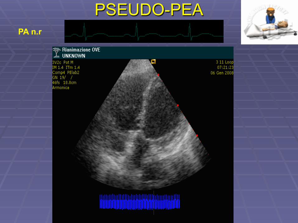
PSEUDO-PEA PA n.r
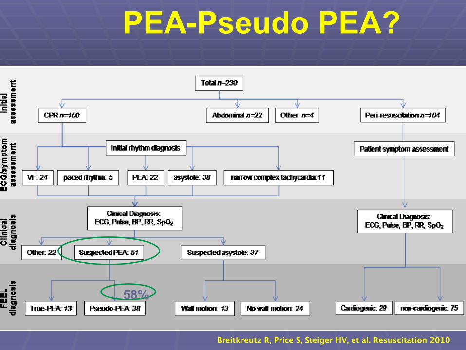
Breitkreutz R, Price S, Steiger HV, et al. Resuscitation 2010.
PEA-Pseudo PEA?
58%
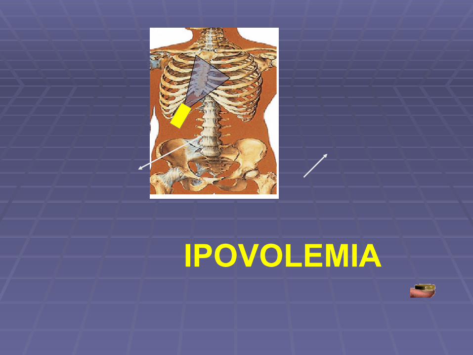
IPOVOLEMIA
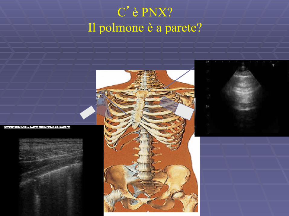
C’è PNX? Il polmone è a parete?
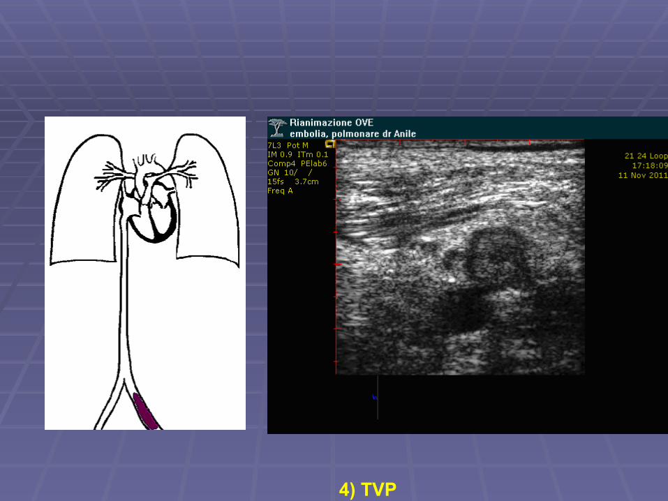
4) TVP
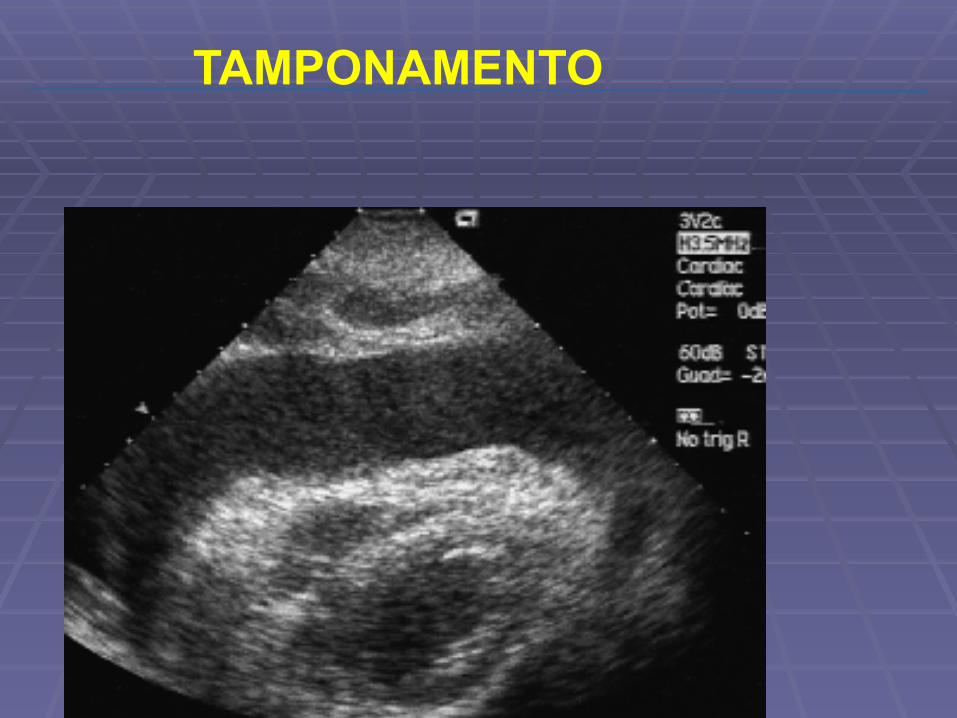
TAMPONAMENTO
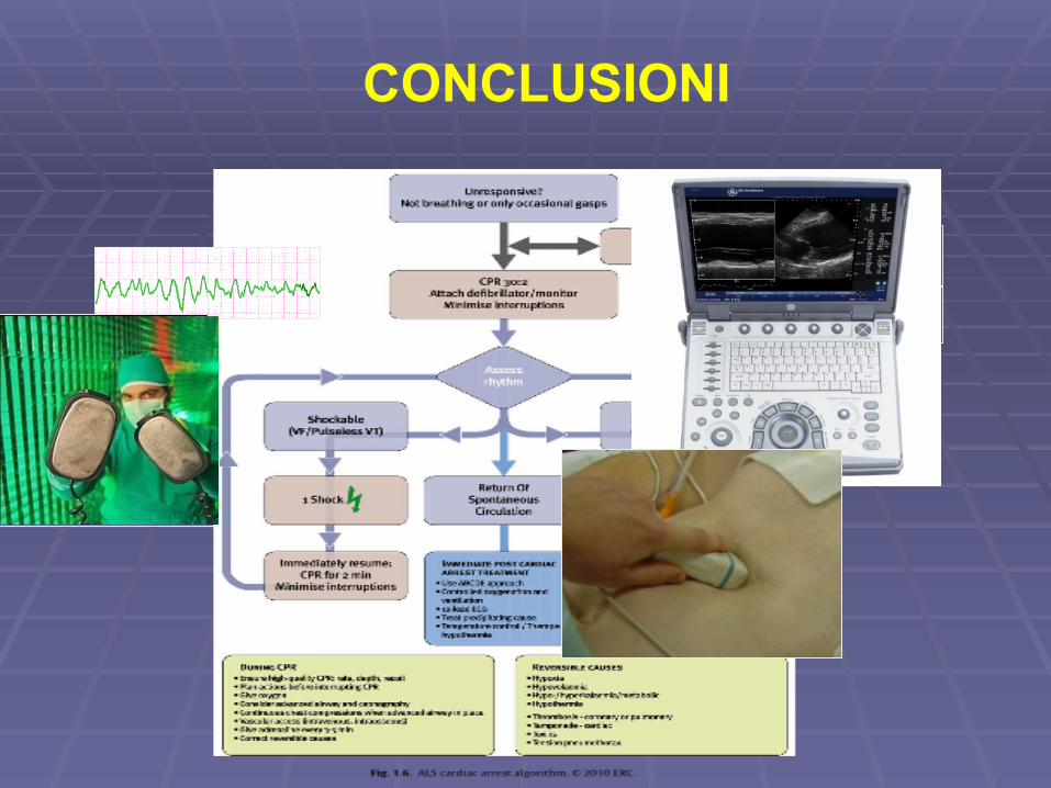
CONCLUSIONI
GRAZIE