Relazione tra: eziologia, patogenesi, manifestazioni … tra: eziologia, patogenesi, manifestazioni...
54
Relazione tra: eziologia, patogenesi, manifestazioni morfo-funzionali e Complicazioni in 4 tipi di malattie.
Transcript of Relazione tra: eziologia, patogenesi, manifestazioni … tra: eziologia, patogenesi, manifestazioni...

Relazione tra: eziologia, patogenesi, manifestazioni morfo-funzionali e
Complicazioni in 4 tipi di malattie.
Infiammazione
•! Che cosa è: processo reattivo (morboso) locale, base delle malattie
•! Sede: vascolo-connettivale (microcircolo)
•! Nomenclatura: suffisso “ITE” con il nome dell’organo interessato,
polmonite, pancreatite, gastrite….
•! Andamento stereotipato. Per effetto dei mediatori, nonostante la diversità degli stimoli.
•! Fenomeni elementari costanti: vasodilatazione, variazione
permeabiltà, migrazione cellulare.
•! Decorso: variabile per quantità e diversità stimoli, e delle reattività
dell’ospite ( danno variabile).

INFIAMMAZIONE
Ippocrate: individua fenomeni quali rossore e gonfiore
Celso: definisce i 4 punti cardinali della infiammazione
Galeno introduce il concetto della “funzione lesa”
Hunter: non si tratta di una malattia ma di una risposta agli stimoli
E. Metchnicoff: scopre i macrofagi
Lewis: scopre il ruolo dell’Istamina
Definizione generale
L’Infiammazione è il più importante processo morboso reattivo
locale che forma la base patologica delle malattie

Infiammazione acuta (sintesi)
•! Classificazione: acuta e cronica
•! Acuta: fenomeni vascolari prevalenti (essudativa), con migrazione
cellulare Neu e Mon , esito benevolo e guarigione completa.
•! Acuta: due fasi
•! Fase 1- Vasodilatazione e aumento permeabilità, con azione sulle
arteriole e sugli endoteli ( azione dell’istamina).
•! Fase 2- i fenomeni cellulari diventano prevalenti, si forma l’essudato
e con il procedere della perdita dei fluidi si arriva al rallentamento
ematico e alla fase di iperemia passiva. Marginazione dei granulociti
neutrofili e formazione di “rouleaux” di globuli rossi

What is inflammation?
Rubor Tumor Calor Dolor
(redness) (swelling) (heat) (pain)
What sort of things induce this inflammation?
Four basic symptoms (known since ancient Greek & Roman times)

EXOGENOUS FACTORS which can induce inflammation:
•! Mechanical (traumatic injury – wounding, fracture etc)
•! Physical (low or high temperature, ionising irradiation, microwaves)
•! Chemical (venoms, caustic agents, poisons etc)
•! Nutritive (deficiency of oxygen, vitamins & basic nutrients)
•! Biological (viruses, microorganisms, unicellular & multicellular parasites)
ENDOGENOUS FACTORS like autoantigens may also induce inflammation.
So, these induce inflammation but what is inflammation?
Inflammation is a general term for the local
accumulation of fluid, plasma proteins, and white
blood cells.
This is also known as an inflammatory response.
It’s a process whereby molecules and cells of the
immune system are concentrated at a site of tissue damage or infection.
These molecules and cells come from the blood.
How is the blood supply to the damaged site improved?
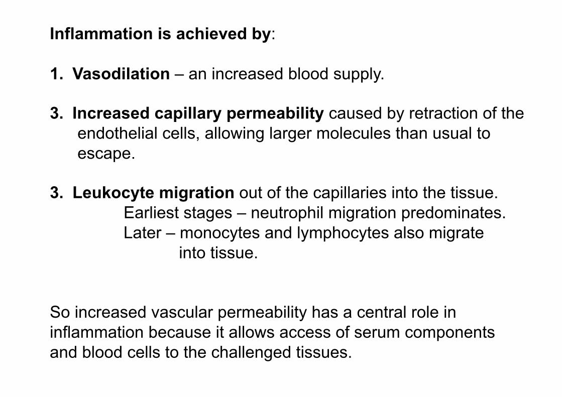
Inflammation is achieved by:
1.! Vasodilation – an increased blood supply.
3.! Increased capillary permeability caused by retraction of the
endothelial cells, allowing larger molecules than usual to
escape.
3.! Leukocyte migration out of the capillaries into the tissue.
Earliest stages – neutrophil migration predominates.
Later – monocytes and lymphocytes also migrate
into tissue.
So increased vascular permeability has a central role in
inflammation because it allows access of serum components
and blood cells to the challenged tissues.

Vascular permeability (leakiness) is promoted by many
inflammatory mediators.
Endothelial cells
lining blood vessel
Complement
Immunoglobulin
What’s the advantage in leaking complement and immunoglobulin at the
inflamed site?
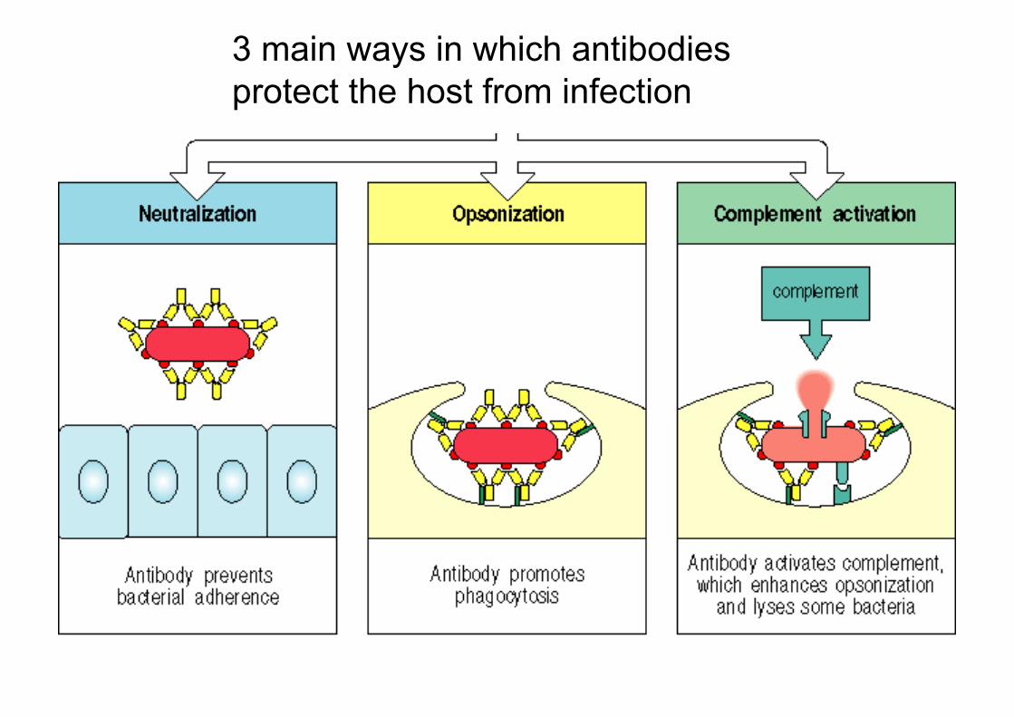
3 main ways in which antibodies
protect the host from infection
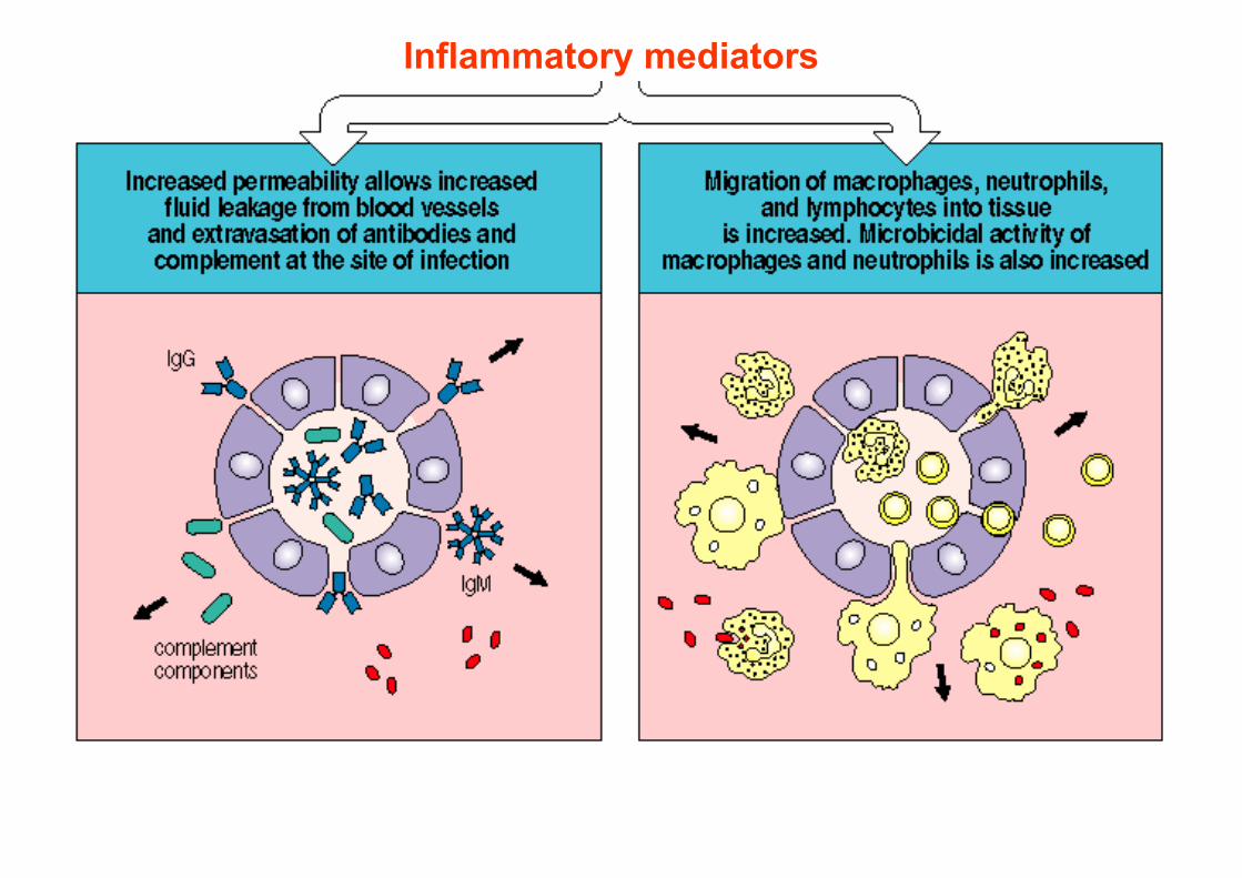
Inflammatory mediators

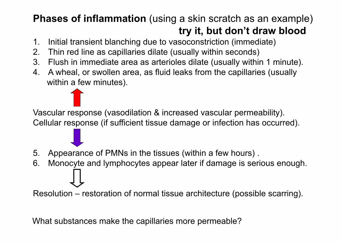
Phases of inflammation (using a skin scratch as an example)
try it, but don’t draw blood 1.! Initial transient blanching due to vasoconstriction (immediate)
2.! Thin red line as capillaries dilate (usually within seconds)
3.! Flush in immediate area as arterioles dilate (usually within 1 minute).
4.! A wheal, or swollen area, as fluid leaks from the capillaries (usually
within a few minutes).
Vascular response (vasodilation & increased vascular permeability).
Cellular response (if sufficient tissue damage or infection has occurred).
5.! Appearance of PMNs in the tissues (within a few hours) .
6.! Monocyte and lymphocytes appear later if damage is serious enough.
Resolution – restoration of normal tissue architecture (possible scarring).
What substances make the capillaries more permeable?

Many agents increase vascular permeability, including:
C3a C5a
Ba
C2 kinin
Histamine Leukotrienes
Serotonin
Most release occurs from the post capillary venules.
Complement agents (several produced by MØs)
Released from mast cells
What’s in the initial inflammatory fluid exudate?
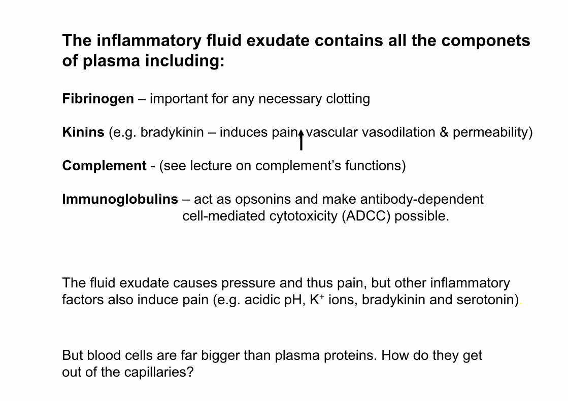
The inflammatory fluid exudate contains all the componets
of plasma including:
Fibrinogen – important for any necessary clotting
Kinins (e.g. bradykinin – induces pain, vascular vasodilation & permeability)
Complement - (see lecture on complement’s functions)
Immunoglobulins – act as opsonins and make antibody-dependent
cell-mediated cytotoxicity (ADCC) possible.
The fluid exudate causes pressure and thus pain, but other inflammatory
factors also induce pain (e.g. acidic pH, K+ ions, bradykinin and serotonin).
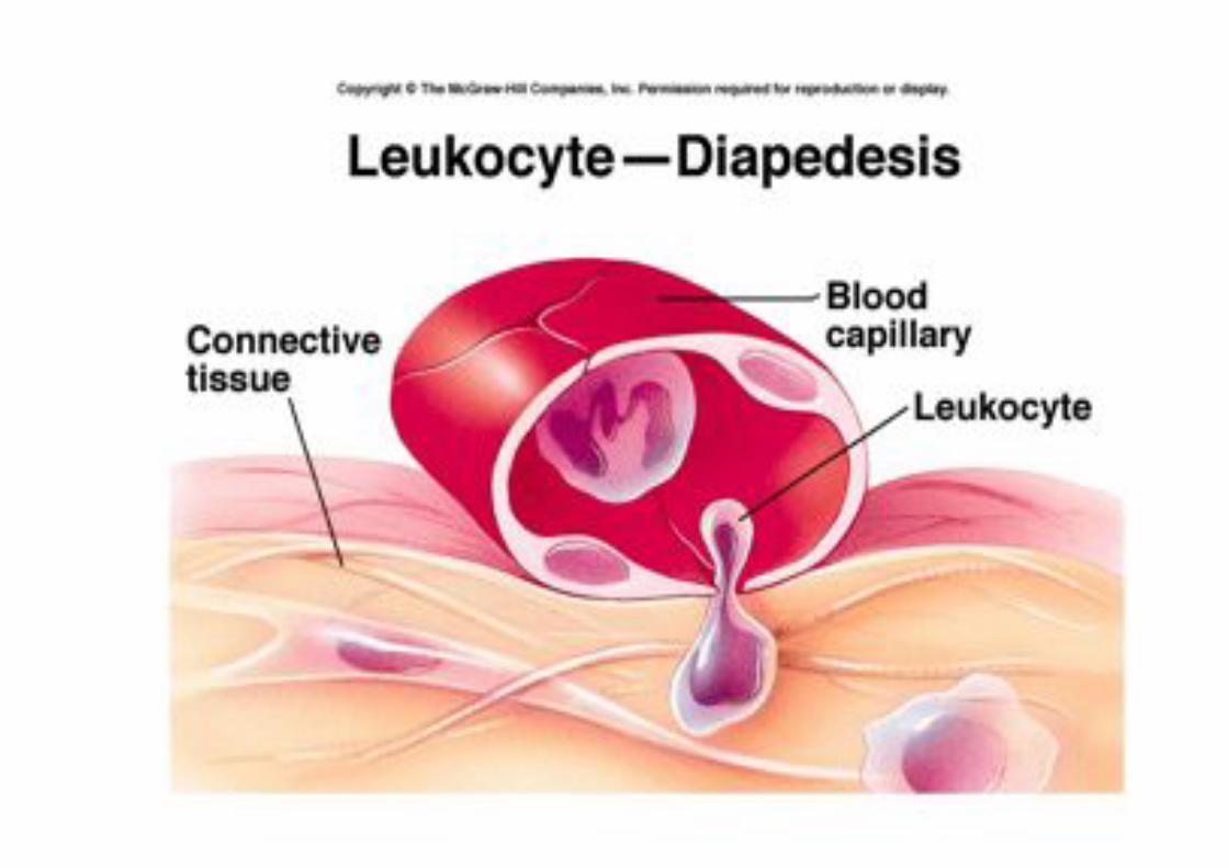
But blood cells are far bigger than plasma proteins. How do they get
out of the capillaries?

Leukocyte extravasation
In inflamed regions white blood cells are caught
“like flies on fly-paper” and escape from the blood by squeezing between endothelial cells and
crawling through the basement membrane with
the aid of digestive enzymes.
What makes the moving leukocytes stick to the endothelial cells?


One of the first effects of inflammation is to increase P-
selectin expression on endothelial cells which binds Sialyl-
Lewisx on the PMNs causing rolling.
How do the leukocytes come to a halt?

Activated endothelial cells express:
Intercellular adhesion molecule (ICAM-1)
Endothelial leukocyte adhesion molecule (ELAM-1) Vascular cell adhesion molecule (VCAM-1)
Human leukocytes express
integrins as receptors which bind to these molecules:
Leukocyte function associated antigen 1 (LFA-1)
Very late antigen 4 (VLA-4)
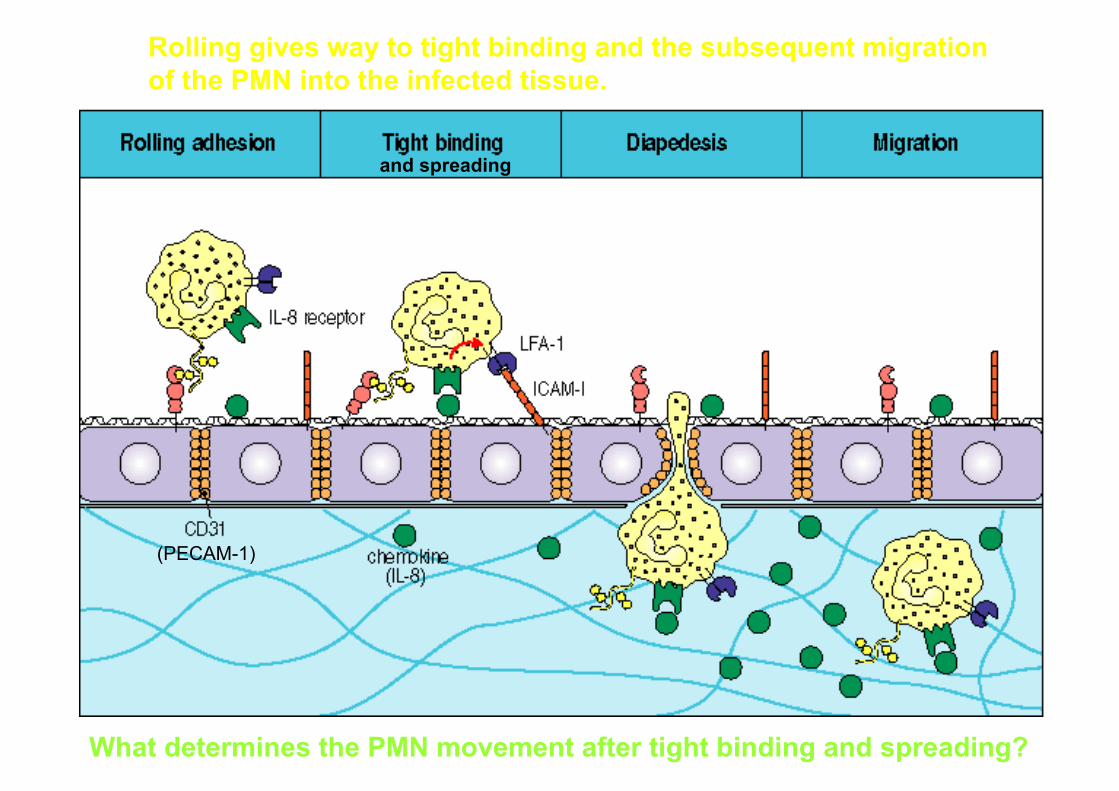
Rolling gives way to tight binding and the subsequent migration
of the PMN into the infected tissue.
What determines the PMN movement after tight binding and spreading?
(PECAM-1)
and spreading


Leukocytes can detect a concentration gradient of
chemotactic factors (e.g. of C3a, C5a, leukotrienes or IL-8) and move towards it.
This movement of leukocytes (e.g. PMNs, monocytes
and lymphocytes) towards a site of inflammation is
called Chemotaxis.
Chemotaxis differs from chemokinesis (a random
increase in movement).
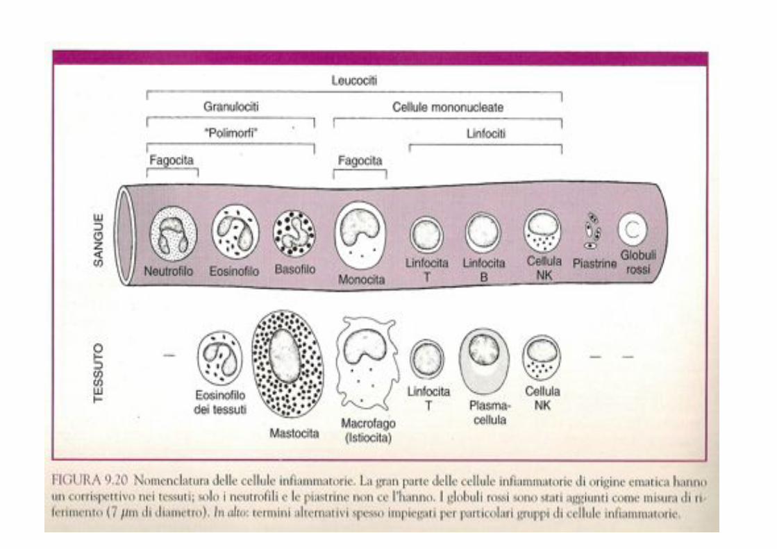
What are the main cell types involved in the inflammatory response?

PMN
T CELL Neutrophils, Monocyte and T cells in the
blood extravasate into the tissues to contribute to the inflammatory response.
Macrophages and mast cells resident in
the tissue are of central importance to the inflammatory response, releasing
inflammatory mediators when activated.
MAST
CELL MACRO-
PHAGE
Cells of most tissues can themselves
secrete inflammatory mediators (prostaglandins) when damaged
or challenged.
Endothelial cells lining local blood
vessels, respond to secretions from MØs, mast cells and tissue cells, by
allowing serum to leak between them and by trapping leukocytes and
promoting their migration into the tissues.
Some of the most important cells of the inflammatory response
Monocyte
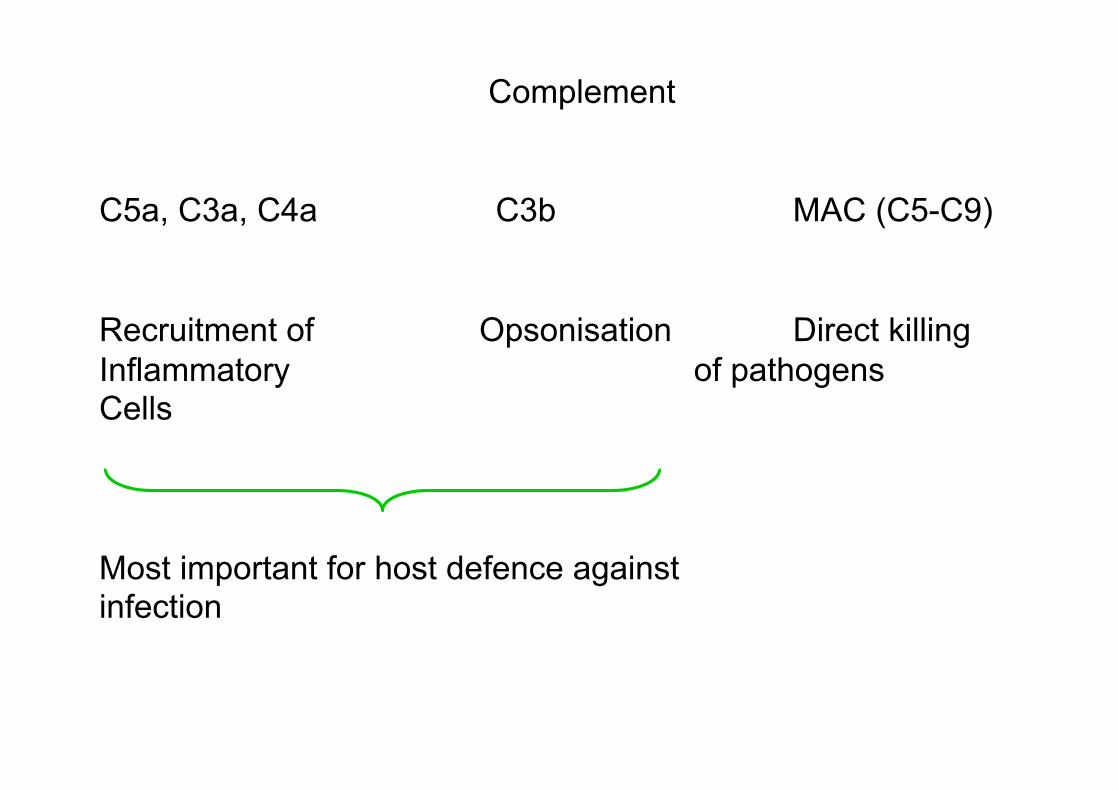
Complement
C5a, C3a, C4a C3b MAC (C5-C9)
Recruitment of Opsonisation Direct killing
Inflammatory of pathogens Cells
Most important for host defence against infection

Neutrophils (polymorphonuclear leukocytes - PMNs)
Bone marrow makes 1011 PMNs / day (1012 in acute Inflammation).
4-10hr !-life in circulation; 1-2 days survival in tissue.
Each day about 3 x 10⁹ neutrophils enter tissues of the oral cavity (‘the most contaminated site of the body’).
At infected sites activated macrophages release
cytokines that attract neutrophils which soon become the
dominant phagocytic cell type.
PMNs are specialised for working under anaerobic
conditions which prevail in damaged tissues.
Neutrophil arrival - 1st event of inflammatory response.
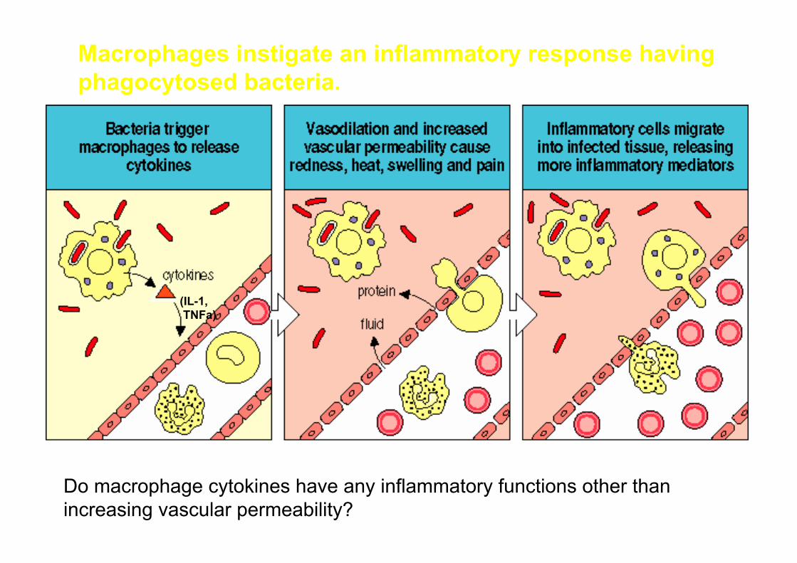
Macrophages instigate an inflammatory response having
phagocytosed bacteria.
(IL-1, TNFa)
Do macrophage cytokines have any inflammatory functions other than
increasing vascular permeability?
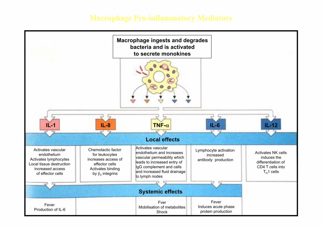
Macrophage ingests and degrades bacteria and is activated
to secrete monokines
Activates vascular endothelium
Activates lymphocytes Local tissue destruction
increased access of effector cells
Chemotactic factor for leukocytes
increases access of effector cells
Activates binding by !2 integrins
Activates vascular endothelium and increases vascular permeability which leads to increased entry of IgG complement and cells and increased fluid drainage to lymph nodes
Lymphocyte activation increased
antibody production Activates NK cells
induces the differentiation of CD4 T cells into
TH1 cells
Fever Production of IL-6
Local effects
Systemic effects
Fver Mobilisation of metabolites
Shock
Fever Induces acute phase
protein production
IL-1 IL-8 TNF-" IL-6 IL-12
Macrophage Pro-inflammatory Mediators
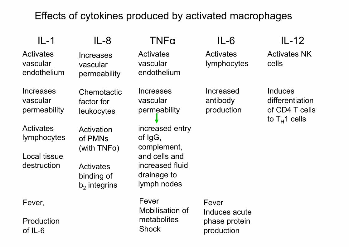
Effects of cytokines produced by activated macrophages
IL-1 IL-8 TNF" IL-6 IL-12
Activates
vascular endothelium
Increases
vascular
permeability
Activates lymphocytes
Local tissue destruction
Increases
vascular permeability
Chemotactic
factor for
leukocytes
Activation of PMNs
(with TNF")
Activates
binding of b2 integrins
Activates
vascular endothelium
Increases
vascular
permeability
increased entry of IgG,
complement,
and cells and increased fluid
drainage to lymph nodes
Activates
lymphocytes
Increased
antibody
production
Activates NK
cells
Induces
differentiation
of CD4 T cells to TH1 cells
Fever,
Production
of IL-6
Fever
Mobilisation of metabolites
Shock
Fever
Induces acute phase protein
production
LOCAL
EFFECTS
SYSTEMIC EFFECTS

Cytokines from activated macrophages coordinate the
body’s response to infection.
FEVER
What are acute phase proteins?

The acute
phase
response increases
the
supply of
recognition
molecules of innate
immunity.

Acute-phase proteins (CRP and MBP) produced by the liver can each
bind structural features of bacterial cells.
Upon binding they act as opsonins and also activate complement
(lysis – dotted bacterial margin).
CRP MBP
(found as far back as invertebrates)

Encapsulated bacteria are more efficiently engulfed by phagocytosis
when the bacteria are coated with antibody, C3b, CRP or MBP.

Cytokines from activated macrophages coordinate the
body’s response to infection.
FEVER
Has fever any benefits?
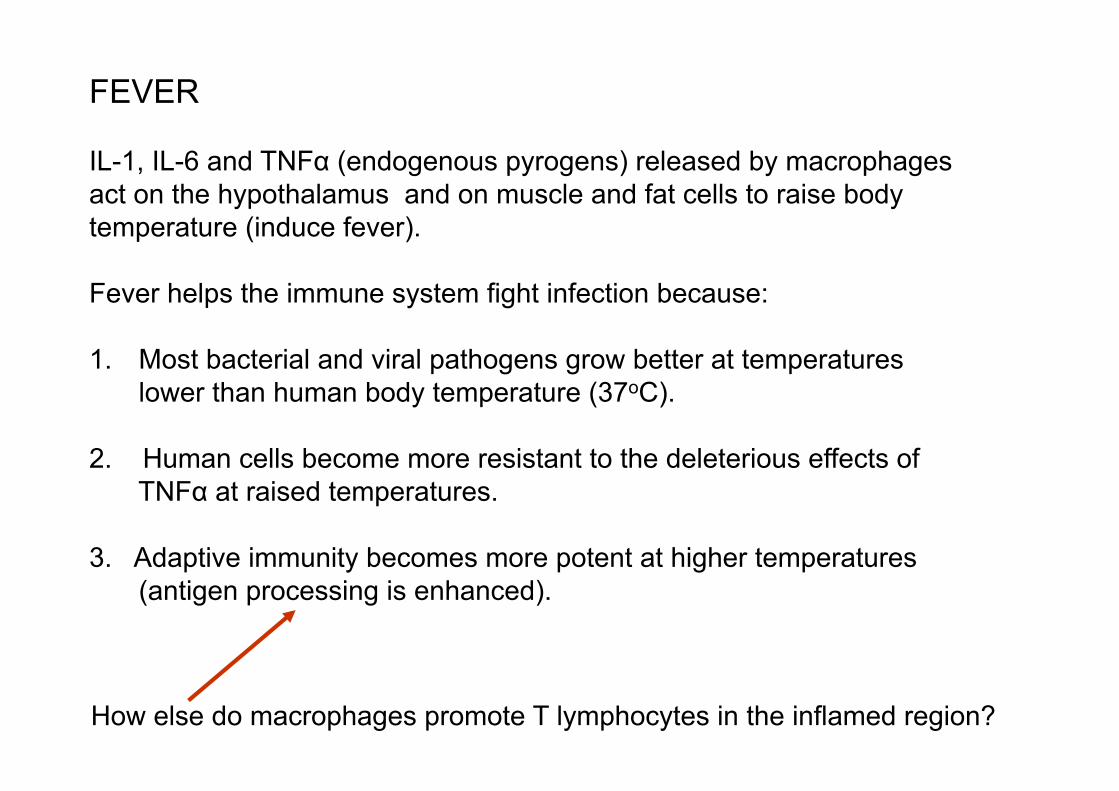
FEVER
IL-1, IL-6 and TNF" (endogenous pyrogens) released by macrophages
act on the hypothalamus and on muscle and fat cells to raise body
temperature (induce fever).
Fever helps the immune system fight infection because:
1.! Most bacterial and viral pathogens grow better at temperatures
lower than human body temperature (37oC).
2. Human cells become more resistant to the deleterious effects of
TNF" at raised temperatures.
3. Adaptive immunity becomes more potent at higher temperatures
(antigen processing is enhanced).
How else do macrophages promote T lymphocytes in the inflamed region?

Macrophage cytokines influencing T cells:
IL-1 A general activator of all T cells
IL-6 T cell growth and differentiation
IL-12 Preferentially activates TH1 cells
TH1 cytokines released in response to macrophage (MØ) cytokines:
Interferon-# (IFN- #) expression of MHC on MØs and other local cells
MØ antigen processing
induces MØ maturation
NK cell activity
inhibits TH2 cells
causes antiviral effects
Tumour necrosis factor (TNF) several roles in inflammation but high local
levels can cause tissue destruction and has
potent systemic effect of causing weight loss.

Th1 cell cytokine effects on MØs

IN CONCLUSION, molecular control of inflammation is by :
•! Cytokines
•! Plasma enzyme systems: Complement
Clotting (kinin & fibrinolytic) pathways
•! Lipid mediators: Prostaglandins & leukotrienes
•! Vasoactive mediators (e.g. histamine) from mast cells, basophils & platelets
•! The antigen itself – when Ag cannot be cleared the inflammation is chronic
and different from sites where it is rapidly cleared.
Acute inflammation is the term used to describe early and often transient
episodes. What types of antigen cannot be cleared in chronic inflammation?
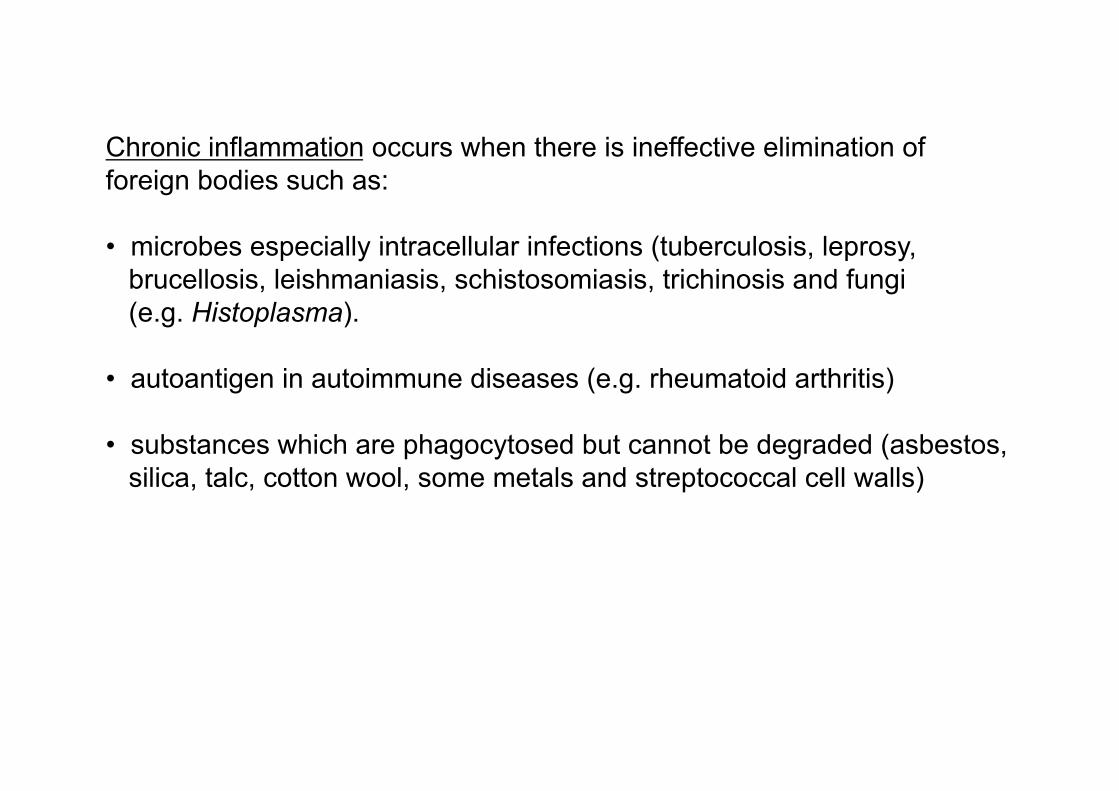
Chronic inflammation occurs when there is ineffective elimination of
foreign bodies such as:
•! microbes especially intracellular infections (tuberculosis, leprosy,
brucellosis, leishmaniasis, schistosomiasis, trichinosis and fungi
(e.g. Histoplasma).
•! autoantigen in autoimmune diseases (e.g. rheumatoid arthritis)
•! substances which are phagocytosed but cannot be degraded (asbestos,
silica, talc, cotton wool, some metals and streptococcal cell walls)

infezione L’infezione è rimossa
L’Infezione
non è rimossa
L’infezione permane
Tuberculosis
Hepatitis B Schistosomiasis
INFIAMMAZIONE ACUTA Risposta Innata (PMNs, complemento, IFN)
Risposta adattativa
INFIAMMAMMAZIONE CRONICA
formazione del Granuloma
INFIAMMAZIONE ACUTA E CRONICA
What is a granuloma?
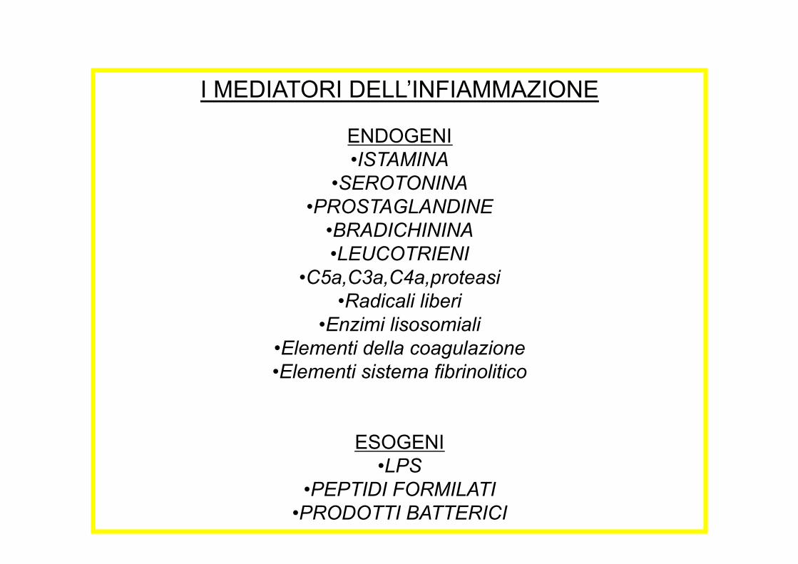
I MEDIATORI DELL’INFIAMMAZIONE
ENDOGENI
•!ISTAMINA
•!SEROTONINA
•!PROSTAGLANDINE
•!BRADICHININA
•!LEUCOTRIENI
•!C5a,C3a,C4a,proteasi
•!Radicali liberi
•!Enzimi lisosomiali
•!Elementi della coagulazione
•!Elementi sistema fibrinolitico
ESOGENI
•!LPS
•!PEPTIDI FORMILATI
•!PRODOTTI BATTERICI
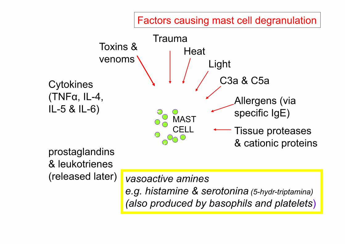
MAST
CELL
vasoactive amines
e.g. histamine & serotonina (5-hydr-triptamina)
(also produced by basophils and platelets)
Trauma
Heat
Light
C3a & C5a
Allergens (via
specific IgE)
Factors causing mast cell degranulation
prostaglandins
& leukotrienes
(released later)
Substances released by mast cells
Tissue proteases
& cationic proteins
Toxins &
venoms
Cytokines
(TNF", IL-4,
IL-5 & IL-6)
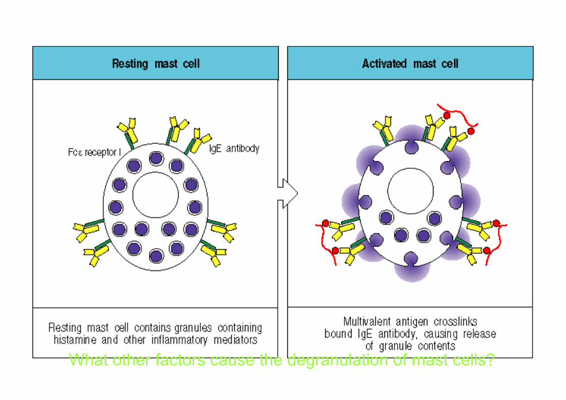
What other factors cause the degranulation of mast cells?