Fondamenti d anatomia e istologia - UniBG · Ingegneria delle tecnologie per la salute Fondamenti d...
116
Ingegneria delle tecnologie per la salute Fondamenti d anatomia e istologia Sistema nervoso
Transcript of Fondamenti d anatomia e istologia - UniBG · Ingegneria delle tecnologie per la salute Fondamenti d...

Ingegneria delle tecnologie per la salute
Fondamenti d anatomia e istologia
Sistema nervoso
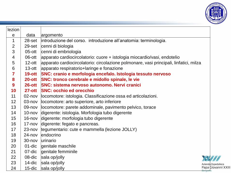
lezion
e data argomento
1 28-set introduzione del corso. introduzione all’anatomia: terminologia.
2 29-set cenni di biologia
3 05-ott cenni di embriologia
4 06-ott apparato cardiocircolatorio: cuore + istologia miocardio/vasi, endotelio
5 12-ott apparato cardiocircolatorio: circolazione polmonare, vasi principali, linfatici, milza
6 13-ott apparato respiratorio+laringe e fonazione
7 19-ott SNC: cranio e morfologia encefalo. Istologia tessuto nervoso
8 20-ott SNC: tronco cerebrale e midollo spinale, le vie
9 26-ott SNC: sistema nervoso autonomo. Nervi cranici
10 27-ott SNC: occhio ed orecchio
11 02-nov locomotore: istologia. Classificazione ossa ed articolazioni.
12 03-nov locomotore: arto superiore, arto inferiore
13 09-nov locomotore: parete addominale, pavimento pelvico, torace
14 10-nov digerente: istologia. Morfologia tubo digerente
15 16-nov digerente: morfologia tubo digerente
16 17-nov digerente: fegato e pancreas.
17 23-nov tegumentario: cute e mammella (lezione JOLLY)
18 24-nov endocrino
19 30-nov urinario
20 01-dic genitale maschile
21 07-dic genitale femminile
22 08-dic sala op/jolly
23 14-dic sala op/jolly
24 15-dic sala op/jolly

hystology

Ectodremic origin
From the neural crest
3 vescicles:
•Prosencephaln
•Mesencephalon
•rhomboencepahlon
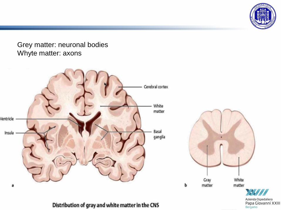
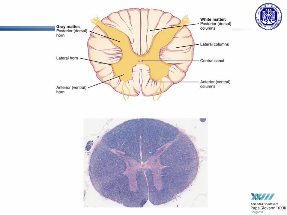
Grey matter: neuronal bodies
Whyte matter: axons


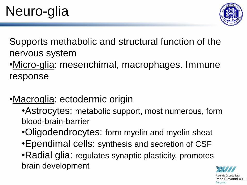
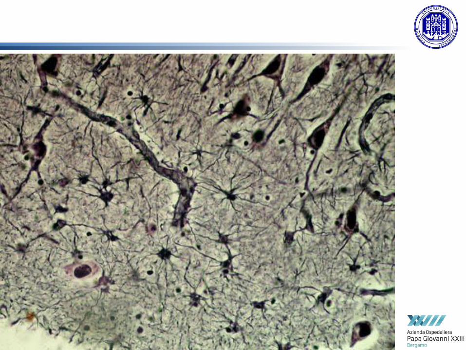
Neuro-glia
Supports methabolic and structural function of the
nervous system
•Micro-glia: mesenchimal, macrophages. Immune
response
•Macroglia: ectodermic origin
•Astrocytes: metabolic support, most numerous, form
blood-brain-barrier
•Oligodendrocytes: form myelin and myelin sheat
•Ependimal cells: synthesis and secretion of CSF
•Radial glia: regulates synaptic plasticity, promotes
brain development

csf

Blood brain barrier - BBB
Hyperselective
permeability
Protective function

Oligodendrocyes- myelin sheat

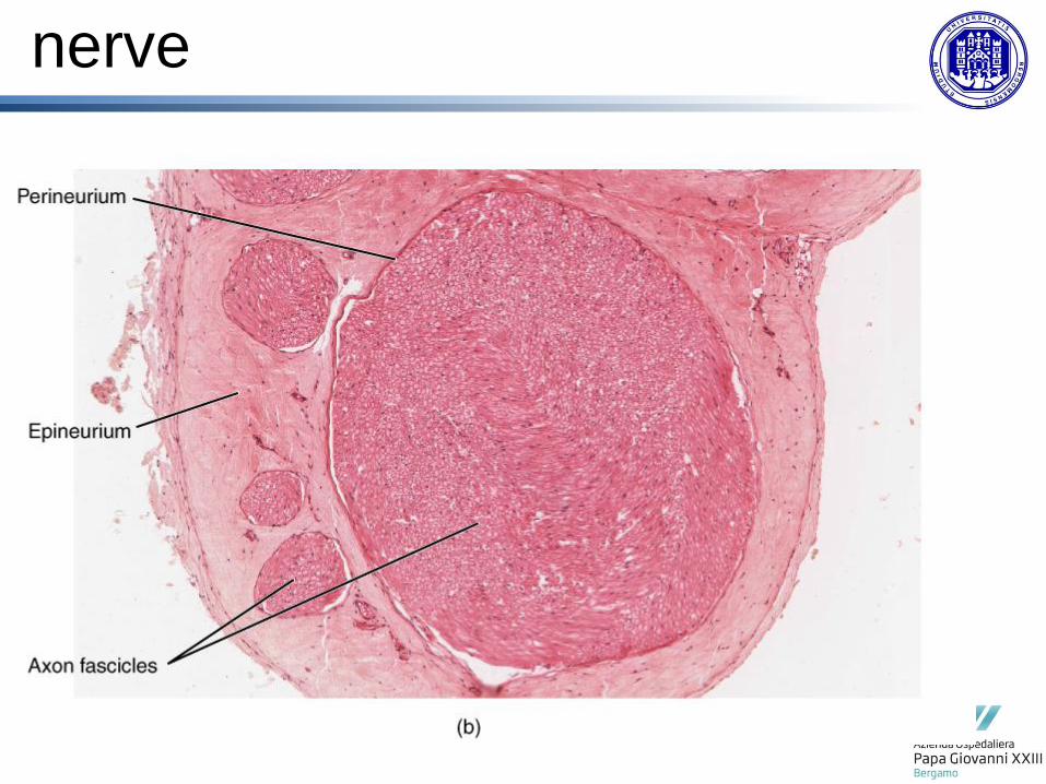
peripheral nerve fibers
A group: Fibers of the A group have a large diameter, are myelinated,
and have the highest conduction velocity of all the nerves in the
body.
The A group consists of four types of nerve fibers (alpha, beta
gamma, delta). afferent and efferent. Aα Ø 13-20 μm 80-120 m/s
B group: Nerve fibers in this group are myelinated with a small
diameter. Generally, they are the preganglionic fibers of the
autonomic nervous system and have a low conduction velocity. B
Ø 1-5 μm 3-15 m/s
C group: The C group fibers are unmyelinated and as the B group
fibers have a small diameter and low conduction velocity. These
fibers include postganglionic fibers of ANS and dorsal root fibers
(teperature, pain, touch, pressure) C Ø 0.2-1.5 μm 0.5-2 m/s

synapsis
Connection between neurons
Elettrochimic signal
Chimic signal
(neurotransmitters)
Elettrochimic signal
neurotrasmitters
Classification of neurons based on nurotransmitter:
•Glutamatergic (Glutamate)
•GABAergic (GABA)
•Adrenergic (Epinephrine)
•Noradrenergic (Norepinephrine)
•Dopaminergic (dopamine)
•Colinergic (acetilcholine)
•Serotoninergic (serotonine)
Different functions: eccitation/inhibition
Metabotropic/ionotropic receptors
nerve


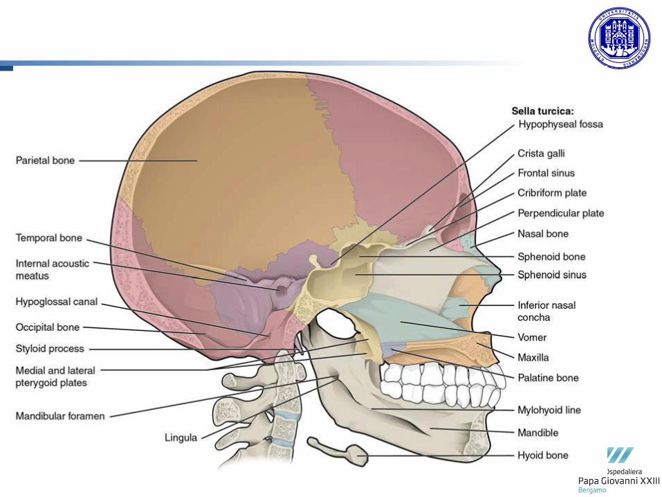
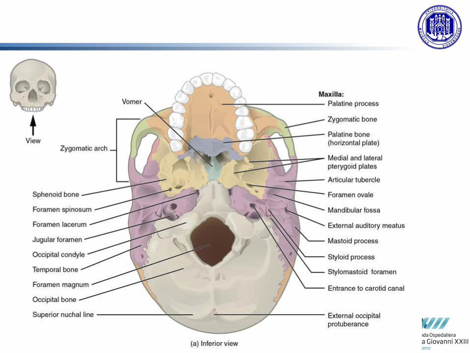
The Skull
and
Vertebral Column

neurocranium
Facial skeleton
(splancnocranium)
twenty two bones:
Eight bones of the neurocranium (occipital bone, 2 temporal bones,
2 parietal bones, sphenoid bone, ethmoid bone,frontal bone),
and fourteen bones of the viscerocranium (vomer, 2 conchae, 2 nasal
bones, 2 maxilla, mandible, 2 palatine bones, 2 zygomatic bones, 2 lacrimal
bones)
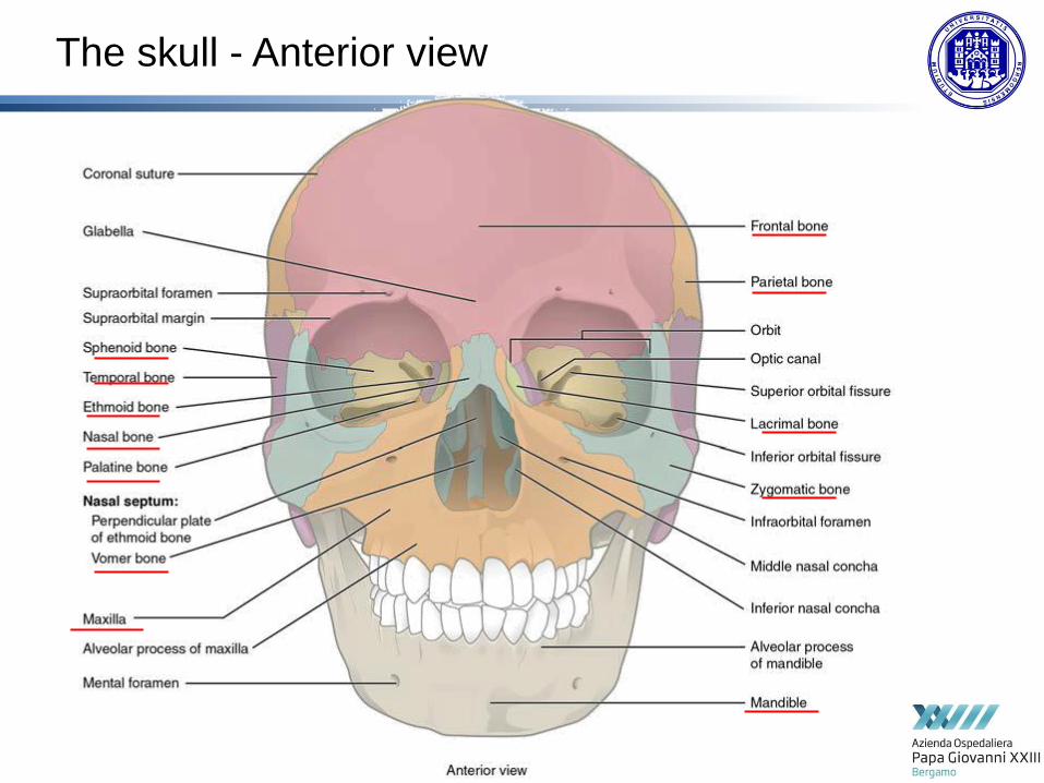
The skull - Anterior view

The skull - Lateral view

The skull – inner


Vertebral column
33-34 vertebre
•7 cervical
•12 dorsal
•5 lumbar
•5 sacral
•4-5 coccygeal
1 cifosys (skull)
1 lordosys
(cervical)
2 lordosys
(lumbar)
2 cifosys (dorsal)
3 cifosys (sacral)

vertebre


vertebre


meninges
cortex
Epidural space
Subarachnoideal space
subdural space

meninges
Dura mater: adhese to the skull, dense connettive tissue,
fibrous.
Arachnoid: thin trasparent membrane, not follow cortex
convolutions. Flat cells
Subarachnoideal space: trabecolae, contains CSF, brain’s
blood vessels and cranial nerves.
Pia mater: adhese to the brain cortex, follows its
convolutions. Rich in capillaries, very thin. Flat cells
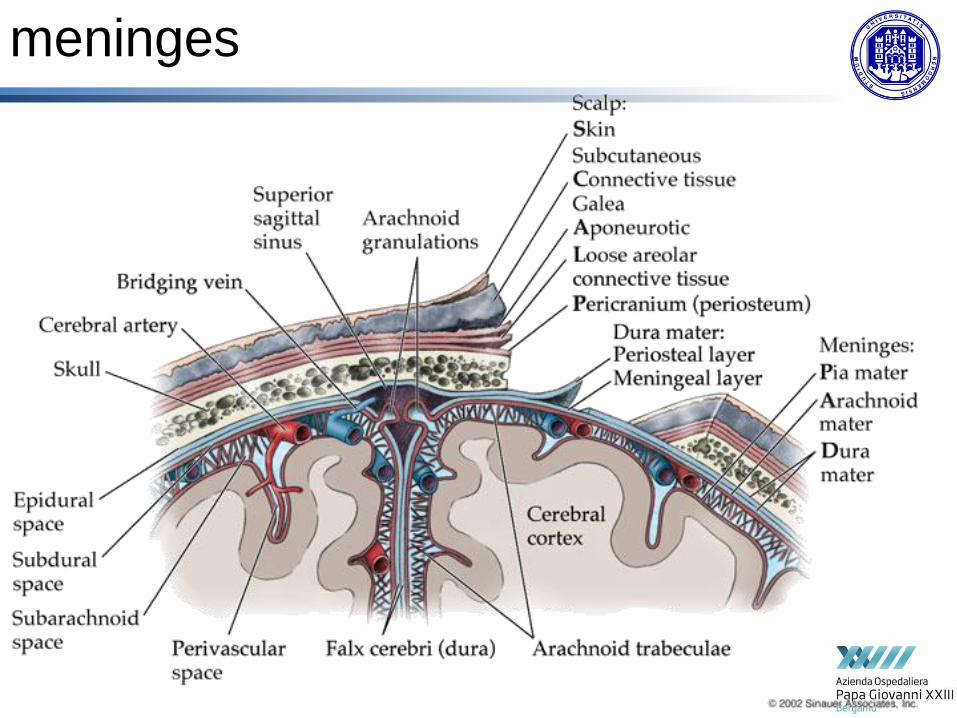
meninges

The brain

Brain’s anatomical regions

Cortex: grey matter

Cortical areas
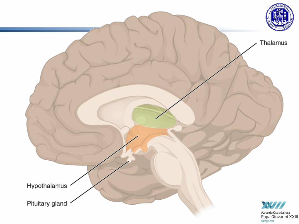


Basal Ganglia
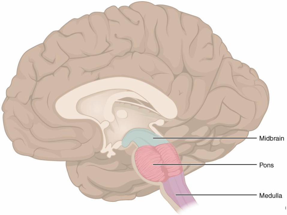

Pituitary gland

Brainstem
midbrain
medulla


Limpid
60-200 ml
15-20 cmH2O
No proteins
8 cell/mm3 (leucytes)
Same concentration of serum glucose
Product by choroid plexus, 500 ml every day
Contained in lateral ventricles, third and fourth ventricles,
spinal canal and sub-arachnoideal space
Reabsorption by arachnoideal granulation
Cerebro-spinal fluid CSF
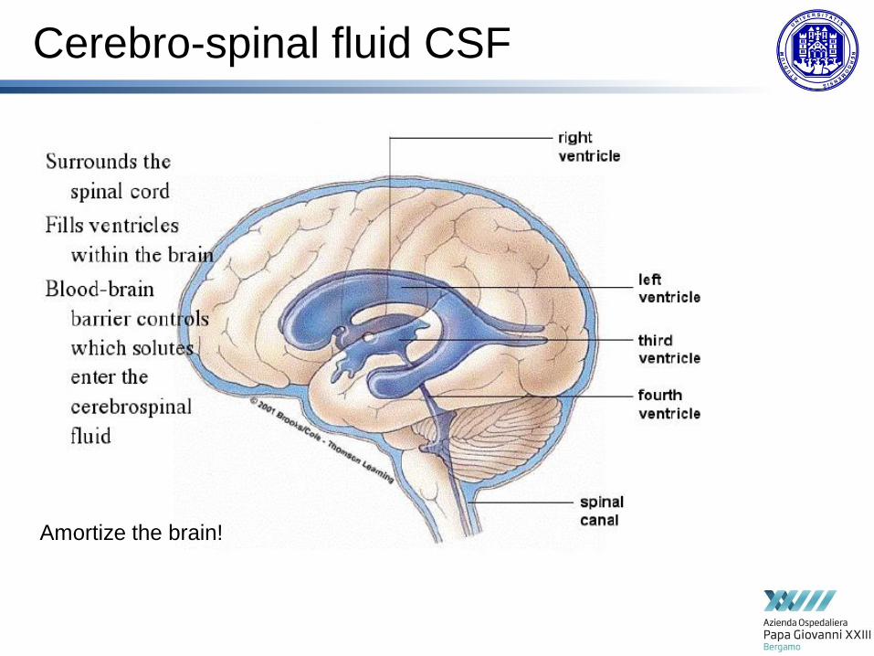
Cerebro-spinal fluid CSF
Amortize the brain!

CSF circulation
X2, lushka magendie

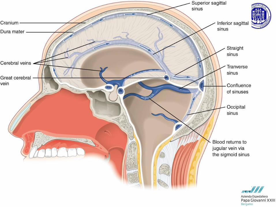
vascularization

Middle meningeal

Blood supply from internal carotid artery and
vertebral artery
Anastomosys between these two arteries: Willie’s
circle
After this anastomotic system the circulation is a
TERMINAL CIRCULATION
stroke


Vie nervose


moto


senso


Autonomous nervous system
(ANS)
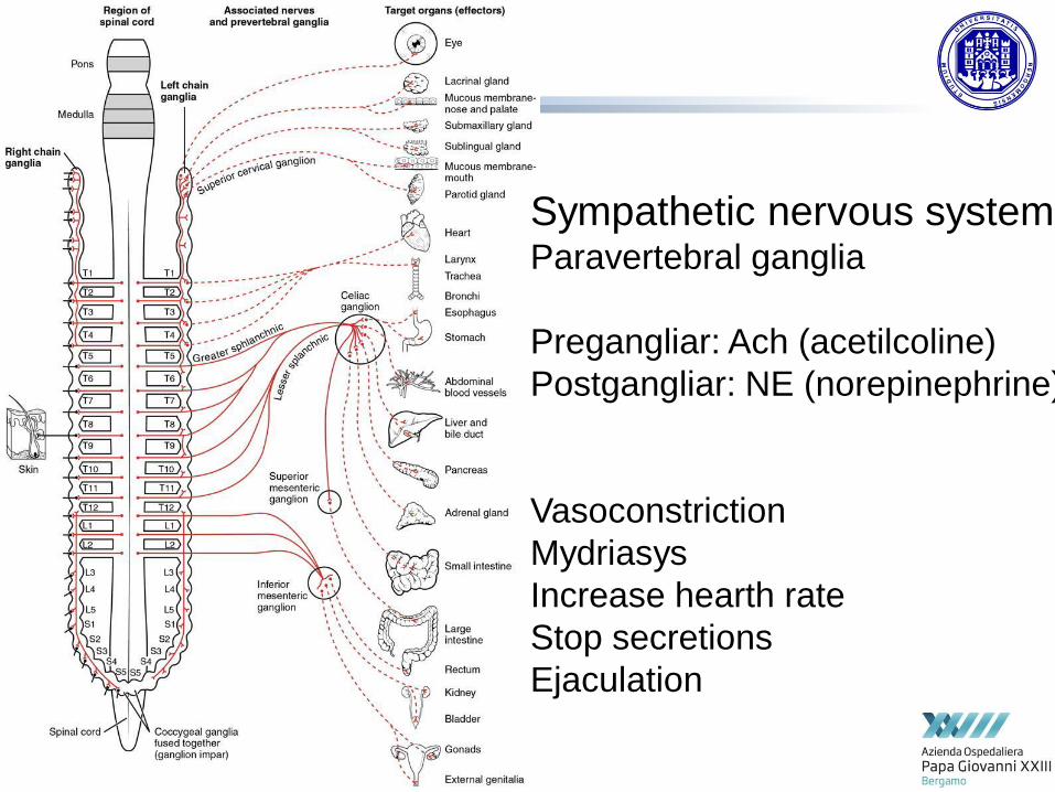
Sympathetic nervous system Paravertebral ganglia
Pregangliar: Ach (acetilcoline)
Postgangliar: NE (norepinephrine)
Vasoconstriction
Mydriasys
Increase hearth rate
Stop secretions
Ejaculation
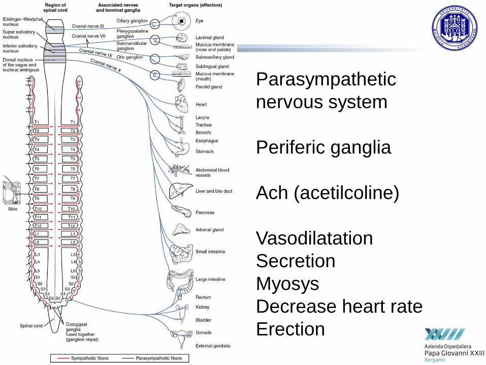
Parasympathetic
nervous system
Periferic ganglia
Ach (acetilcoline)
Vasodilatation
Secretion
Myosys
Decrease heart rate
Erection

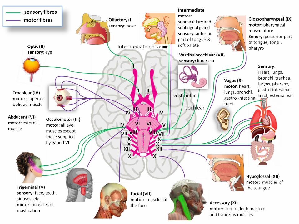
Cranial nerves

Cranial nerves


Ocular reflexes

VII cranial nerve – facial
Upper third: bilateral
innervation. No palsy

Organi di senso

I cranial nerves

Auditory system

Eardrum

Eardrum
Pars flaccida
Pars tensa

Eardrum
The pars flaccida consists of two layers (skin and mucosa), is relatively
fragile, and is associated with eustachian tube dysfunction
and cholesteatomas.
The larger pars tensa region consists of three layers: skin, fibrous tissue,
and mucosa. It is comparatively robust, and is the region most commonly
associated with perforations.[2]
The pars tensa forms most of the tympanic membrane. Its periphery is
thickened to form a fibrocartilaginous ring called the anulus tympanicus. The
central part of the pars tensa is tented inwards at the level of the tip
of malleus and is called the umbo.
A cone of light can be seen radiating from the tip of the malleus to the
periphery in the antero-inferior quadrant.

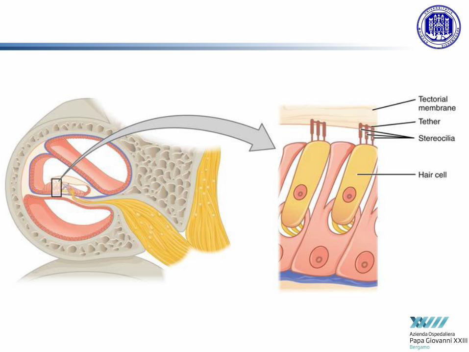
Auditory system

Auditory system
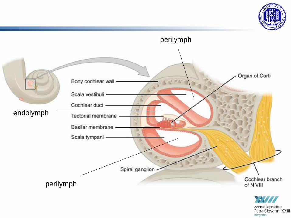
perilymph
perilymph
endolymph


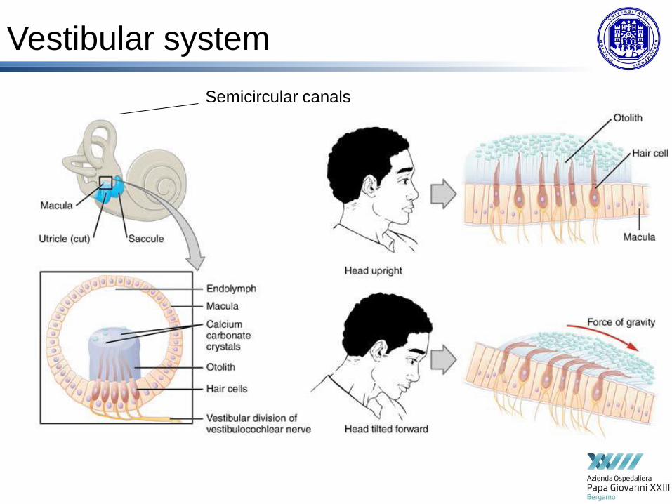
Vestibular system
Semicircular canals
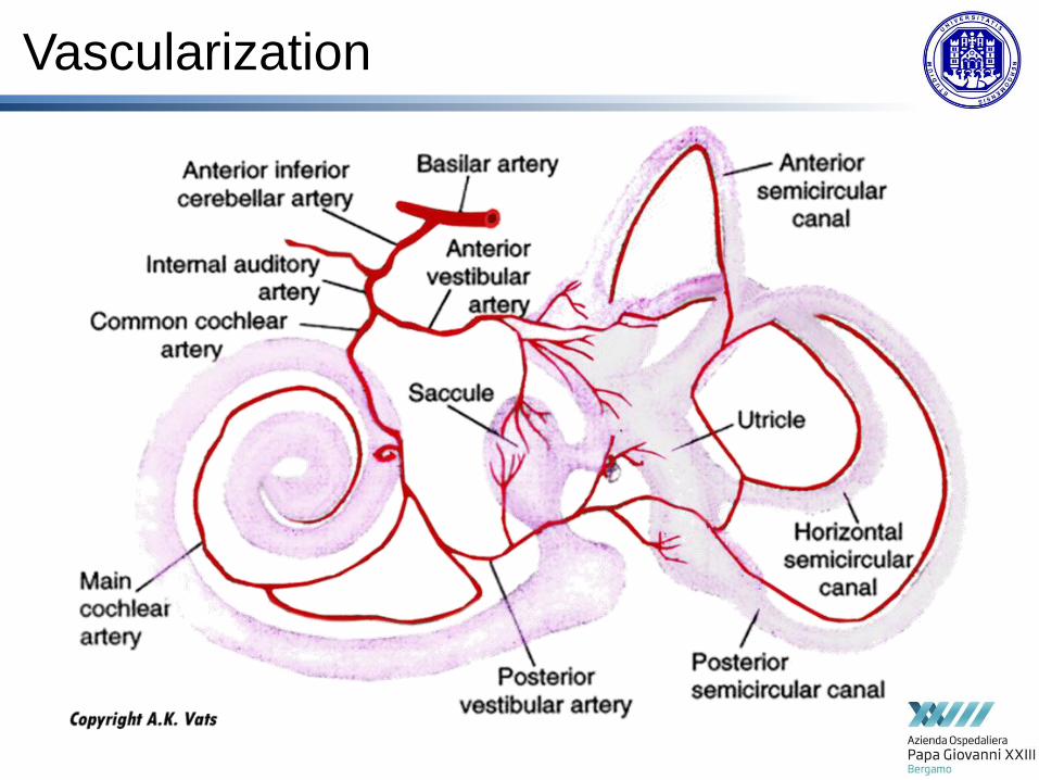
Vascularization

The eye
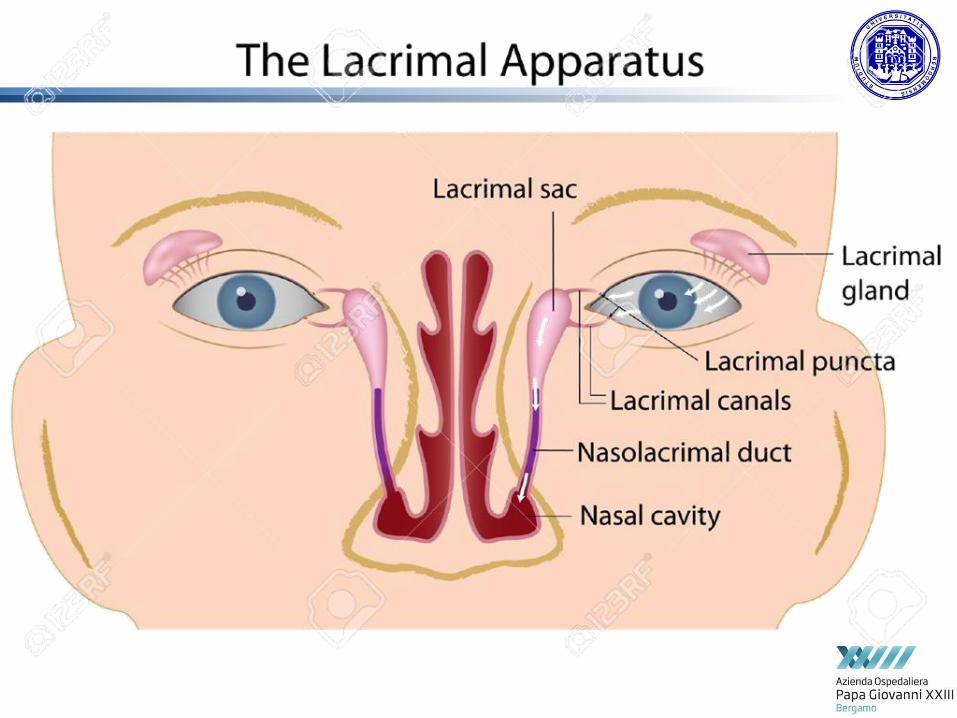

Lacrimal film
In humans, the tear film coating the eye, known as the precorneal film,
has three distinct layers, from the most outer surface
Name Container(s) Secretors Functions
Lipid
layer Oils
Meibomian
glands (or
tarsal
glands)
Coats the aqueous layer, provides
a hydrophobic barrier that envelops
tears and prevents their spilling onto
the cheek. These glands are found
among the tarsal plates. Thus, the tear
fluid deposits between the eye proper
and oil barriers of the lids.[3]
Aqueou
s layer
Water, electrolytes, and
other substances such as
proteins
(e.g., antibodies,[2] lipocali
n,lactoferrin, lysozyme,[4] a
ndlacritin)
Lacrimal
gland
Promotes spreading of the tear film, the
control of infectious agents and osmotic
regulation.
Mucous
layer Mucins
Conjunctiv
algoblet
cells
Coats the cornea, provides a
hydrophilic layer and allows for even
distribution of the tear film.


III
III
III
III
VI
IV
Extrinsic eye muscles


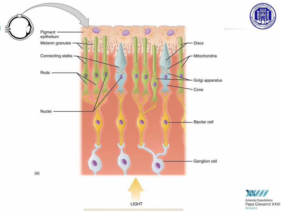


photoreceptors
photoreceptor proteins in the cell absorb photons, triggering a
change in the cell's membrane potential.
Rods Cones
Used for scotopic vision (vision under low light conditions)
Used for photopic vision (vision under high light conditions)
Very light sensitive; sensitive to scattered light
Not very light sensitive; sensitive to only direct light
Loss causes night blindness Loss causes legal blindness
Low visual acuity High visual acuity; better spatial resolution
Not present in fovea Concentrated in fovea
About 120 million rods distributed around the retina[12]
About 6 million cones distributed in each retina[12]
One type of photosensitive pigment Three types of photosensitive pigment in humans
Confer achromatic vision Confer color vision

photoreceptors

Fundus oculi
Macula lutea and
Fovea centralis (high concentration of rodes)
Optic disc
(no receptor)