
Atassie eredodegenerative ad esordio precoce: descrizione ...
91
0 Sede Amministrativa: Università degli Studi di Padova Dipartimento di SALUTE DELLA DONNA E DEL BAMBINO ___________________________________________________________________ DOTTORATO DI RICERCA IN MEDICINA DELLO SVILUPPO E SCIENZE DELLA PROGRAMMAZIONE CICLO XXVII TITOLO TESI Atassie eredodegenerative ad esordio precoce: descrizione del pattern di alterazione patologica mediante neuroimaging avanzato e studio neuropsicologico per la definizione di indicatori paraclinici utili al monitoraggi o dell’evoluzione o alla verifica di efficacia di trattamento. Coordinatore : Ch.mo Prof. Giuseppe Basso MD Supervisore :Ch.mo Prof. Andrea Martinuzzi MD PhD Dottoranda : Dr.ssa Marinela Vavla MD MSc
Transcript of Atassie eredodegenerative ad esordio precoce: descrizione ...
0
Sede Amministrativa: Università degli Studi di Padova
Dipartimento di SALUTE DELLA DONNA E DEL BAMBINO
___________________________________________________________________
DOTTORATO DI RICERCA IN
MEDICINA DELLO SVILUPPO E SCIENZE DELLA PROGRAMMAZIONE
CICLO XXVII
TITOLO TESI
Atassie eredodegenerative ad esordio precoce: descrizione del
pattern di alterazione patologica mediante neuroimaging
avanzato e studio neuropsicologico per la definizione di
indicatori paraclinici utili al monitoraggio dell’evoluzione o alla
verifica di efficacia di trattamento.
Coordinatore : Ch.mo Prof. Giuseppe Basso MD
Supervisore :Ch.mo Prof. Andrea Martinuzzi MD PhD
Dottoranda : Dr.ssa Marinela Vavla MD MSc
1
UNIVERSITY OF PADUA
DIPARTMENT OF WOMEN'S AND CHILDREN'S HEALTH
_________________________________________________________
SUBMITTED FOR THE COMPLETION OF THE
PHD DEGREE IN DEVELOPING MEDICINE AND PROGRAMMING SCIENCES
XXVII CYCLE
TITLE OF THE THESIS:
Early onset hereditary neurodegenerative ataxias: a description of the pathologic modification pattern using advanced neuroimaging techniques and a
neuropsychological study for the definition of paraclinical indicators in monitoring the disease progression and verification of treatment efficacy.
Coordinator: Prof. Giuseppe Basso MD Supervisor: Prof. Andrea Martinuzzi MD, PhD Candidate: Dr. Marinela Vavla MD, MSc
Started on January 2012.

2
Expected termination by March 2015
SUMMARY OF THE THESIS
Early onset Hereditary ataxias represent a group of genetic ally and clinically
heterogeneous conditions. The most important clinical pre sentation
symptoms are gait and limb ataxia, dysarthria and eye moveme nt
impairment, in addition to other non-neurological involve ment. Friedreich's
ataxia (FRDA) is the most common autosomal recessive ataxia in terms of
frequency and as a form with early onset.
Friedreich's ataxia (FRDA) is a progressive hereditary neu rodegenerative
condition caused by an autosomal recessively inherited GAA repeat in the
FXN gene.
We performed a multidisciplinary overview of FRDA integrat ing it with an
extensive cognitive and neuropsychological assessment. I n addition, we
used clinical measures and advanced tractography combined to functional
MRI (fMRI) to explore white matter (WM) connectivity and mot or dysfunction
in a cohort of FRDA patients. This study is intended to provid e a
multidisciplinary overview of the clinical condition inte grating it with a
comprehensive MRI protocol on FRDA patients compared to con trols. We
have designed an ongoing longitudinal study in order to be ab le to describe
the disease progression and to search for any poten tial biomarkers.
METHODS: Twenty one patients with a molecularly confirmed diagnosis of FRDA
were recruited. The patients were aged >12 years of age and had an early onset
and molecularly defined diagnosis of FRDA. All participants gave their written
informed consent. All patients underwent a full clinical (neurological and ataxia
scoring scales) and neuropsychological assessment (WISC III, WAIS R), specific
tests for the attentive, executive and memory functions and MMPI A for the
2
personality assessment. Seventeen FRDA patients and 13 healthy controls
underwent a neuroimaging study protocol on a 3T MRI scanner that included
advanced neuroimaging DTI and fMRI. After the pre-processing, a nonlinear
monoexponential model was used to calculate fractional anisotropy (FA), mean,
radial and axial diffusivity (MD, RD, AD) maps. Non-parametric voxel-based
permutations were performed on the WM maps regions of interest (ROI),
considering age and sex via a general linear model (GLM) with critical threshold
0.05 while correcting for multiple tests. An fMRI sequence was acquired during a
simple block design finger-tapping task. After a standard pipeline pre-process,
intra- and intergroup GLM analysis were conducted, considering age and sex
variables and also p < 0.001 threshold.
RESULTS: The cohort presents with an early age at onset (AAO) (10.6 ± 4.6,
range 4-20). The F:M ration was 16:5. The age at the visit was .9 ±10.3 years
(range 12-50) and disease duration was 16.3 ± 8.8 years (range 3-32). FRDA
cohort presented as homozygous for the GAA repeat expansion in 96%. The mean
GAA repeat expansion in the short allele was 653.7 ± 221 (range 170-946) that
correlated negatively with AAO. In most cases the onset was with ataxia, gait
clumsiness, and scoliosis, but few with asymptomatic cardiomyopathy and pes
cavus. Vibratory sense was impaired in all the patients, with milder deficits in the
other senses. Dysarthria was present in all patients. Muscle strength and tone
were impaired in almost all the patients. One of them presented with a spastic
ataxia with retained DTR. The pyramidal signs were present in 57%. Nystagmus
was present in 61.9%. Half of the patients were wheelchair bound. Few patients
developed diabetes mellitus. Cardiac involvement was registered in 76.2%, mostly
presented as ventricular, septal or apical hypertrophy, but few with arrhythmias
and valve prolapsed. The pulmonary system was involved in 28.6% of the patients
3
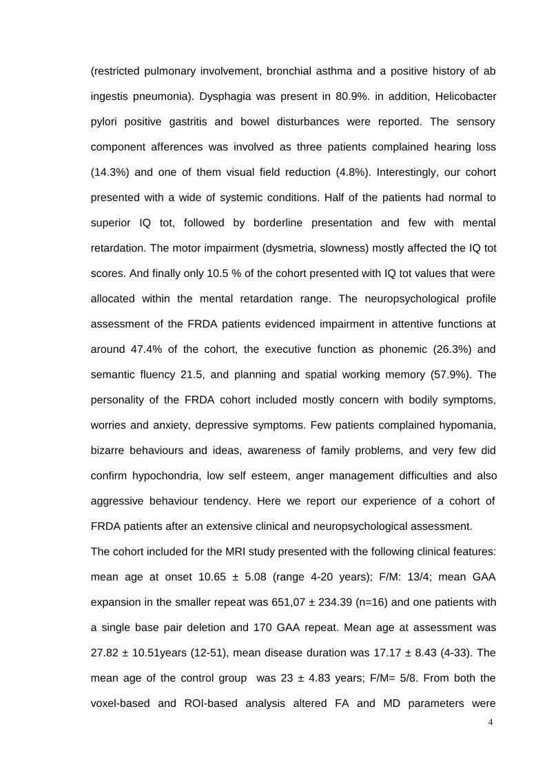
(restricted pulmonary involvement, bronchial asthma and a positive history of ab
ingestis pneumonia). Dysphagia was present in 80.9%. in addition, Helicobacter
pylori positive gastritis and bowel disturbances were reported. The sensory
component afferences was involved as three patients complained hearing loss
(14.3%) and one of them visual field reduction (4.8%). Interestingly, our cohort
presented with a wide of systemic conditions. Half of the patients had normal to
superior IQ tot, followed by borderline presentation and few with mental
retardation. The motor impairment (dysmetria, slowness) mostly affected the IQ tot
scores. And finally only 10.5 % of the cohort presented with IQ tot values that were
allocated within the mental retardation range. The neuropsychological profile
assessment of the FRDA patients evidenced impairment in attentive functions at
around 47.4% of the cohort, the executive function as phonemic (26.3%) and
semantic fluency 21.5, and planning and spatial working memory (57.9%). The
personality of the FRDA cohort included mostly concern with bodily symptoms,
worries and anxiety, depressive symptoms. Few patients complained hypomania,
bizarre behaviours and ideas, awareness of family problems, and very few did
confirm hypochondria, low self esteem, anger management difficulties and also
aggressive behaviour tendency. Here we report our experience of a cohort of
FRDA patients after an extensive clinical and neuropsychological assessment.
The cohort included for the MRI study presented with the following clinical features:
mean age at onset 10.65 ± 5.08 (range 4-20 years); F/M: 13/4; mean GAA
expansion in the smaller repeat was 651,07 ± 234.39 (n=16) and one patients with
a single base pair deletion and 170 GAA repeat. Mean age at assessment was
27.82 ± 10.51years (12-51), mean disease duration was 17.17 ± 8.43 (4-33). The
mean age of the control group was 23 ± 4.83 years; F/M= 5/8. From both the
voxel-based and ROI-based analysis altered FA and MD parameters were
4
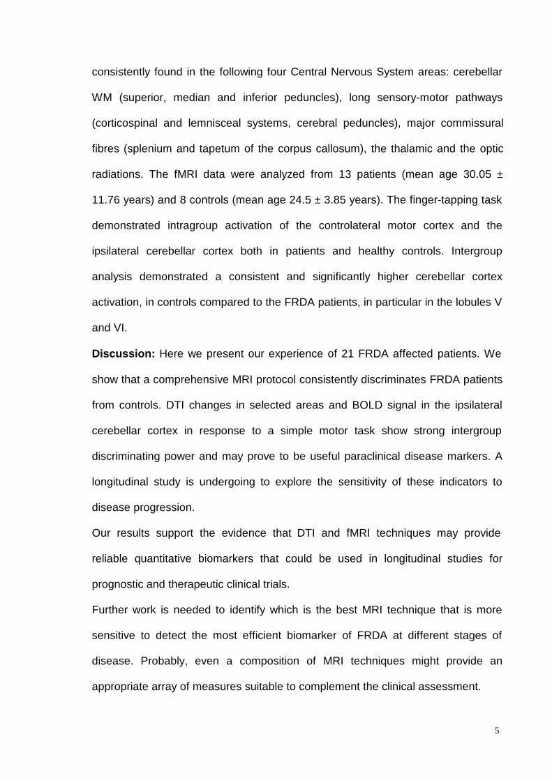
consistently found in the following four Central Nervous System areas: cerebellar
WM (superior, median and inferior peduncles), long sensory-motor pathways
(corticospinal and lemnisceal systems, cerebral peduncles), major commissural
fibres (splenium and tapetum of the corpus callosum), the thalamic and the optic
radiations. The fMRI data were analyzed from 13 patients (mean age 30.05 ±
11.76 years) and 8 controls (mean age 24.5 ± 3.85 years). The finger-tapping task
demonstrated intragroup activation of the controlateral motor cortex and the
ipsilateral cerebellar cortex both in patients and healthy controls. Intergroup
analysis demonstrated a consistent and significantly higher cerebellar cortex
activation, in controls compared to the FRDA patients, in particular in the lobules V
and VI.
Discussion: Here we present our experience of 21 FRDA affected patients. We
show that a comprehensive MRI protocol consistently discriminates FRDA patients
from controls. DTI changes in selected areas and BOLD signal in the ipsilateral
cerebellar cortex in response to a simple motor task show strong intergroup
discriminating power and may prove to be useful paraclinical disease markers. A
longitudinal study is undergoing to explore the sensitivity of these indicators to
disease progression.
Our results support the evidence that DTI and fMRI techniques may provide
reliable quantitative biomarkers that could be used in longitudinal studies for
prognostic and therapeutic clinical trials.
Further work is needed to identify which is the best MRI technique that is more
sensitive to detect the most efficient biomarker of FRDA at different stages of
disease. Probably, even a composition of MRI techniques might provide an
appropriate array of measures suitable to complement the clinical assessment.
5
Acknowledgements
Dedicated to all my patients
Thank you to the supervisor, Prof. Andrea Martinuzzi, for your expertise and
mainly for the mentoring modalities, for the constructive feedbacks, for providing a
guided supervision and letting me work independently.
Thank you to Drs Petacchi and Montanaro, for your costant advice,
encouragement and expertise.
Thank you to all my collaborators from Conegliano, Pieve di Soligo and Bosisio
Parini (colleagues, psychologists, phyosiotherapists, occupational therapists,
speech and language therapists, CUP-programming centre, CED, nurses,
engineers and technicians) for patiently putting up with the requests of this project,
timelines and for your valuable feedbacks.
Thank you to all the patients that participated in this project.
Thank you to the “Ogni Giorno per Emma – ONLUS” association for funding, and
especially to Mrs Bertazzon for your help with the recruitment, for your costant
presence and for persistently believing in our efforts.
Thank you to Claudia, Giulia, Pellegrina, Laura, Silvia B, you know why.
Thank you to Egrina, Tanja, Valentina, Tixhe for you were there when I needed the
most and I didn't need to ask.
Thank you to my parents and Klajdi, for you didn't complain about my absences,
for you gave me strength to keep going.
6
CHAPTER 1: INTRODUCTION TO
HEREDODEGENERATIVE ATAXIAS
Early onset Hereditary ataxias represent a group of genetically and clinically
heterogeneous conditions. The most important clinical presentation symptoms are
gait and limb ataxia, dysarthria and eye movement impairment, in addition to other
non-neurological involvement. The mode of inheritance may be autosomal
dominant (AD), autosomal recessive (AR), X-linked or diaginic inheritance.
Friedreich's ataxia (FRDA) is the most common autosomal recessive ataxia in
terms of frequency and as a form with early onset, is followed by Ataxia-
Telenagectasia (A-T), ataxia with Oculomotor Apraxia type 1 (AOA1) and type 2
(AOA2) (Ruano et al., 2014). FRDA is the object of this study.
History of Friedreich’s Ataxia
FRDA was initially described by Nicholaus Friedreich. He described a cohort of 8
patients, members of 3 families. He published three papers in a cohort of 14 years
(1863a, 1863b, 1863c, 1876, 1877). Later on, Brousse proposed to name this
condition after Nicholaus Friedreich (Brousse, 1882). In 1890 Ladame presented a
review on a very large cohort of patients (n=165) pointing out the difficulty of
diagnosis (Ladame, 1890). There had been several controversies when diagnosing
and reporting FRDA. The atypical forms of FRDA had been described since 1897,
when Hodge described 3 siblings with increased DTR (Hodge, 1897), while
Sherman (1934) described a spastic component in patients presenting with
FRDA. Wilson firmly reported the diagnosis of FRDA with retained DTR (Wilson,
1940). But it was Bell and Carmichael (1939) that had introduced the idea that a
loss of DTR was an early sign in FRDA. In 1981, Harding described a large cohort
7
of 115 patients (Harding, 1981) presenting an extensive elaboration of clinical
presentation and course of FRDA patients and two years later (1983) presenting a
refined classification and diagnostic criteria.
Epidemiologic studies
The global prevalence of AR ataxias ranges from 0.0-7.2: 100.000, with an
average of 3.3: 100.000 (1.8-4.9: 100.000) and highlighting FRDA as the most
frequent form (Ruano et al., 2014).
The geographical distribution of FRDA patients is situated mostly in Caucasians
areas, rare in sub-Saharan African and very rare in the far East. There is the
hypothesis of a founding mutation origin in Western Europe that appears to be to
approximately 682 ± 203 generations ago corresponding to the Paleolythic period
(Vankan, 2013).
The prevalence values of FRDA are variable, they range in Caucasian population
1:20.000 to 1:50.000. The highest prevalence is reported in Spain (1:21.000);
secondly in North Ireland (1:23.000) and the third highest frequency is in France
(1:43.000). In East Europe, the prevalence values are extremely small, and they
record as follows: Russia 1:330.000; the Scandinavia records are in the range of
the minimun FRDA prevalence with Sweden 1:420.000 and Finland 1: 750.000
(Vankan, 2013). The Italian prevalence was estimated in 3 major epidemiological
publications that were conducted in North Italy with 1:83.000 (Leone et al., 1990),
South Italy 1:90.000 (Filla et al., 1981) and for the whole Italian patients 1:90.909
(Romeo et al., 1983) with an incidence of around 1:25.449 new cases per year.
The carrier frequency is reported to be 60-1:100. 8
The observed FRDA distribution in Europe co-localizes with the a chromosomal
marker gradient related to R1b that is believed to be present within West Europe.
The chromosomal gradient is apparently due to the paleolithic migrations out of
Franco-Cantabrian Ice age refugees and Neolithic migration entering West Europe
with the advance of agriculture (Vankan 2013).
Genetics
In 1996 there was a publication by Campuzano et al (1996) reporting a linkage of
FXN gene to the FRDA. The FXN gene was reported in the critical region for the
FRDA locus on chromosome 9q13-q21.1. The gene contains 5 exons (1, 2, 3, 4,
5a) and two splicing form (exons 5b and 6), and also 4 introns. It codifies for a
protein called frataxin, expressed in 2 isomers according to whether either exon 5a
(210 amino acids) or exon 5b (170 amino acids) are transcribed. This protein
apparently has a high level of expression in the heart, an intermediate level of
expression in the liver, skeletal muscle and pancreas, and very low level in the
spinal cord, cerebellum and cerebral cortex. Campuzano et al. reported, after a
screening of 184 FRDA patients, three point mutations from three families of
French, Spanish and Southern Italian origin (1996). They found that in 79
molecularly defined FRDA patients, including 5 with point mutations, there were
GAA repeat expansion that appeared to be disease correalated. About 98% of
FRDA had GAA repeat expansions. The sizes of GAA repeat were between 200
and 900, mostly containing 700-800 repeats. In the following year, 1997 the latter
research group (Campuzano et al., 1997) published the functional work on FXN
gene by reporting the reduction of FXN protein levels in FRDA patients and also
localizing the protein within the cell giving rise to hypothesis on the probable
function of the protein. FRDA appears to be due to a loss of function of the protein.
9
The FXN gene has its homologous in nematode, yeast and mouse (Campuzano et
al., 1996; Koutnikova et al., 1997). The FXN gene exons 3-5 encode for the
domain of the protein with the highest level of evolutionary conservation
(Campuzano et al., 1996).
Themolecular weight of frataxin is a 18 kDa. The N-terminal epitope is not found
within the mitochondria, suggesting that the protein goes through a pre-processing
through a proteolytic cleavage into its mature form containing the C-terminal that enters
the mitochondria. An important finding of the same group, was that the FXN protein was
found in the inner mitochondrial membrane, suggesting a rationale for the impairment of
cells with a high energy consumption level such as neurons. This finding derived from
ransfected and non-transfected cells that were everexpressing FXN gene.
These important findings opened the door to a classification of FRDA under the umbrella
of mitochondrial disorders, that appeared to be caused by a loss of function of a nuclear
encoded protein (Campuzano et al., 1997) due to instabilityof normal repeats (NR) from
which new expanded repeats are generated.
Pathological changes
The clinico-anatomic correlations in FRDA are representedas a combination of the
developmental and degenerative processess in dorsal root columns and the sensory nerves,
progressive destruction of the dentate nucleus (DN), atrophy of Betz cells and
degenerationof the corticospinal tracts (Figure 1) (Koeppen and Mazurkiewitcz, 2013).
The frataxin protein is synthesized as a precursor of 210 aminoacids imported into the
mitochondrion. The mature form is fully functional for cell survival.
Frataxin is an iron-binding and an aggregate formation protein. In addittion, frataxin
apparently interacts with ferrochelatase, that is involved in the enzymatic reaction that
leads to the final step of heme byosyntheisis by inserting iron into the porphyrin (Foury
10
and Cazzalini, 1997; Lesuisse et al., 2003). Lastly, frataxin appears to be linked to the
mitochondrial aconitase, subunits of complex II of the respiratory chain and several
chaperones (Bulteau et al., 2004; Gonzales-Cabo et al., 2005; Shan et al. 2007).
Frataxin binds iron and is required for the synthesis of iron-sulphur clusters and, thereby,
for the synthesis of enzymes in the respiratory chain complexes I – III and aconitase
(Pastore and Puccio, 2013).
The CNS involvement is viewed as a dying-back neuropathy of the following: long
ascending and descending tracts of spinal cord, large sensory fibres of peripheral nerves,
posterior sensory fibres of peripheral nerves. In addition, dentate nucleus and optic nerve
tracts are involved.
Classification and diagnosis
The first effort in presenting diagnostic criteria was madefrom Geoffroy and collaborators
(1976). They divided their 50 patients with a diagnosis of FRDA into 4 groups as follows:
complete typical FRDA, incomplete atypical FRDA, atypicalFRDA and no FRDA. They
proposed some commendable diagnostic criteria, but since they were not fully applicable
to all the FRDA patients due to the early onset of the cohort and the homogeneity of the
study population (10 French-Canadian families from Quebec), were subsequently refined
(Harding, 1983). She elaborated a classification from her previous works on 90 families
with a total of 115 FRDA diagnosed patients (Harding, 1981).
Harding reported early onset (before 25 years of age) FRDA patients with presentation
symptoms mainly limb and truncal ataxia, and absent DTR as consistent diagnostic
criteria. In addition, she described other symptoms that would eventually develop during
the disease progression such as dysarthria, pyramidal signs, and sensory impairment (sense
of position and vibration). With the advent of the gene discovery, the suspected FRDA
diagnosis was confirmed by genetic detection of pathogenicvariants in the FXN gene in
11
both alleles. This allowed the confirmation of the cases that presented with atypical
symptoms such as very early or very late onset, retained or brisk reflexes spasticity or
limited progression (Durr et al., 1996; Filla et al., 2000; McCabe et al., 2000).
The clinical diagnosis of FRDA should be suspected in the presence of the combination of
the following findings: progressive ataxia (gait and limbs), absent muscle DTR in LL
(inconstant finding), dysarthria, an onset generally before 25 years of age and in an
autosomal recessive inheritance manner. In addition, skeletal deformities (scoliosis, pes
cavus), corticospinal tract (CST) involvement (LL weakness, Babinski), diabetes mellitus
or glucose intollerance, hypertrophyc cardiomyopathy, optic atrophy or deafness. In
addition, a series of instrumental examinations are important in order to complete the
diagnostic process such as cerebral magnetic resonance (MRI) visual evoked potentials,
motor and sensory nerve conduction velocities.
Clinical features
The residual amount of FXN protein is reported in the range of4-29% in patients as
compared to the levels of healthy controls, and that these levels were inversely correlated
to the GAA repeat size of the short allele (Campuzano et al., 1997).
Early studies reported that the GAA repeat expansion is negatively correlated with the age
at onset (AAO) and positively correlated with disease progression (Campuzano et al.,
1997, Filla et al., 1996; Durr et al., 1996; Montermini et al., 1997; Lamont et al., 1997;
Monros et al., 1997) suggesting a role of GAA repeat expansion in the FXN protein
residual levels and subsequently in the disease severity implication. Similarly, positive
correlation have been shown between GAA repeat expansion and incidence of
cardiomyopathy.
The normal chromosomes have fewer than 33 GAA repeat expnasion. The smallest
syntomatic GAA repeat expansion has been reported to be 44. FRDA patients present
12
usually with 600-900 GAA repeat expansion, with minimum andmaximum pathogenic
repeat expansion reported to be 70 and 1700, respectively (Pandolfo, 2001).
Regarding the AAO, there have been reported two ages of onsetin FRDA. Most of the
reports allocate the onset to be early, thus before the age of25 years old. However, the late
onset cases have been reported as well.
Generally, FRDA is classified in two different phenotypic representation, the classical and
atypical phenotype. The latter incorporates the low-onsetFRDA (LOFA) and very-low-
onset FRDA (VLOFA), Acadian type and early onset FRDA.
Classical Phenotype.
The AAO is generally around puberty, reported to be 10.5 ± 7.4years and 15.5 ± 8 years
(Harding, 1981; Filla et al., 1990). The same authors reported a modal age at onset at
around 10-12 years and 12-15 years, respectively. Harding (1981) described cases with a
very early onset, before age of 5 years old, describing thosecases of FRDA patients as the
ones that rapidly deteriorate, while other associated early AAO with a larger size of the
short allelle (GAAsr), a more severe phenotype, a faster progression of disability and
higher incidence of non-neurological features such as cardiomyopathy, diabetes mellitus
(DM) and pes cavus (Durr et al., 1996; Schols et al., 1997).
The presenting symptoms are usually associated with the gait and limb ataxia, clumsiness
(Harding, 1981; Filla et al., 1990; Durr et al., 1996; Delatycki et al., 1999). Nevertheless,
scoliosis and pes cavus might be the first symptom to be observed by the clinician, leading
to a necessary further neurological assessment.
The neurological features are mainly represented by gait and lower limb (LL) ataxia. The
ataxic signs are of mixed origin, such as spinocerebellar degeneration, peripheral sensory
neuropathy, cerebellar and vestibular pathology (Corben and Delatycki, 2012). The upper
limb (UL) ataxia is reflected in the impairment of the manualdexterity, difficulty on
13
handwriting, use of cutlery, washing, carrying objects. The LL ataxia is observed as
impossibility or difficulty in the heel-to-shin task performance. Weakness and muscle
wasting are usually noted later in life.
DTR are usually absent in the LL, and mostly absent in the UL. The extensor plantar
reaction is an early pyramidal sign. The muscle tone is initially normal or reduced,
progressively decreasing. While spasticity appears to be associated to LL and is
responsible for associated symptoms, such as pain, contractures and discomfort. The
sensory system is almost always involved, predominantly with the vibration and joint
position sense impairment. Visual system appears to be involved, with mostly fixation
instability, less commonly nystagmus and smooth pursuit movements impairment,
decreased visual acuity and increased pattern visual-evoked potential latency (Durr et al.,
1996; Fortuna et al., 2009). Dysarthria is a common and earlysymptom, while dysphagia
develops in advanced stages and hearing loss appears to be a common but understated
problem. The bladder hyperactivity is common in FRDA, on thecontrary the bowel
problems cause fewer problems (Parkinson et al., 2013).
The non-neurological features involve heart and pancreas (Figure 2). Hypertrophic
cardiomyopathy or left ventricle hypertrophy (LVH) eitherconcentric or asymetric septal
hypertrophy (Goeffroy et al., 1974; Filla et al., 1990; Durret al., 1996; McCabe et al.,
2000). Some patient present with EKG alterations such as T wave inversion, ST-segment
abnormalities or arrhythmias (Dutka et al., 1999; Bourke and Keane, 2011).
Diabetes mellitus (DM) is another non-neurological feature in FRDA that appears to be
either due to insulin-resistance or decrease insulin secretion (Finocchiaro et al., 1988). The
incidence of DM in FRDA cohorts was reported to be around 6-32% (1976; Harding,
1981; Filla et al., 1990; Schols et al., 1997; Delatycki et al., 1999; McCabe et al., 2000;
Durr et al., 1996).
14
Importantly, skeletal deformities are present in FRDA. Scoliosis is a common feature,
occasionally the presenting symptom. It is commonly mild when the AAO is relatively late
(Parkinson et al., 2013).
Foot abnormalities are present in about 55-90% of the cases reported and consist in pes
cavus, talipes equinovarus and pes planus as well (Harding,1981; Geoffroy et al. 1976;
Ackroyd et al. 1984; Filla et al. 1990; Durr et al. 1996; Schols et al. 1997; Delatycki et al.
1999; McCabe et al., 2000).
Non classical phenotype
There have been, however, described cases with AAO after 25 years of age. In particular,
“Late onset FRDA” (LOFA) form has been reported to have a meanage at onset at around
28.8 years (range of 25.5–48) (Bhidayasiri et al., 2005; Arnold et al., 2006). LOFA
appeared to have a milder phenotypic representation with retained LL DTR. The latest
AAO reported have been allocated at the seventies (Gallimanet al, 2008; Stolle et al.,
2008) and to our knowledge at 82 years old (Alvarez et al., 2013). This form is usually
classified as “Very late onset FRDA” (VLOFA) with a AAO after40 years old. Another
atypical phenotype is FRDA with retained reflexes, known asFARR (Klockgether et al.,
1996; Coppola et al., 1999).
Another atypical clinical representation of FRDA was reported in from Richter et al.
(1996) in a series of patients deriving from 10 Acadian families in Canada. Their clinical
presentation overlapped the classical phenotype, but lacking cardiomyopathy and DM,
and eventually displayed retained or increased DTR.
About 98% of FRDA patients have a GAA repeat expansion in a homozigous pattern
(Campuzano et al., 1996). In addition 2-4 % of FRDA patients present with either FXN
point mutation or deletion. The former mutations might be either truncating or missense,
and appear to be responsible for a milder phenotype (Cossee et al., 1999). The latter
15
presentation is a rare presentation and usually associatedwith an earlier onset and a more
severe phenotype (Zulke et al., 2004; Anheim et al., 2012).
NPS studies
In 1976, Geoffroy et al. (1976) mentioned decreased IQ as a clinical criteria for FRDA.
However, despite slowed information processing in FRDA, cognition does not appear to be
affected (Corben et al., 2006). From a study of 13 FRDA patients (Mantovan et al., 2006),
the IQ profile was characterised by concrete thinking associated to impairment in concept
formation and visuospatial reasoning. Other findings of DeNobrega et al. (2007) that
related to impairment in phonemic and action fluency, led the authors to claim primary
prefrontal or cerebello-prefrontal dysfunction. Furtherstudies, conducted by Corben and
collaborators (Corben et al 2010, 2011a, b, c), hypothesised the disruption of cerebro-
ponto-cerebello-thalamo-cerebral loops to explain the cerebellar impairmet that were
probably causative of the difficulty in accommodating unexpected movements, difficulty
in the movement initiation without a direct visual cue, and impairments in the reaction
time to incongruent stimuli. Sustained volitional attention and working memory is
impaired in FRDA (Klopper et al., 2011). Lately, findings in36 FRDA patients, confirmed
motor and mental speed, conceptual thinking, verbal fluency, acquisition of verbal
information, use of semantic strategies in retrieval and action naming deficits. These
findings were suggestive of parieto-temporal dysfunction(Nieto et al., 2012). In summary,
the cerebro-cerebellar circuits may be functionally important in FRDA, and the eventual
interruption is to be regarded as causative in FRDA.
MRI studies
Magnetic resonance imaging (MRI) studies reflec the clincial features. MRI findings show
cervical cord atrophy, posterior column atrophy. In early stages, there might be either no
involvement of cerebellum or brainstem or minimal atrophy of the superior vermis and
16
medulla oblongata (De Michele et al., 1995). Additional MRIstudies, such as VBM
studies conducted by Della Nave et al., (2008) report symmetrical volume loss in the
dorsal medulla, inferomedial cerebellar hemispheres, rostral vermis and dentate regions,
which appeared to correlated with disease duration and severity. Another study, reported
correlations between superior cerebellar peduncle atrophy and clincal severity, AAO and
DD (Akhalagi et al., 2011).
Treatment in FRDA
There is no cure for FRDA. There have been many clinical trials that have tried different
molecules ( Fig. 3). Among them antioxidants have been attempted.
Coenzyme Q10 and vitamin E were used in association, demonstrating an improvement in
ATP production for either 6 months (Lodi et al., 2001) and 47 months (Hart et al., 2005).
The former findings were oriented toward ATP improvement inthe heart and skeletal
muscle; the latter confirmed these findings by reporting improvement in bioenergetics and
in cardiac function. Conversely, another study high dosageof coenzyme Q10 and vitamine
E controlled to low dosage coenzyme Q10 failed to demonstrate any intergroup differences
in ICARS score (Cooper et al., 2008).
Idebenone, a short chain analog of coenzyme Q10, was thoughtlead to left ventricular
hypertrophy reduction in some studies (Hausse et al., 2002,Buyse et al., 2003; Mariotti et
al., 2003) but it was not confirmed by others (Lagedrost et al., 2011). Likewise, the
neurological benefits of idebenone compared to placebo andmeasured by ICARS from a
phase II clinical trial were reported by Di Prospero et al. (2007) and rejected by Lynch et
al. (2010) conducted by a phase III study.
Controversial findings were reported in three case reportstreated with intra muscle
injection of thiamine (Costantini et al., 2013).
17
Iron chelators, as deferiprone, have been used in an open label study showing apparently
reduction of iron in the dentate nucleus (DN) and neurological benefits (Boddaert et al.,
2007). Consistently, intraventricular septum thickness reduction was observed from an
open label study combining deferiprone and idebeone (Velasco-Sanchez, et al., 2011) but
no ataxia score significant change was observed. Attempts to increase the levels of frataxin
protein have included studies in cellular models trying different molecules such as hemin,
butyric acid, and erythropoietin (Sturm et al., 2005; Sarsero et al., 2003). Clinical studies
with erythropoietin as an open label study (Boesch et al., 2007) led to significant decrease
levels of oxidative markers, while a six-months placebo-controlled study did not identify
any clinical benefit (Mariotti et al., 2012).
An upregulation of FXN expression was tried by using histonedeacetylase inhibitors
(HDACi) (Herman et al., 2014) as Rai and colleagues (Rai et al., 2008) demonstrated how
compound 106, HDACi analogue led to restored frataxin levels in heart and central
nervous system (CNS) in FRDA mouse model. A phase I clinical trial of RG2833 was
concluded (Gottesfeld et al 2013). Similarly, another molecule, class III HDACi led to
increase in frataxin expression in FRDA cell and mouse models (Chan et al., 2013) while a
clinical open label study reported findings of increase levels of FXN transcript
approximately equivalent to the asymptomatic carriers (Libri et al., 2014). Soragni et al
(2014) demonstrated that class I HDACi can induce epigenetic changes, such as increase
in FXN mRNA and acetylation of a key residue either in the blood of FRDA patients or in
the iPSC-derived neuronal cells and PBMC of treated patients providing proof of concept
for epigenetic therapy.
Finally, interferon gamma (yIFN) seems another molecule important in upregulating
frataxin levels in cellular and mouse models of FRDA with prevention of dorsal root
ganglion (DRG) in dorsal root ganglia and motor performanceimprovement (Tomassini et
18
al., 2012), with ongoing phase I clinical trials. Seyer et al. (2014) published their findings
of an open label trial of yIFN for 4 months in 12 children with FRDA, demonstrating small
significant changes of frataxin levels in erythrocytes, PBMC and platelats and FARS score
changes equivalent to a 18 months improvement.
An attempt to try gene therapy in conditional cardiac FXN deletion mouse model,
demonstrated the FXN delivered intravenously via an adeno-associated virus vector
prevented and reversed cardiomyopathy (Perdomini et al., 2014).
Lately, Corben et al. (2014) have published recommendations addressing almost all the
areas of health issues (neurological, heart, scoliosis, diabetes mellitus, genetic issues,
pregnancy and quality of life issues) in patients with FRDA.These recommendations are
generated from the evidence of systematic reviews, from randomized clinical trials (RCT),
from comparative studies with control group or historical control and from case series.
The purpose of this study
This study is intended to provide a multidisciplinary overview of the clinical condition
integrating it with a comprehensive MRI protocol on FRDA patients compared to controls.
We have designed a longitudinal study in order to be able to describe the disease
progression and to search for any potential biomarkers.
19
CHAPTER 2: Clinical and neuropsychological assessment in the
cohort of Friedreich's Ataxia patients
Abstract
Friedreich's ataxia (FRDA) is an autosomal recessive (AR) progressive hereditary
neurodegenerative disorder. The prevalence is reported 2-5:100.000 in the Caucasian
populations. Around 98% of FRDA patients present with GAA repeat expansion. The
clinical diagnosis of FRDA should be suspected in the presence of the progressive ataxia,
absent muscle deep tendon reflexes, dysarthria, early onset and an AR transmission. In
addition, skeletal deformities, pyramidal involvement, diabetes mellitus, cardiac
hypertrophy, optic atrophy atrophy or deafness can be found. Specific neuropsychological
profiles including executive and memory deficits, have been detected in FRDA. We
performed a multidisciplinary overview of the clinical condition integrating it with an
extensive cognitive and neuropsychological assessment. Twenty one patients with a
molecularly confirmed diagnosis of FRDA were recruited. The patients were aged >12
years of age and had an early onset and molecularly defined diagnosis of FRDA. All
participants gave their written informed consent. All patients underwent a full clinical
(neurological and ataxia scoring scales) and neuropsychological assessment (WISC III,
WAIS R), specific tests for the attentive, executive and memory functions and MMPI A
for the personality assessment. The cohort presents with anearly age at onset (AAO) (10.6
± 4.6, range 4-20). The F:M ration was 16:5. The age at the visit was .9 ±10.3 years (range
12-50) and disease duration was 16.3 ± 8.8 years (range 3-32). FRDA cohort presented as
homozygous for the GAA repeat expansion in 96%. The mean GAA repeat expansion in
the short allele was 653.7 ± 221 (range 170-946) that correlated negatively with AAO. In
most cases the onset was with ataxia, gait clumsiness, and scoliosis, but few with
20
asymptomatic cardiomyopathy and pes cavus. Vibratory sense was impaired in all the
patients, with milder deficits in the other senses. Dysarthria was present in all patients.
Muscle strength and tone were impaired in almost all the patients. One of them presented
with a spastic ataxia with retained DTR. The pyramidal signswere present in 57%.
Nystagmus was present in 61.9%. Half of the patients were wheelchair bound. Few
patients developed diabetes mellitus. Cardiac involvement was registered in 76.2%, mostly
presented as ventricular, septal or apical hypertrophy, but few with arrhythmias and valve
prolapsed. The pulmonary system was involved in 28.6% of thepatients (restricted
pulmonary involvement, bronchial asthma and a positive history of ab ingestis
pneumonia). Dysphagia was present in 80.9%. in addition, Helicobacter pylori positive
gastritis and bowel disturbances were reported. The sensory component afferences was
involved as three patients complained hearing loss (14.3%)and one of them visual field
reduction (4.8%). Interestingly, our cohort presented with a wide of systemic conditions.
Half of the patients had normal to superior IQ tot, followed by borderline presentation and
few with mental retardation. The the motor impairment (dysmetria, slowness) mostly
affected the IQ tot socres. And finally only 10.5 % of the cohort presented with IQ tot
values that were allocated within the mental retardation range. The neuropsychological
profile assessment of the FRDA patients evidenced impairment in attentive functions at
around 47.4% of the cohort, the executive function as phonemic (26.3%) and semantic
fluency 21.5, and planning and spatial working memory (57.9%). The personality of the
FRDA cohort included mostly concern with bodily symptoms, worries and anxiety,
depressive symptoms. Few patients complained hypomania, bizarre behaviours and ideas,
awareness of family problems, and very few did confirm hypochondria, low self esteem,
anger management difficulties and also aggressive behaviour tendency. Here we report our
21
experience of a cohort of FRDA patients after an extensive clinical and
neuropsychological assessment.
Introduction
Friedreich's ataxia (FRDA) is a hereditary neurodegenerative disorder trasmitted in an
autosomal recessiva (AR) manner. FRDA was initially described by Nicholaus Friedreich
(1863a). The prevalence is reported 2-5:100.000 in the Caucasian populations. The
prevalence in the whole Italian population is reported to be1:90.909 (Romeo et al., 1983)
with an incidence of around 1:25.449. The carrier frequencyis 60-1:100. Campuzano et al.
(1996) discovered that FXN gene is linked to FRDA. Around 98%of FRDA patients
present with GAA repeat expansion in both alleles. The pathological repeat size ranges
from 66 to 1700. The FXN gene encodes for the frataxin proteinwhich is entangled in the
synthesis of enzymes involved in the respiratory chain complexes I – III and aconitase
(Pastore and Puccio, 2013). The neuropathological findings are in line with a degeneration
in the dorsal root ganglia (DRG), sensory nerves, progressive destruction of the dentate
nucleus, atrophy of Betz cells and degeneration of the corticospinal tracts (CST) Koeppen
and Mazurkiewitcz, 2013). Harding (1981) described a largecohort of patients and
subsequently refined the diagnostic criteria for FRDA (Harding et al.,1983).
The clinical diagnosis of FRDA should be suspected in the presence of the combination of
the following findings: progressive ataxia of gait and limbs, absent muscle deep tendon
reflexes (DTR) in the lower limbs (LL), dysarthria, onset before 25 years and an AR
transmission. In addition, skeletal deformities (scoliosis, pes cavus), CST involvement (LL
weakness, Babinski sign), diabetes mellitus (DM) or glucose intolerance, hypertrophyc
cardiomyopathy, optic atrophy or deafness can be found. With the gene discovery, the
FRDA can be molecularly confirmed.
22
Specific neuropsychological profiles including executive and memory deficits, have been
detected in FRDA (Mantovan et al. 2006; Nieto et al. 2012) indicating parieto-temporal
dysfunctions. The personality of FRDA patients has been characterized by increased
irritability, poor impulsive control, reduced defensiveness and a poor-self-presentation
(Mantovan et al. 2006).
The aim of this study was to explore the clinical presentation of the FRDA cohort afferent
into our centres. We investigated our cohort via a thorough neurological and
neuropsychological assessment. This study is intended to provide a multidisciplinary
overview of the clinical condition integrating it with an extensive cognitive and
neuropsychological assessment.
Methods
Participants
Twenty two patients with a molecularly confirmed diagnosisof FRDA were recruited at
the Research Centres “Eugenio Medea” in Conegliano/Pieve di Soligo (Treviso) and
Bosisio Parini (Lecco, Drs. Grazia D'Angelo, Erika Brighina) and in Bologna (Dr Valerio
Carelli) between 2011 and December 2014. The patients were aged >12 years of age and
had an early onset and molecularly defined diagnosis of FRDA. All participants, but two,
were native Italians, mostly originating from Central and North Italy. The other two
patients were of non-Italian nationality (Albanian and German). One patient had to be
excluded from the study due to difficulties in clinical protocol administration as the she
had undergone orthopaedic surgery for feet deformities.
Ethic committee approval and patients consent
The study has been reviewed and approved by the Institutional Review Board (IRB) on
07/07/2011 (Prot. No 051/11-CE). All participants gave their written informed consent in
accordance with the 1964 Declaration of Helsinki.
23
Measurement tools
All patients underwent a full clinical assessment including neurological examination from
a trained neurologist. Disease severity was assessed by three different ataxia rating scales.
The Scale for the Assessment and rating of Ataxia (SARA) (Subramony et al., 2005)
includes 8 items regarding upright posture, speech and limb kinetic function (range 0-40).
The International Cooperative Ataxia Rating Scale (ICARS)(Trouillas et al., 1997)
consists of 19 items divided into 4 subgroups assessing posture and gait, limb movements,
speech and oculomotor disturbance (range 0-100). The Friedreich Ataxia rating Scale
(FARS) (Schmitz-Hubsch et al., 2006) consists of the following sub-scales: functional
staging (0-6), activities of daily living (ADL) (0-36), neurological examination (0-117),
PATA rate and 9-hole peg test (9-HPT) for the manual dexterity.
Motor function and movement were examined with the 6-MinuteWalking Test (6MWT)
(Laboratories, 2002), the modified Ashworth scale (MAS) (Bohannon & Smith, 1987),
muscle strength assessed with Medical Research Council (MRC) (Compston, 2010) and
also articular and sensory assessment was completed.
The independence in activities of daily living was exploredwith the functional
independence measure (FIM) (Keith et al., 1987) (range 7-126).
NEUROPSYCHOLOGICAL PROTOCOL
The patients and healthy controls underwent a complete neuropsychological assessment
weighted specifically for the group ages: 12-16 and 16-50 years (and over). The cognitive
functions in the subjects aged 12-16 years were studied through the Wechsler Intelligence
Scale for Children (WISC) III (Orsini e Picone, 2006) and in the group of adults and older
adolescents was used the Wechsler Adult Intelligence Scale (WAIS) R (Wechsler, 1997).
The neuropsychological protocol was designed to specifically investigate the attentive,
executive and memory functions. The attentive functions were explored via a software for
24
the attention and concentration and the Trail Making Test A-B (Mondini et al., 2011).
Memory was tested by direct and inverse span tests, memory prose tests and also the recall
of the Rey complex figure for the memory functions. In addition, the semantic and
categorical verbal fluency and the Tower of London test (Shallice, 1982) were used to
detect the executive functions. Finally, the Minnesota Multiphasic Personality Inventory A
(MMPI A) was used to test the personality and to explore eventual psychopathological
indexes in the adults.
Statistical analysis
Statistical analysis was undertaken using SPSS V. 21. Descriptive variables were presented
as means, medians, modes and standard deviation. Bivariatecorrelations were used by
performing Spearman test.
Results
Clinical data
Twenty one patients with a molecularly defined diagnosis of FRDA were examined.
The mean age at the time of the visit (AAV) was 26.9 ±10.3 years(range 12-50, mode 26
years) (Table 2.1). The mean disease duration was 16.3 ± 8.8 years (range 3-32). The
gender ration was F:M 16:5. Patients declared an AAO of about10.6 ± 4.6 (range 4-20),
with a bimodal distribution (10 and 11 years).
All the patients had a molecularly definite diagnosis of FRDA. Twenty of them were
heterozygous for the GAA repeat expansion, but one of them had 170 GAA repeat
expansion on one allele and a point mutation on the other. Themean GAA repeat
expansion in the short allele was 653.7 ± 221 (range 170-946), while the long allele
counted for 809.5 ± 245.1 (range 350-1230). GAA repeat expansion correlated negatively
with AAO (r2 -0.709, p= 0.001).
25
The symptoms at onset are as follows: ataxia, gait clumsiness, and scoliosis (7 patients,
33.3%). One patient reported to have had an onset with asymptomatic cardiomyopathy
(4.8%), another one pes cavus (4.8%) and one of them with tendency to fall (4.8%).
The clinical features of the patients derived from the administration of a series of ataxia
scales, from the neurological examination and other complementary assessments. Table
2.3 presents a summary of the ataxia rating scales. The variables include SARA, ICARS
and FARS neurological examination full score. In addition,the subitems of ICARS and
FARS are reported for a better characterization of each itemor group of subitems. The
manual dexterity was assessed through 9-HTP. All the patients but two were right handed.
Vibratory sense was impaired in 100% of the patients. Other forms of sensory impairments
regarded tactile (n=10, 47.6%), proprioception (n=7, 33.3%), pain (n=5, 23.8) and
temperature (n=4, 19%).
Dysarthria was present in all patients but with a range of various severity. Most of the
patients had a mild dysarthria as measured by FARS ADL score 0.5 to 1 (n=16, 76%).
Tremor and dysmetria were present and not always associatedin the same case index.
Almost all patients had muscle weakness, mainly in LL. Muscle tone was reduced in the
majority of patients, but one who presented with a spastic ataxia. Almost 57% presented
with pyramidal signs (Babinski positive in 12 of them) and 3 of them had increased deep
tendon reflexes (DTR). Nystagmus was present in 61.9%(n=13).
Twelve patients were wheelchair bound. They started using the wheelchair at a mean age
of 22.3 ± 10.9 years with the earliest and the latest at 11 and 49 respectively. Five patients
were autonomous in performing the 6 MWT with a mean length distance of 241 ± 147.5
meters (range 51-390). One of the patients performed the 6MWT with the use of a walking
aid. Three of them were not able to perform the 6MWT as they were not autonomously
deambulant, and another one preferred to avoid it due to tendency of falls.
26
Regarding the different aids used in their everyday life, asmentioned in the previous
paragraph, n=13 were regular wheelchair users (57.1%). Five of them used foot plantar
(23%). The orthopaedic corset was used from 5 FRDA patients (23%). Ankle foot
orthotics (AFO) was used in 3 cases (14.3%) and 1 patient useda walking aid (4.7%) and
another one used an index splint to facilitate herself for the computer use.
Functional independence was measured either with FIM or with ADL section of the FARS.
The former presented with a mean of 99.6, ranging from a full autonomy in everyday
functionality (126) to an almost complete dependence in all the daily activities (53).
Two patients presented with a diagnosis of Diabetes Mellitus (DM) with years after onset
21 and 14 and were both in treatment with oral hypoglycaemic.
Cardiac involvement was registered in 16 patients (76.2%).Patients mostly presented left
ventricular hypertrophy (n=5, 23.8%), septal or apical hypertrophy (n=2, 9.5%),
arrhythmias (n=2, 9.5%) and mitral prolapsed in one of them (4.8%).
The respiratory system was involved in 6 patients. Two of them had a restrictive
pulmonary condition, and another had reduced FVC. One had a diagnosis of bronchial
asthma and another had positive history of ab ingestis pneumonia.
The gastrointestinal system was largely involved as 17 FRDA(80.9%) patients complaints
dysphagia, mainly for liquids. One patient had a history of Helicobacter pylori positive
gastritis (4.8%) and two of them had bowel disturbances (9.5%) (one stipsi and the other
bowel incontinence episodes).
The cohort of patients presented other conditions, systematic involvement. One of them
presented a D4-D9 hernia (4.8%), two patients had had trauma(hand and fibular epiphysis
fractures) (9.5%). Seven patients had undergone surgery for dorsal lumbar arthrodesis
(n=2, 9.5%), knee arthroscopy (n=1, 4.8%), appendicectomy(n=2, 9.5%), ovarian cyst
removal and tonsillectomy (n=1, 4.8%).
27
Pain was common and it was complained as headache by 3 patients (14.3%) and as LL
pain due to muscle contractures in another (4.8%).
The sensory component afferences appeared affected as three patients complained hearing
loss (14.3%) and one of them visual field reduction (4.8%).
Systemic involvement was observed due to the presence of celiac disease (n=1, 4.8%),
kidney stone (n=1, 4.8%), acne vulgaris and seborrhoic dermatitis (n=2, 9.5%). In
addition, other patients had been diagnosed with rheumatoid arthritis (n=1, 4.8%),
autoimmune thyroiditis (n=2, 9.5%) and iron-deficiency anaemia (n=1, 4.8%).
Interestingly, one patient complained sleepwalking (4.8%), another one presented with a
congenital VII cranial nerve palsy and 3 of them with mood swings, apathy and anxiety
(14.3%).
The patients were in treatment with various drugs such as idebenone (n=11, 52.4%),
deferiprone (n=2, 9.5%), vitamins (group B, D, E, n=8, 38.1%), antispastics (n=2, 9.5%),
oral hypoglycaemics (n=2, 9.5%), acetyl salicylic acid (n=2, 9.5%). In addition, they were
in therapy with metothrexate (n=1, 4.8%), levotyroxine (n=1, 4.8%) and citalopram,
sertraline and amantadine (n=3, 14.3%).
Nineteen patients underwent the NPS protocol assessment. Two of the patients of the
cohort did not undergo the NPS protocol for their first language was not Italian as they
were of Albanian and German nationality.
Neuropsychological data
The IQ assessment of the FRDA patients was administered from3 qualified psychologists.
The distribution of the IQ components is evidenced in the figure 2.1. Five patients out of
19 (26.3%) had normal values of the three IQ components: verbal (IQ v), performance (IQ
p) and total (IQ tot). Other four patients (21.5%) presentedwith superior to high cognitive
potential IQ v; normal (2), border (1) and superior (1) IQ p, whereas the IQ tot ranged
28
from normal (1) to superior (3) values. Other two patients (10.5%) had normal IQ tot but
disarmonic verbal and performace values. Another 10.5 % of the cohort (2) presented with
IQ tot values that were allocated within the mental retardation range, both with border
level of IQ v and IQ p <70. Finally, four patients (2) (1.5%) had border IQ tot, with
dysarmonic IQ v and IQ p.
The neuropsychological profile assessment of the FRDA patients evidenced impairment in
attentive functions CPT in 2 patients (10.5%) and TMT-A and TMT-B in 9 patients
(47.4%). Executive functions were impaired as demonstrated from altered results in
phonemic in 5 patients (26.3%) and semantic fluency in 4 (21.5%), in addition with 57.9%
of ToL impairment values. Memory functioning was impaired as measured from altered
direct span in 4 (21.5%), inverse span in one (5.3%), and Rey recall figure in two (10.5%).
From the MMPI A administration, we observed that 21% of patients (4) had concerns
related to bodily symptoms. Worries and anxiety was found present in 21% (4).
Depressive aspects of FRDA patients personality were noticed in 16% of the cohort (3). In
addition, the following findings characterize our cohort:hypomania in 11% (2), bizarre
behaviours and ideas in 11% (2), family problems awareness in 11% (2), hypochondria in
5% (1), low self esteem in 5% (1), anger management difficulties in 5% (1) and aggressive
behaviour tendency in 5% (1).
Discussion
The cohort presents with an early AAO. 10.6 ± 4.6 (range 4-20), with a bimodal
distribution (10 and 11 years). This findings are in line either for the AAO or for the mode
of onset with the previously published literature (Harding, 1981; Filla et al., 1990). The
cases that presented the lowest age at onset had larger GAA repeat expansion in the short
allele, a severe phoenotype, a fast progression and an important functional impairment as
previously reported (Harding, 1981; Durr et al., 1996; Schols et al., 1996). The gender
29
distribution is wheighted more in towards the female end. Although, there is differences in
gender prevalence in other reports. The mean AAV was 26.9 ±10.3 years (range 12-50)
and DD was 16.3 ± 8.8 years (range 3-32). All the patients had amolecularly definite
diagnosis of FRDA. Almost 96% of them were homozygous for theGAA repeat
expansion, but one of them (4%) was heterozygous for GAA repeat expansion and a carrier
of point mutation. Interestingly, Campuzano reported thatabout 98% of FRDA were
homozygous for GAA repeat expansion (Campuzano et al., 1996), whereas the remaining
of 2-4 % of FRDA patients present with either FXN point mutation or deletion. The
heterozygous case index had had an onset at around 18 years old with a wheelchair bound
age at around 49 years. The milder point mutation is in fact a good explanation for the
milder phenotypes (Cossee et al., 1999). The mean GAA repeatexpansion in the short
allele was 653.7 ± 221 (range 170-946), while the long allelecounted for 809.5 ± 245.1
(range 350-1230) and it correlated negatively with AAO. TheGAA repeat correlated
negatively with AAO as reported previously (Campuzano et al., 1997, Filla et al., 1996;
Durr et al., 1996; Montermini et al., 1997; Lamont et al., 1997; Monros et al., 1997). Most
of the FRDA patients had an onset with ataxia, gait clumsiness, and scoliosis, but few of
them with asymptomatic cardiomyopathy and pes cavus. Thesefindings are in line with
what has been previously published (Harding, 1981; Filla etal., 1990; Durr et al., 1996;
Delatycki et al., 1999).
Vibratory sense was impaired in all the patients, whereas inother studies this loss was
estimated be around 73-88% (Harding, 1981; Durr et al., 1996; Schols et al., 1997;
Delatycky et al., 1999). Other types of sensory deficits such as tactile, temperature and
proprioception, were observed but with a smaller impact.
Dysarthria was present in all patients but with a range of various severity, this was
reportedly to be in around 90% of previous works (Parkison etal., 2013). Poole et al.
30
(2015) report a study on nasality in 37 FRDA patients compared to a control group, their
findings suggest variability of nasality in FRDA with either hyper- or hyponasality, how
perceptual ratings of hypernasality correlate with GGA2 repeat length suggesting probable
genetic influence on nasality profile. Vogel et al. (2014) conclude in their review that there
is insufficient evidence to determine the effectiveness ofany treatment for speech
disorder.
Muscle strength and tone were impaired in almost all the patients. One of them presented
with a form of spastic ataxia with retained DTR. This was an atypical phenotype known as
FARR (Klockgether et al., 1996; Coppola et al., 1999).
The pyramidal signs were present in 57% (Babinski positive)and absent DTR in all but in
14% of them. Other works report DTR presence in ranges from 1-33% (Harding,1981;
Delatycky et al., 1999; Durr et al., 1996).
Nystagmus was present in 61.9%(n=13), and it is usually a common early sign (Parkinson
et al., 2013).
Almost half of the patients were wheelchair bound by mean ageof 22.3 ± 10.9 years. This
is quite similar to the age reported in literature (25 years)(Harding, 1981; Parkinson et
al.,2013). Despite the wheelchair, patients used other aids in order to comply with a better
residual functioning such as foot plantar, orthopaedic corset, AFOs and finger splints.
Only 9.5% had developed DM. The DM incidence is known to account for 10-30% of the
FRDA patients (Parkinson et al., 2013).
Cardiac involvement was registered in 76.2%, mostly presented as ventricular hypertrophy,
septal or apical hypertrophy, and also few arrhythmias and valve prolapsed. Usually, the
FRDA patients develop hypertrophic cardiomyopathy or LVH (Goeffroy et al., 1974; Filla
et al., 1990; Durr et al., 1996; McCabe et al., 2000), in addition to some EKG alteration
(Dutka et al., 1999; Bourke and Keane, 2011).
31
The pulmonary system was involved in 28.6% of the patients. The patients presented with
restricted pulmonary involvement, bronchial asthma and a positive history of ab ingestis
pneumonia.
Dysphagia was present in 80.9%. in addition, other interesting findings such as
Helicobacter pylori positive gastritis and bowel disturbances were reported and no bladder
involvement. Conversely, Parkinson et al. (2013) reports that bladder hyperactivity as
comer and rarer bowel problems.
The sensory component afferences appeared affected as three patients complained hearing
loss (14.3%) and one of them visual field reduction (4.8%). Dag et al. (2013) studied OCT
in 10 FRDA patients reporting a retinal thinning, a generalized reduction of visual field
testing and correlation of ICARS score to the retinal nerve fibre thickness. Fortuna et al.
(2009) reported an extensive study in 26 FRDA patients, reporting visual pathway
involvement as optic radiation ADC impairment in DTI, different patterns of visual field
impairment, reduced retinal nerve fibre layer (RNFT) thickness, abnormal VEP and also
correlation with clinical variables, ICARS scores, GAA triplet expansion, AAO and DD.
Interestingly, our cohort presented with a wide systemic involvement such as celiac
disease, acne vulgaris and seborrhoic dermatitis, rheumatoid arthritis, autoimmune
thyroiditis, iron-deficiency anaemia and sleepwalking.
We assessed nineteen patients NPS protocol. Around 47.8% ofthe cohort presented with
normal to superior IQ total. Six patients had disarmonic IQ values, nevertheless 2 of them
had normal IQ tot and 4 were within the border range. We observed, that many of the
patients had very good scores in the verbal components, but it was the motor impairment
(dysmetria, slowness) that affected the IQ tot socres. And finally only 10.5 % of the cohort
presented with IQ tot values that were allocated within the mental retardation range. There
are few studies that have dealt with cognitive function in FRDA. Initially, it was mention a
32
decrease in IQ (Geoffroy et al. 1976), but it was not sustained by Harding in her extensive
115 patients in 1981 (Harding, 1981). Conversely, Wollman et al (2002) reported reduced
motor functioning and mental retardation.
The neuropsychological profile assessment of the FRDA patients evidenced impairment in
attentive functions around 47.4% of the cohort. The executive function appeared altered in
26.3% and 21.5% in the phonemic and semantic fluency respectively. In addition, 57.9%
presented with impaired ToL test values. Memory functions were affected as measured by
direct span in 21.5%, inverse span in 5.3% and Rey figure in 10.5%. Previous studies, have
reported reduced processing speed of information (Mantovan et al., 2006; White et al.,
2000). The latter paper reported in addition visuospatial deficits, impaired verbal learning
and executive dysfunctions. A reduced verbal spam and deficit in letter fluency, impaired
acquisition and consolidation of verbal information was reported (Wolman et al. 2000), as
well as differential impairments in semantic verbal, phonemic, and action fluency
performances (De Nobrega et al.,2007).
The personality traits were variably affected. Mostly theyinterested concern with bodily
symptoms, worries and anxiety. Nevertheless, 16% of the cohort complained depressive
symptoms. In addition, few patients complained hypomania,bizarre behaviours and ideas,
awareness of family problems, and very few did confirm hypochondria, low self esteem,
anger management difficulties and also aggressive behaviour tendency. Flood et al. (1987)
reported major depression in FRDA patients, whereas Leclercq et al. (1985) and White et
al (2000) failed to show any psycho-organic symptoms and mood disorders. Mantovan et
al. (2006) reported that the personality traits of FRDA patients were characterized by
increased irritability, poor impulsive control, reduced defensiveness and a poor-self-
presentation. Ciancarelli et al (2010) reported 29% of her cohort to have mood disorders.
33
The same authors report their experience of 1 year neuropsychological rehabilitation in the
FRDA cohort which apparently contributed to the reduction of cognitive decline.
Da Silva et al. (2013) investigated 22 FRDA patients and reported reduction of grey matter
(GM) volumes in medial and orbital region of frontal lobe andanterior cingulated gyri.
The Berg Depression Inventory scores inversely correlatedwith the GM volume of right
superior frontal gyrus. Akhalaghi et al. (2013 cognitive deficits) studied 12 FRDA patients
found reduced FA, increased ADC and RD in the dentate-thalamic, thalamo-cortical and
dentate-rubral tracts. The white matter (WM) changes in thelatter correlated with
cognitive impairments as assessed by Simon effect.
Actually, the whole range of cognitive impairments in the FRDA patients could be due to
the disruption of different neural circuit that provide connection between cerebellum and
other central nervous system (CNS) structures. The cerebellar circuitry consists of
prevalently of corticoponto-pontocerebellar tracts, cerebellothalamic-thalamocortical
tracts, and also of parieto-cerebellar, prefrontal-cerebellar and hypothalamus-cerebellar
connection. Cerebellum is known so far to be an important CNScomponent involved in
neurocognitive development, language function, working memory, executive function and
the cerebellar internal control models (Koziol et al., 2014).
34
CHAPTER 3: NEUROIMAGING FINDINGS IN A COHORT
OF FRIEDREICH PATIENTS: DTI AND FUNTIONAL
MAGNETIC RESONANCE
Abstract
Background: Friedreich's ataxia (FRDA) is a progressive hereditary neurodegenerative
condition caused by an autosomal recessively inherited GAArepeat in the FXN gene. In
this study we used clinical measures and advanced tractography combined to functional
MRI (fMRI) to explore white matter (WM) connectivity and motor dysfunction in a cohort
of FRDA patients. Methods: Molecularly defined FRDA patients (n=17) were clinically
assessed with the specific ataxia scales. Patients and age matched healthy controls
underwent a neuroimaging study protocol on a 3T MRI scanner that included advanced
neuroimaging DTI and fMRI. After the pre-processing, a nonlinear monoexponential
model was used to calculate fractional anisotropy (FA), mean, radial and axial diffusivity
(MD, RD, AD) maps. Non-parametric voxel-based permutations were performed on the
WM maps regions of interest (ROI), considering age and sex via a general linear model
(GLM) with critical threshold 0.05 while correcting for multiple tests. An fMRI sequence
was acquired during a simple block design finger-tapping task. After a standard pipeline
pre-process, intra- and intergroup GLM analysis were conducted, considering age and sex
variables and also p < 0.001 threshold. Results: Our cohort included early onset FRDA
patients, mean age at onset 10.65 ± 5.08 (range 4-20 years); F/M: 13/4; mean GAA
expansion in the smaller repeat was 651,07 ± 234.39 (n=16) and one patients with a single
base pair deletion and 170 GAA repeat. Mean age at assessmentwas 27.82 ± 10.51years
(12-51), mean disease duration was 17.17 ± 8.43 (4-33). The mean age of the control
group was 23 ± 4.83 years; F/M= 5/8. From both the voxel-basedand ROI-based analysis
35
altered FA and MD parameters were consistently found in the following four Central
Nervous System areas: cerebellar WM (superior, median and inferior peduncles), long
sensory-motor pathways (corticospinal and lemnisceal systems, cerebral peduncles), major
commissural fibres (splenium and tapetum of the corpus callosum), the thalamic and the
optic radiations. The fMRI data were analyzed from 13 patients (mean age 30.05 ± 11.76
years) and 8 controls (mean age 24.5 ± 3.85 years). The finger-tapping task demonstrated
intragroup activation of the controlateral motor cortex and the ipsilateral cerebellar cortex
both in patients and healthy controls. Intergroup analysisdemonstrated a consistent and
significantly higher cerebellar cortex activation, in controls compared to the FRDA
patients, in particular in the lobules V and VI. Discussion:We show that a comprehensive
MRI protocol consistently discriminates FRDA patients from controls. DTI changes in
selected areas and BOLD signal in the ipsilateral cerebellar cortex in response to a simple
motor task show strong intergroup discriminating power andmay prove to be useful
paraclinical disease markers. A longitudinal study is undergoing to explore the sensitivity
of these indicators to disease progression.
INTRODUCTION
Friedreich ataxia (FRDA) is characterized by a set of motor and sensory deficits which
result in ataxic behaviour. The disease is caused by the lackof frataxin protein due to
intronic GAA trinucleotide repeat expansion in the FXN geneon chromosome 9
(Campuzano et al., 1996). Age of onset, clinical progression and severity are not uniform
among patients, but correlate in various ways with the expansion size (Montermini et al.
1997).
In the last years, some in vivo MRI studies have provided information relative to the
damage of cerebellar, cerebral and spinal cord areas involved in FRDA and other
genetically determined ataxias (Akhalaghi et l. 2012; Akhlaghi et al. 2013; Jayakumar et
36
al. 2008; Ormerod et al. 1994; Villanueva-Haba et al. 2001) which could be useful to
monitor disease progression.
With the advent of the VBM, it was possible to quantify the degree of atrophy, to monitor
it in time and to identify various patterns typical of a specific form of ataxia (Della Nave et
al. 2008a). Various studies have evidenced a significant correlation between the degree of
the cerebellar atrophy, the severity of the clinical picture and also the duration of the
disease (Della Nave et al., 2008a; Della Nave et al. 2008b; Mantovan et al. 2006; Ormerod
et al. 1994; Prakash et al. 2009).
There are no quantitative objective biomarkers that show strong and reliable correlation
with progression rate and severity. To date there is no effective treatment available for
FRDA and the few clinical trials carried out so far reveal theweakness of the poor
capacity to detect and document promptly and objectively meaningful changes (Di
Prospero et al., 2007). Markers of oxidative damage such as 8-hydroxy-2’-deoxyguanosine
(Schulz et al., 2000) have been proposed to document the efficacy of treatment, but
showed poor correlation with the clinical variables (Di Prospero et al., 2007). On the other
side validated and commonly used clinical severity scales don’t show the sufficient
sensitivity to capture changes in the short term (6 months - 2years), making them unfit to
reliably monitor any expected treatment-induced changes and in a sufficiently short term.
These problems, coupled with the rarity of FRDA, are obstacles that make assessment of
treatment efficacy slow and inefficient, thus resulting infurther procrastination in the
development of an effective therapy.
The advanced neuroimaging techniques such as Voxel-Based Morphometry (VBM),
Susceptibility Weighted Imaging (SWI), Diffusion Tensor Imaging (DTI) and functional
Magnetic Resonance Imaging (fMRI) could offer on one hand the necessary complement
to the description of the neuropathological basis of the disease, and on the other could also
37
represent an objective indicator of the disease progression that could be used even as
paraclinical end-point in therapeutic trials. Surrogate end-points based on neuroimaging
indicators have been extensively used in other neurological diseases such as Multiple
Sclerosis, and their introduction speeded up significantly the recognition of effective
treatments and their longitudinal evaluation (Sormani and Bruzzi, 2013).
Modifications of the fMRI pattern in response to specific tasks involving both the motor
and the planning ability have also been demonstrated in FRDApatients, and fMRI based
protocols could offer an adjunctive indicator of disease progression or of therapy induced
modification (Jayakumar et al. 2008; Mantovan et al.2006).Previously, has been reported
a heterogenous pattern of cortical activation following a finger tap motor task in fMRI
(Mantovan et al.2006). In additions, another study using a cognitive task (Simon effect)
demonstrated a reduction of the BOLD effect in FRDA patients(Georgiou-Karistianis et
al., 2012).
Neither of these studies however included any follow-up assessment to demonstrate
clinical progression.
DTI is a non invasive neuroimaging technique that allows thestudy of diffusion process in
the brain tissue, in particular to sensitize the MRI signal intensity in relation to water
diffusion. The pulsed magnetic field gradient is used principally and the precession of the
protons is proportional to the magnet field gradient which is in turn related to precession
of the protons (Qiu et al., 2015). The final step of events leads to pulsed magnetic field
gradient which leads to signal loss due to the amount of waterdiffusion derived at each of
the location of the spatial domains.
The structural MRI is used to study brain structure, and fMRIis used to study brain
function. The fMRI studies the blood oxygenation level dependent (BOLD effects). It
measures the deoxyemoglobin levels that lead to a perturbation in the local magnetic field.
38
The result is the brightness which in turn is an fMRI image linked to the level of local
magnetic perturbation. fMRI measures the local increase inbrain activity as a sign of the
initial use of the local pool of oxygen, which is then followed by a larger increase in
regional oxygen delivery than needed due to local area flooded by oxyhaemoglobin, less
deoxyhaemoglobin, less magnetic perturbation than at restwith a brighter image as an
outcome. The data are preprocessed in order to be cleaned up and to increase the signal to
noise ratio (SNR). This process is important for the removalof detrimental effects of head
motion, background noise, physiological noise, brain anatomy variability. The latter is
corrected by smoothing and normalisation to standard template. The noise is usually
related to breathing, heartbeat, machine artefacts and also movements that are induced by
movement of the subject (or body segments) when the stimulus appear.
The main objectives of this study regard an attempt to To establish an efficient protocol to
obtain, from neuroimaging, objective and quantitative biomarkers for FRDA useful to
monitor disease progression and response to treatment. This objective was designed to be
pursued through: longitudinal analysis of the patterns of cerebral and cerebellar damage in
FRDA using advanced neuroimaging techniques (VBM, DTI, fMRI); and eventual
correlation of the clinical (motor and cognitive) data with the neuroimaging ones.
The motor task was selected considering the motor impairment in FRDA patients. The
finger tapping task was administered to study the motor cortex activation, motor
coordination and precision. The cognitive task consists ofthe Stroop test (color reading
frame) which is useful to study the selective attention, theability to ignore irrelevant
details and the conceptual thinking, in addition to be able to measure the reaction time.
Therefore, this study is intended to provide a multidisciplinary overview of the clinical
condition integrating it with a comprehensive MRI protocolin FRDA patients compared to
39
controls. The initial proposal of the project was to comply with a longitudinal design in
order to study the disease progression and to search for any potential biomarkers.
METHODS
Participants, informed consensus and clinical assessments are described in chapter 2.
A control group of healthy controls was considered, matching age and gender
characteristics. The control group underwent the MRI protocol and neuropsychological
one. They were all free of any significant pathology that would interfere with the study
protocol.
Neuroimaging protocol
All the MRI scans were performed with a Philips Achieva system equipped with a 3.0
Tesla magnet. The MRI protocol included diffusion tensor imaging (DTI), fMRI with
motor and cognitive task.
DTI data were acquired by means of a 2D T2-weighted EPI sequence (slice thickness =
2mm, acquired matrix=112x112, field of view= 224x224 mm2, final voxel
size=2.2x2.2x2.2 mm3, TR=8,645s ,TE=63ms, flip angle= 90°) along 6-15-32 non-
collinear directions with repeated acquisitions and multiple b-values (0, 300, 1100
sec/mm2). DTI data were used to characterize the diffusion parameters in the white matter
(WM) Moreover, a T2W structural volume was acquired with a 2DTurbo Spin-Echo
(TSE) sequence to correct DTI data for the susceptibility artefacts (slice thickness=1.7mm,
acquired matrix=112x112, field of view=224x224 mm2, finalvoxel size=2x2x1.7 mm3,
TR=3s ,TE=100ms).
DTI data were analysed using TORTOISE software V.2.0.1 (Pierpaoli et al. 2010)
(http://www.tortoisedti.org), a free set of tools developed by the NIH paediatric
neuroimaging group. The analysis pipeline can be divided inthree phases: preprocessing,
tensor estimate and postprocessing.
40
In the preprocessing phase the acquired data are prepared for the analysis. Firstly, all
images are reoriented to the AC-PC plane in order to achieve acommon system of
reference. Secondly, a motion correction procedure is performed to eliminate
misalignment among volumes due to patient motion during theacquisition. Then, images
are corrected for B0 susceptibility and EPI distortion artefacts applying a robust
registration analysis (Rohde et al., 2004). In this step thestructural T2W image is also
used. Finally, a visual inspection of the corrected data is performed to detect remaining
artefacts and data corruption. Corrupted data may be discarded from the subsequent
analyses.
The diffusion tensor estimate is performed on the preprocessed data using the method
described in Change et al. (2005). This method allows a robust estimate of the diffusion
tensor by iteratively identifying outliers on the data and accordingly updating the fit
weights.
In the postprocessing phase the diffusion data are preparedfor the comparison among
subjects and for the statistical analysis. In particular, astudy template is built according to
Chang et al (2005) and all subject tensors are moved to the study template using the tensor
based registration algorithm included in DTI-TK (Zhang et al. 2007) (http://dti-
tk.sourceforge.net). Once the subject tensor is moved to the template space, several
diffusion parameters are derived, such as the Fractional Anisotropy (FA), the Mean
Diffusivity (MD), the Axial and Radial Diffusivities (AD, RD). Diffusion maps are
analysed using both voxel- and ROI-based approaches. Moreover, the deformation fields
computed to move each subject tensor to the template are usedto perform a diffeomorphic
analysis of the white matter (WM) driven from the diffusion data.
In order to quantify eventual statistical differences between two groups, and to investigate
eventual disease-related structural differences, the diffusion data of all the subjects were
41
aligned in a common space. In order to perform this operation, a reference diffusion tensor
atlas was created by use of DTI-TK software, leading to the alignement of all diffusion
tensors in all the subjects. Notably, the DTI-TK performs non linear rigid, affine and
diffeomorphic registrations in a succession manner on the diffusion tensors. By using the
spatial transformations calculated on the diffusion tensors, the FA maps were aligned.
Three types of tests were performed: Tract Based Spatial Statistics (TBSS) (Smith et al.,
2006), voxel-based statistics on permutations and statistical analysis of the regions of
interest (ROI) on WM tracts by means of general linear model (GLM).
TBSS follows an hypothesis on two samples based on permutations using the
“Randomise” tool (Winkler et al., 2014) of FSL (Jenkinson etal., 2012) comparing the FA
values of both groups of the WM. The test was performed with multiple correction tests
and with the “Threshold-Free Cluster Free Enhancement” (TFCE) (Smith & Nichols,
2009), that leads to the automatic elimination of eventual less significative clusters. The
significative threshhold accepted was 0.05 after 10.000 casual permutations.
For the regional statistics, we used definite ROIs derivingfrom the WM of the John
Hopkins University atlant including FSL (Moriet al., 2005), measuring the mean FA value
for every single person. The ROIs used correspond to the principal WM tracts either above
or below the cerebellar tentorium. The mean FA values calculated were used as a Y vector,
therefore a linear regression was performed following the linear model (Matlab, The
MathWorks, Inc., Natick, Massachusetts, United States):
The intergroup statistical significant difference for ROIs values were considered when p-
values reached the level of <0.005 with multiple test corrections.
fMRI
42
Functional data were acquired by means of a T2-weighted EPI sequence of 178 volumes
(TR = 2 sec, FOV = 128 x 128 x 40, voxel size 1.875 x 1.875 x 3.5 mm3) covering the
whole brain and cerebellum. Functional images were acquired both during cognitive and
motor tasks, and also during rest.
The cognitive task was used to investigate brain activationduring executive and attentive
functions (Stroop test), while the motor stimulus was selected to study brain activation
during a task that implies manual coordination and precision (Finger tapping test). Resting
state data were processed in order to measure functional connectivity.
During the Finger-tapping task, subjects were asked to press the buttons of an hand-shaped
response-pad with a precise order, from thumb to small finger, and with the best possible
accuracy. A block-design paradigm with 6 repetitions (20s of stimulus per each hand + 16s
of rest) for each hand were used, for a total duration of 5 minutes and 40 seconds. The
Endinburg inventory was administered to all patients and healthy controls in order to test
the handedness. (Oldfield, 1971).
The block-design task of the Stroop-test consisted in 30 alternating blocks of colour
identification trials such as congruent and incongruent colour/word blocks. During colour
identification blocks, subjects would view a series of 10 stimuli (‘XXXX’) and would be
instructed to identify the font colour of each stimulus as quickly as possible (e.g., ‘XXXX’
in blue). Four colours were – red, blue, yellow and green – andmapped to response keys
for, respectively, the index and middle fingers of the rightand left hand. During congruent
and incongruent colour/word blocks, subjects would view a series of 20 colour names
presented in a congruent or incongruent font colour and would be asked to identify the
font colour. Each stimulus was displayed for 1000 ms; a 12-second rest interval occured
half-way through the task.
43
Colour identification requests and congruent/incongruent stimuli were randomly presented
through MRI compatible goggles. Subjects registered theirresponses using response pads
with their left and right hands. The total paradigm would last 7 minutes.
Before the exam each subject was instructed about how to perform tasks in the correct way
with a short training.
The functional data were analysed using Matlab 7.11 (The Mathworks Inc., Natick,
MA/USA), and Statistical Parametric Mapping (SPM8) software, Welcome Department of
Imaging Neuroscience, London, UK). Preprocessing of fMRI data will include different
steps. A slice timing correction of the shift between slicesin the range of (0, TR) seconds,
to obtain the time-course desired. In the next step we performed the realignment of the
volumes obtaining also a mean volume of the whole sequence; this volume was
normalized on the Montreal Neurological Institute (MNI) space using the standard EPI
template included in the SPM package. Then we obtained a transformation matrix to apply
to all the single volumes to normalize the entire sequence onthe standard space, re-
sampled at the voxel size of 2x2x2 mm3. To check the activations on the morphological
T1-weighted volume we also performed the segmentation process with the use of
segmentation parameters to normalize morphological data.Finally we smoothed functional
data with 6-mm isotropic FWHM Gaussian kernel, in order to attain and to compensate for
the residual macro-anatomical variations among the subjects. A random effect analysis
was used (single-case analysis) at the point when we collected a sufficient number of
patients. The experimental conditions were specified as interest regressors. Linear
contrasts to the parameter estimates of the experimental conditions was applied at the level
of each single subject in order to obtain a t-statistic for every voxel. The Random Effects
Analysis at the level of the group analysis occured, when thelinear contrast images were
inserted in a one-sample t-test analysis in order to create SPM{T} maps, indicating the
44
specific and significant activations for every contrast atthe level of group analysis. A
statistical cut off value of p<0.05 was used, corrected for multiple contrasts at cluster
levels with a height threshold at the level of each voxel of p<0.001 (not corrected).
T1-WEIGHTED IMAGES
Anatomical images were acquired with a T1-weighted 3D TurboField Echo (TFE)
sequence (TR=8.3 ms, TE=3.9 ms, 150 sagittal slices with no gap, FOV=240x240 mm2,
voxel size 1x1x1 mm3) as anatomical reference for fMRI data and for gray/white matter
(GM/WM) segmentation and volumes calculation.
T1 image analysis include several preprocessing and quantification steps. Firstly, intensity
artefacts due to the bias field inhomogeneities are corrected using N4ITK tool (Tustison et
al., 2010) included in ANTs (http://stnava.github.io/ANTs/, Advanced Normalization
Tools). Then, the brain is extracted from the images and it issegmented into WM, GM and
cerebro-spinal fluid (CSF) using the ATROPOS (Avants et al., 2011) tool included in
ANTs. From the segmented images of the brain, WM and GM volumes are derived.
Subsequently, images are elaborated using the tools included in FreeSurfer software suite
(http://surfer.nmr.mgh.harvard.edu). More precisely, the following steps are performed:
surface generation, topology correction, surface inflation, registration to a spherical atlas,
cortical parcellation and thickness calculation (Fischl et al. 2000).
Voxel-based morphometry (VBM)
The VBM was performed to assess eventual intergroup differences in the cortical
thickness of patients and healthy controls. We have faced some problems due to the
volume registration and segmentation, which le to difficulties in preliminary analysis.
RESULTS
45
In this study we report the MRI findings regarding the MRI data acquired from 30 subjects
who voluntarily underwent MRI scan at the Scientific Institute “Eugenio Medea” in
Bosisio Parini (Lecco).
Our cohort included early onset FRDA patients with a mean ageat onset 10.65 ± 5.08
years (range 4-20 years); F/M gender ratio: 13/4; mean GAA expansion in the smaller
repeat was 651,07 ± 234.39 (n=16) and one patients with a single base pair deletion and
170 GAA repeat. The mean age at assessment was 27.82 ± 10.51 years (range 12-50),
mean disease duration was 17.17 ± 8.43 years (range 4-33). The mean age of the control
group was 23 ± 4.83 years; F/M gender ratio = 5/8. The fMRI datawere analyzed from 13
patients (mean age 30.05 ± 11.76 years) and 8 controls (mean age 24.5 ± 3.85 years).
ROI-based analysis
From an initial analysis of the FA values, significant differences in FRDA patients
compared to healthy controls were found (Table 3.1). The FA values of controls are
significantly higher when compared to the FRDA patients.
We further analysed the WM of the bundles by considering the MD values. Significant
differences found in FRDA patients compared to healthy controls are demonstrated in
table 3.2. The statistical analysis has taken into account the differences in age and gender
distribution of both cohorts.
In summary, the areas that showed FA and MD impairment are assembled as follows:
- Cerebellar WM (superior, midldle and inferior peduncles)
- Motor and sensory long tracts (lemniscus, CST and cerebral peduncles)
- Major commissural bundles (splenium of corpus callosum, tapetum)
- Thalamic and optic radiations
Voxel-based analysis
46
The voxel-based analysis has confirmed the ROI-based analysis, demonstrating FA and
MD variations corresponding to the above mentioned bundle voxels. The principal
differences were evidenced at the corpus callosum, that demonstrated diffusive WM
structural alterations. However, this finding should be cautiously analysed due to the
possible partial volume influx on the data. Figures 3.1-3.5demonstrate voxels
corresponding to the FA impaired areas such as optic radiation, the CST, and cerebellar
involvement (middle and superior peduncles).
Finger tapping data
In this section of the study, the two groups consisted of 13 FRDA patients (mean age =
30.05 ± 11.76 years) and 8 healthy controls (mean age = 24,5 ± 3,85 years).
An initial Fixed Effect analysis was performed on each subject via GLM by using the
movement parameters as confounds. These data were subsequently used to perform the
group Random Effect Analysis. Age and gender were used as regressors. The intergroup
differences were calculated through a two-tailed test.
By considering the FRDA group we have demonstrated that the finger tapping task led to
the expected and evident cortical activation in line with the healthy control group, the
controlateral motor cortex and ipsylateral cerebellar cortex. The figures demonstrates
activation of controlateral motor cortical and omolateralcerebellar areas activated during
Right (Figure 3.6 and 3.7) and Left hand (Figure 3.8 and 3.9) finger tapping task in FRDA
patients and healthy controls.
By considering an intergroup analysis of the differences incortical activation between
FRDA patients and healthy controls, emerged that the cerebellar cortex activation, in
particular lobules V and VI, was higher in the patients groupwhen compared to the
healthy control one (Figure 3.10 and 3.11).
Stroop test
47
We performed the analysis of data derived from Stroop test with analogous methods to the
previous task.
Unfortunately, the small number of patients and due to the presence of movement artefacts
in a consistent part of the cohort did not allow to generate any data. The good quality data
were distributed as follows: 9 FRDA patients and 6 healthy controls. Both groups were
small enough to produce significant results. Therefore, itis necessary to provide a larger
sample in order to be able to perform valid intra- and intergroup analysis.
Resting state data
The resting state fMRI data analysis is ongoing by means of ICA.
TBSS
The TBSS statistical analysis are ongoing.
Voxel-Based Morphometry (VBM)
The main hypothesis to test is whether there is any differences in the intergroup cortical
thickness. The VBM analysis is ongoing.
DISCUSSION
We report the DTI and fMRI data from a cohort of FRDA patients confronted to a control
group. The patients were all homozygous for GAA triplet repeat, but one that was
heterozygous for the expansion and presented with a deletion.
The cohort of patients had an early onset of FRDA, with an age at MRI that ranged from
12 to 50 years but with a mean age at around 27 years old. The disease duration ranged
from 4 to 33 years. The control group younger than the patients had a mean age of 23
years. Regarding the DTI, we analysed 17 FRDA patients, whereas for the fMRI analysis
only 13 of them. The control group size was 8 adult subjects.
From the DTI analysis (either ROI-based or voxel-based) emerged that there is a
significant reduction of FA and MD values in FRDA patients in4 major CNS areas. There
48
is a prevalent involvement of cerebellar peduncles (SCP, MCP and ICP) (FA/MD). The
long sensory tracts FA is prevalently impaired in the mediallemniscuses, CST and cerebral
peduncles, with MD additionally impaired in the medial lemniscuses. The FA and MD are
impaired as well in the major commissural bundles such as corpus callosum (CC) (body,
splenium and tapetum). And finally, FA and MD values regarding thalamic and optic
radiation are impaired in FRDA patients compared to healthycontrols. Prakash et al.
(2009) studied a group of SCA1 patients finding out significantly decreased FA in all the
three cerebellar peduncles and this decrease correlated with disease severity. Akhalaghi et
al., 2011 demonstrates that the cross-sectional area of SCPwas significantly reduced in
FRDA patients and correlates positively to AAO, and negatively to FARS score and DD.
Other studies reported atrophic CNS areas in FRDA patients.Pagani et al. (2010) reported
WM atrophy in their 16 FRDA patients in the following areas: central portion of the
medulla oblongata, dorsal upper pons, SCPs, the central portion of the midbrain, the
medial portion of the right cerebral peduncle, the peridentate region, bilaterally, and the
optic chiasm. These findings were found to correlate with the clinical status of the
patients. Chevis et al. (2013) demonstrated that spinal cord area in their 33 FRDA patients
was smaller than the healthy controls and negatively correlated with the FARS scores.
Della Nave et al (2008) by mean of TBSS found decreased FA in medulla, cerebellar
hemispheres and small segments of occipitofrontal and inferior longitudinal fasciculus in
14 FRDA patients. In addition, Della Nave et al. (2011) reported increased MD in 14
FRDA patients in the decussation of the SCPs. Zalesky et al. (2013) reported an extended
study in 13 FRDA patients indicating the WM connectivity disruptions of the cerebello-
cerebral circuitry, either in motor areas (supplementary motor area, putamen and pallidum)
or in non-motor areas (cingulated cortex, hippocampus and frontal cortex). Additionally,
they imply for the disruption of the connectivity between brainstem and cerebellum. While
49
Fortuna et al. (2009) reported an increased ADC in the optic radiations. Rizzo et al. (2011)
reported increased MD in medulla, cerebellar hemispheres,vermis and peduncles,
brainstem and optic radiations. Corben et al. (2014) reported that the reduction in the
magnetization transfer imaging ratio in the SCP and no alteration in the CC in their cohort
of 10 FRDA patients, was indicative of SCP myelination scarcity. Synofzik et al (2011)
reports hyperechogenicity of the dentate nucleus in a cohort of 34 FRDA in most of the
patients and observe this finding even in the patients with a short DD.
The fMRI findings with the finger tapping task evidenced, from an intragroup analysis, a
high activation of the controlateral motor cortex and ipsilateral cerebellar cortex. This
finding was consistent either with left or right hand, and also consistently present in both
cohorts. An intergroup analysis compared the activated areas in both groups during the
finger tapping task. From this analysis emerges that there is a higher cerebellar cortex
activation in FRDA patients, and in particular in the lobules V and VI. The lobule V is
involved in sensorymotor tasks, in motor activation and sematosensory activation. In
addition, the lobule VI is involved in sensorymotor tasks, language, spatial tasks, executive
functions, emotions (Grimaldi and Manto, 2012).
Previously published functional works report a variety of findings.
Jayakumar et al. (2008) have performed a fMRI study with a setof supination/pronation
tasks in SCA1 patients, suggesting a decoupling of sensorimotor cortical and cerebellar
areas, therefore a probable rupture of
cortico-cerebellar loops. Interestingly, from an fMRI study in healthy volunteers ( Liu et
al., 2011) with a finger tapping task found out three regionsinvolved in sustained negative
BOLD response, mainly frontal, somatosensory and occipital. They suggested that the
findings imply more of a suppression of neuronal activity rather than blood steal event.
Mantovan et al. (2006) report heterogeneous cortical activation in FRDA during self-paced
50
finger movements. Other groups have provided even cognitive tasks. In particular,
Georgiou-Karistianis et al. (2012) by using the Simon effect in fMRI implied a reduction
in functional brain activation, reduced functional connectivity between cortical and
subcortical regions. This implies a possible disruption ofcortico-cerebellar loops and
ineffective engagement of cognitive and attention regions.
A limitation of this study is the sample size. This is due to the fact that FRDA is a rare
condition and despite the not indifferent number of patients recruited (n=22), some of
them were in a very advanced stage of their disease which explains why their MRI scan
data were almost “dirty” of movement artefacts. An insufficient sample size did not allow
a statistical analysis for the fMRI cognitive task data. In addition, the cognitive task
provided within the MRI protocol appeared to be not easy to all the patients. Some of them
stated that they did not understand the task despite they were provided with a pre-MRI
training. The recruited patients ranged either in severityof disease (FARS stage 2-6) or in
AAV (12-50 years). In addition, the DTI and fMRI analysis lack of the clinical scales
correlation.
Von Hohenberg et al. (2013) performed DTI in 12 FRDA and foundout significant
correlations between radial diffusivity (RD) and FARS scores and also with the number of
GAA repeat expansion, suggesting the DTI as an informative biomarker in this condition.
Mascalchi, (2013) in Letters writes in response to Vedolin et al. (2012) pointing out that
visually assessed MRI in early onset FRDA are normal, no atrophy or reduction in
cerebellar size can be found and this is confirmed by Della Nave et al. 2008 (Brain
WM....) by VBM study. Nevertheless, Mascalchi points out points out the occurrence of
microstructural changes in FRDA. These changes could be figured out with
morphommetry or DTI computational measures which lead to the findings that superior
cerebellar peduncles and this correlate to neurological severity, in addition damage in the
51
deep cerebellar nuclei such as dentate nuclei are observed and confirmed. Probably SCP
could be a good biomarker for FRDA but however it required proper computational tools,
and in addition even the iron deposition in the dentate nuclei observed in T2 acquisition
and postprocessing. Santner et al. ( 2014) have tested the effect of two months treatment
rhuEPO in 9 FRDA patients with scans pre- and post-treatment. They found out an
increase in VBM in the grey matter of the thalami (pulvinar) in post when compared to
pre, and this correlated with ataxia scores. In addition there was an increase in the
posterior parietal cortex. But this study has a small sampleand it is still difficult to
generalize or compare these findings to others due to the fact that images are acquired in
different scanners. Solbach et al. (2014) suggest to cautiously consider iron content in DN
as a biomarker in FRDA trials, due to the fact that they found atrophy of the cerebellum
and DN in their cohort (14FRDA/14controls) but normal iron content.
Our results support the evidence that DTI and fMRI techniques may provide reliable
quantitative biomarkers that could be used in longitudinalstudies for prognostic and
therapeutic clinical trials.
Further work is needed to identify which is the best MRI technique that is more sensitive
to detect the most efficient biomarker of FRDA at different stages of disease. Probably,
even a composition of MRI techniques might provide an appropriate array of measures
suitable to complement the clinical assessment.
52
REFERENCES
Ackroyd RS, Finnegan JA, Green SH. Friedreich's ataxia. A clinical review with
neurophysiological and echocardiographic findings. ArchDis Child. 1984 Mar;59(3):217-
21.
Arnold P, Boulat O, Maire R, Kuntzer T (2006) Expanding view of phenotype and
oxidative stress in Friedreich’s ataxia patients with and without odebonone. Schweiz Arch
Neurol Psychiatr 157:169–176.
Akhlaghi H, Yu J, Corben L, Georgiou-Karistianis N, Bradshaw JL, Storey E, Delatycki
MB, Egan GF. Cognitive Deficits In Friedreich Ataxia Correlate with Micro-structural
Changes in Dentatorubral Tract. Cerebellum. 2013 Oct 2.
Akhlaghi H, Corben L, Georgiou-Karistianis N, Bradshaw J, Delatycki MB, Storey E,
Egan GF. A functional MRI study of motor dysfunction in Friedreich's ataxia. Brain Res.
2012 Aug 30;1471:138-54.
Akhlaghi H, Yu J, Corben L, Georgiou-Karistianis N, Bradshaw JL, Storey E, Delatycki
MB, Egan GF. Cognitive deficits in Friedreich ataxia correlate with micro-structural
changes in dentatorubral tract. Cerebellum. 2014 Apr;13(2):187-98. doi: 10.1007/s12311-
013-0525-4. PMID: 24085646
53
Akhlaghi H., Corben L., Georgiou-Karistianis N., BradshawJ., Storey E., Delatycki M. B.
and Egan G. F. Superior cerebellar peduncle atrophy in Friedreich’s ataxia correlates with
disease symptoms. Cerebellum (2011) 10, 81–87.
Anheim M, Mariani LL, Calvas P, Cheuret E, Zagnoli F, Odent S,Seguela C, Marelli C,
Fritsch M, Delaunoy JP, Brice A, Dürr A, Koenig M. Exonic deletions of FXN and early-
onset Friedreich ataxia. Arch Neurol. 2012 Jul;69(7):912-6. PMID: 22409940
Alvarez V, Arnold P, Kuntzer T. Very late-onset Friedreich ataxia: later than life
expectancy? J Neurol. 2013 May;260(5):1408-9. doi: 10.1007/s00415-013-6874-6. Epub
2013 Feb 22. No abstract available. PMID: 23430166
Bell J. and Carmichael EA. (1939) On hereditary ataxia and spastic paraplegia. In:
Treasury of Volume IV, Part III, pp. 141-281.
Brousse M. (1882) De l'ataxie hereditaire. These de Montpelier (quoted by Ladame
(1890)).
Bulteau AL, O'Neill HA, Kennedy MC, Ikeda-Saito M, Isaya G, Szweda LI. Frataxin acts
as an iron chaperone protein to modulate mitochondrial aconitase activity. Science. 2004
Jul 9;305(5681):242-5.
Campuzano V, Montermini L, Moltò MD, Pianese L, Cossée M, Cavalcanti F, Monros E,
Rodius F, Duclos F, Monticelli A, Zara F, Cañizares J, Koutnikova H, Bidichandani SI,
Gellera C, Brice A, Trouillas P, De Michele G, Filla A, De Frutos R, Palau F, Patel PI, Di
Donato S, Mandel JL, Cocozza S, Koenig M, Pandolfo M. Friedreich's ataxia: autosomal
recessive disease caused by an intronic GAA triplet repeat expansion. Science. 1996 Mar
8;271(5254):1423-7. PMID: 8596916
Campuzano V, Montermini L, Lutz Y, Cova L, Hindelang C, Jiralerspong S, Trottier Y,
Kish SJ, Faucheux B, Trouillas P, Authier FJ, Dürr A, Mandel JL, Vescovi A, Pandolfo M,
Koenig M. Frataxin is reduced in Dürr A, Cossee M, Agid Y, Campuzano V, Mignard C,
54
Penet C, Mandel JL, Brice A, Koenig M. Clinical and genetic abnormalities in patients
with Friedreich's ataxia. N Engl J Med. 1996 Oct 17;335(16):1169-75.
Delatycki MB, Paris DB, Gardner RJ, Nicholson GA, Nassif N, Storey E, MacMillan JC,
Collins V, Williamson R, Forrest SM. Clinical and genetic study of Friedreich ataxia in an
Australian population. Am J Med Genet. 1999 Nov 19;87(2):168-74. PMID: 10533031.
Delatycki MB, Corben LA. Clinical features of Friedreich ataxia. J Child Neurol. 2012
Sep;27(9):1133-7. doi: 10.1177/0883073812448230. Epub 2012 Jun 29. Review. PMID:
22752493
Filla A, DeMichele G, Caruso G, Marconi R, Campanella G. Genetic data and natural
history of Friedreich's disease: a study of 80 Italian patients. J Neurol. 1990
Oct;237(6):345-51. PMID: 2277267
Filla A, De Michele G, Cavalcanti F, Pianese L, Monticelli A,Campanella G, Cocozza S.
The relationship between trinucleotide (GAA) repeat length and clinical features in
Friedreich ataxia. Am J Hum Genet. 1996 Sep;59(3):554-60.
Filla A, De Michele G, Coppola G, Federico A, Vita G, Toscano A, Uncini A, Pisanelli P,
Barone P, Scarano V, Perretti A, Santoro L, Monticelli A, Cavalcanti F, Caruso G,
Cocozza S. Accuracy of clinical diagnostic criteria for Friedreich's ataxia. Mov Disord.
2000 Nov;15(6):1255-8. PMID: 11104216
Filla A., Carlomagno S, Di Iorio G, Federico A, Guadagnino M.and Campanella G.
Epidemiological study of inherited ataxia in Campania (a region of southern Italy).
Pharmacology (1981) 22, 66–67
Foury F, Cazzalini O. Deletion of the yeast homologue of the human gene associated with
Friedreich's ataxia elicits iron accumulation in mitochondria. FEBS Lett. 1997 Jul
14;411(2-3):373-7.
55

Friedreich N. Uber degenerative Atrophie der spinalen Hinterstrange. Virchow's Archiv fur
Pathologische Anatomie und Physiologie und fur Klinische Medicin, (1863a) 26, 391-419.
Friedreich N. Uber degenerative Atrophie der spinalen Hinterstrange. Virchow's Archiv fur
Pathologische Anatomie und Physiologie und fur Klinische Medicin, (1863b) 26, 433-459.
Friedreich N. Uber degenerative Atrophie der spinalen Hinterstrange. Virchow's Archiv fur
Pathologische Anatomie und Physiologie undfiir Klinische Medicin, (1863C) 27, 1-26.
Friedreich N. Uber ataxie mit besonderer beriicksichtigung der hereditaren formen.
Virchow's Archiv fur Pathologische Anatomie und Physiologie undfiir Klinische Medicin,
(1876) 68, 145-245.
Friedreich N. Uber ataxie mit besonderer beriicksichtigung der hereditaren formen.
Virchow's Archiv fur Pathologische Anatomie und Physiologie undfiir Klinische Medicin,
(1877) 70, 140-152.
Geoffroy G, Barbeau A, Breton G, Lemieux B, Aube M, Leger C, Bouchard JP. Clinical
description and roentgenologic evaluation of patients with Friedreich's ataxia. Can J
Neurol Sci. 1976 Nov;3(4):279-86. PMID: 1087179
González-Cabo P, Vázquez-Manrique RP, García-Gimeno MA, Sanz P, Palau F.Frataxin
interacts functionally with mitochondrial electron transport chain proteins. Hum Mol
Genet. 2005 Aug 1;14(15):2091-8. Epub 2005 Jun 16.
Harding AE. Friedreich's ataxia: a clinical and genetic study of 90 families with an analysis
of early diagnostic criteria and intrafamilial clusteringof clinical features. Brain. 1981
Sep;104(3):589-620. PMID: 7272714.
Harding AE. Classification of the hereditary ataxias and paraplegias. Lancet. 1983 May
21;1(8334):1151-5. No abstract available. PMID: 6133167.
Hodge G. Three cases of Friedreich's disease all presentingmarked increase of the knee
jerk. British Medical Journal, (1897) 1, 1405-1406.
56
Koeppen AH, Mazurkiewicz JE. Friedreich ataxia: neuropathology revised. J Neuropathol
Exp Neurol. 2013 Feb;72(2):78-90. doi: 10.1097/NEN.0b013e31827e5762. Review.
PMID: 23334592
Koutnikova H, Campuzano V, Foury F, Dollé P, Cazzalini O, Koenig M. Studies of
human, mouse and yeast homologues indicate a mitochondrialfunction for frataxin. Nat
Genet. 1997 Aug;16(4):345-51.
Ladame P. Friedreich's disease. Brain, (1890) 13, 467-537.
Lamont PJ, Davis MB, Wood NW. Identification and sizing of the GAA trinucleotide
repeat expansion of Friedreich's ataxia in 56 patients. Clinical and genetic correlates.
Brain. 1997 Apr;120 ( Pt 4):673-80.
Leone M, Brignolio F, Rosso MG, Curtoni ES, Moroni A, TriboloA, Schiffer D.
Friedreich's ataxia: a descriptive epidemiological studyin an Italian population. Clin
Genet. 1990 Sep;38(3):161-9.
Lesuisse E, Santos R, Matzanke BF, Knight SA, Camadro JM, Dancis A. Iron use for
haeme synthesis is under control of the yeast frataxin homologue (Yfh1). Hum Mol Genet.
2003 Apr 15;12(8):879-89. PMID: 12668611.
Monrós E, Moltó MD, Martínez F, Cañizares J, Blanca J, Vílchez JJ, Prieto F, de Frutos R,
Palau F. Phenotype correlation and intergenerational dynamics of the Friedreich ataxia
GAA trinucleotide repeat. Am J Hum Genet. 1997 Jul;61(1):101-10.
McCabe DJ, Ryan F, Moore DP, McQuaid S, King MD, Kelly A, Daly K, Barton DE,
Murphy RP. Typical Friedreich's ataxia without GAA expansions and GAA expansion
without typical Friedreich's ataxia. J Neurol. 2000 May;247(5):346-55.
Montermini L, Richter A, Morgan K, Justice CM, Julien D, Castellotti B, Mercier J,
Poirier J, Capozzoli F, Bouchard JP, Lemieux B, Mathieu J, Vanasse M, Seni MH, Graham
G, Andermann F, Andermann E, Melançon SB, Keats BJ, Di DonatoS, Pandolfo M.
57
Phenotypic variability in Friedreich ataxia: role of the associated GAA triplet repeat
expansion. Ann Neurol. 1997 May;41(5):675-82. PMID: 9153531.
Pandolfo M. Molecular basis of Friedreich ataxia. Mov Disord. 2001 Sep;16(5):815-21.
Pastore A, Puccio H. Frataxin: a protein in search for a function. J Neurochem. 2013
Aug;126 Suppl 1:43-52. doi: 10.1111/jnc.12220.
Romeo G, Menozzi P, Ferlini A, Fadda S, Di Donato S, Uziel G, Lucci B, Capodaglio L,
Filla A, Campanella G. Incidence of Friedreich ataxia in Italy estimated from
consanguineous marriages. Am J Hum Genet. 1983 May;35(3):523-9.
Ruano L, Melo C, Silva MC, Coutinho P. The global epidemiology of hereditary ataxia
and spastic paraplegia: a systematic review of prevalence studies. Neuroepidemiology.
2014;42(3):174-83. doi: 10.1159/000358801. Epub 2014 Mar5. Review. PMID:
24603320.
Shan Y, Napoli E, Cortopassi G. Mitochondrial frataxin interacts with ISD11 of the
NFS1/ISCU complex and multiple mitochondrial chaperones.Hum Mol Genet. 2007 Apr
15;16(8):929-41. Epub 2007 Mar 1.
Sherman I. Friedreich's disease. A report of 2 unusual cases. Archives of Neurology and
Psychiatry Chicago, (1934) 32, 1282-1285.
Schöls L, Amoiridis G, Przuntek H, Frank G, Epplen JT, EpplenC. Friedreich's ataxia.
Revision of the phenotype according to molecular genetics.Brain. 1997 Dec;120 ( Pt
12):2131-40. PMID: 9448568
Vankan P. Prevalence gradients of Friedreich's ataxia and R1b haplotype in Europe co-
localize, suggesting a common Palaeolithic origin in the Franco-Cantabrian ice age refuge.
J Neurochem. 2013 Aug;126 Suppl 1:11-20. doi: 10.1111/jnc.12215.
Wilson SAK. (1940) Neurology. Edited by A. N. Bruce. London: Arnold, pp. 943-946.
58
Bhidayasiri R, Perlman SL, Pulst SM, Geschwind DH. Late-onset Friedreich ataxia:
phenotypic analysis, magnetic resonance imaging findings, and review of the literature.
Arch Neurol. 2005 Dec;62(12):1865-9.
Bourke T, Keane D. Friedreich's Ataxia: a review from a cardiology perspective. Ir J Med
Sci. 2011 Dec;180(4):799-805. doi: 10.1007/s11845-011-0744-y. Epub 2011 Aug 7.
Finocchiaro G, Baio G, Micossi P, Pozza G, di Donato S. Glucose metabolism alterations
in Friedreich's ataxia. Neurology. 1988 Aug;38(8):1292-6. PMID: 3041313.
Fortuna F, Barboni P, Liguori R, Valentino ML, Savini G, Gellera C, Mariotti C, Rizzo G,
Tonon C, Manners D, Lodi R, Sadun AA, Carelli V. Visual systeminvolvement in patients
with Friedreich's ataxia. Brain. 2009 Jan;132(Pt 1):116-23. doi: 10.1093/brain/awn269.
Epub 2008 Oct 18.
Parkinson MH, Boesch S, Nachbauer W, Mariotti C, Giunti P. Clinical features of
Friedreich's ataxia: classical and atypical phenotypes. JNeurochem. 2013 Aug;126 Suppl
1:103-17. doi: 10.1111/jnc.12317. Review. PMID: 23859346
Dutka DP, Donnelly JE, Nihoyannopoulos P, Oakley CM, Nunez DJ. Marked variation in
the cardiomyopathy associated with Friedreich's ataxia. Heart. 1999 Feb;81(2):141-7.
Galimanis A, Glutz L, Spiegel R, Burgunder JM, Kaelin-Lang A. Very-late-onset
Friedreich ataxia with disturbing head tremor and without spinal atrophy--a case report.
Mov Disord. 2008 May 15;23(7):1058-9. doi: 10.1002/mds.21946. PMID: 18361475
Stolle CA, Frackelton EC, McCallum J, Farmer JM, Tsou A, Wilson RB, Lynch DR.
Novel, complex interruptions of the GAA repeat in small, expanded alleles of two affected
siblings with late-onset Friedreich ataxia. Mov Disord. 2008 Jul 15;23(9):1303-6. doi:
10.1002/mds.22012.
59
Klockgether T, Zühlke C, Schulz JB, Bürk K, Fetter M, Dittmann H, Skalej M, Dichgans J.
Friedreich's ataxia with retained tendon reflexes: molecular genetics, clinical
neurophysiology, and magnetic resonance imaging. Neurology. 1996 Jan;46(1):118-21.
Coppola G, De Michele G, Cavalcanti F, Pianese L, Perretti A,Santoro L, Vita G, Toscano
A, Amboni M, Grimaldi G, Salvatore E, Caruso G, Filla A. Why dosome Friedreich's
ataxia patients retain tendon reflexes? A clinical, neurophysiological and molecular study.
J Neurol. 1999 May;246(5):353-7. PMID: 10399865
Richter A, Poirier J, Mercier J, Julien D, Morgan K, Roy M, Gosselin F, Bouchard JP,
Melançon SB. Friedreich ataxia in Acadian families from eastern Canada: clinical
diversity with conserved haplotypes. Am J Med Genet. 1996 Sep 6;64(4):594-601. PMID:
8870928
Cossée M, Dürr A, Schmitt M, Dahl N, Trouillas P, Allinson P, Kostrzewa M, Nivelon-
Chevallier A, Gustavson KH, Kohlschütter A, Müller U, Mandel JL, Brice A, Koenig M,
Cavalcanti F, Tammaro A, De Michele G, Filla A, Cocozza S, Labuda M, Montermini L,
Poirier J, Pandolfo M. Friedreich's ataxia: point mutations and clinical presentation of
compound heterozygotes. Ann Neurol. 1999 Feb;45(2):200-6. PMID: 9989622
Corben L. A., Akhlaghi H., Georgiou-Karistianis N., Bradshaw J. L., Egan G. F., Storey E.,
Churchyard A. J. and Delatycki M. B. Impaired inhibition of prepotent motor tendencies in
Friedreich ataxia demonstrated by the Simon interference task. Brain Cogn. 2011a, 76,
140–145.
Corben L. A., Georgiou-Karistianis N., Bradshaw J. L., Hocking D. R., Churchyard A. J.
and Delatycki M. B. The Fitts task reveals impairments in planning and online control of
movement in Friedreich ataxia: reduced cerebellar-cortico connectivity? Neuroscience
(2011b) 192, 382–390.
60
Corben L. A., Delatycki M. B., Bradshaw J. L., Churchyard A. J. And Georgiou-Karistianis
N. Utilization of advance motor information is impaired in Friedreich ataxia. Cerebellum
(2011c) 10, 793–803.
Corben L. A., Georgiou-Karistianis N., Fahey M., Storey E.,Churchyard A., Horne M.,
Bradshaw J. L. and Delaytcki M. B. Towards an understanding of cognitive function in
Friedreich ataxia. Brain Res. Bull. (2006) 70, 197–202.
Mantovan M. C., Martinuzzi A., Squarzanti F., Bolla A., Silvestri I., Liessi G., Macchi C.,
Ruzza G., Trevisan C. P. and Angelini C. Exploring mental status in Friedreich’s ataxia : a
combined neuropsychological, behavioural and neuroimaging study. Eur. J. Neurol. (2006)
13, 827–835.
De N obrega E., Nieto A., Barrosso J. and Mont on F. Differential impairment in semantic,
phonemic, and action fluency performance in Friedreich’s ataxia: possible evidence of
prefrontal dysfunction. J. Int. Neuropsychol. Soc. (2007) 13, 944–952.
Klopper F, Delatycki MB, Corben LA, Bradshaw JL, Rance G, Georgiou-Karistianis N. J
Int Neuropsychol Soc. 2011 Jan;17(1):196-200. doi: 10.1017/S1355617710001347. Epub
2010 Nov 17. The test of everyday attention reveals significant sustained volitional
attention and working memory deficits in friedreich ataxia.
Nieto A, Correia R, de Nóbrega E, Montón F, Hess S, Barroso J. Cognition in Friedreich
ataxia.
Cerebellum. 2012 Dec;11(4):834-44. doi: 10.1007/s12311-012-0363-9.
De Michele G., Di Salle F., Filla A., D’Alessio G., Ambrosio G., Viscardi L., Scala R. and
Campanella G. Magnetic resonance imaging in “typical” and “late onset” Friedreich’s
disease and early onset cerebellar ataxia with retained tendon reflexes. Ital. J. Neurol. Sci.
(1995) 16, 303–308.
61
Della N. R., Ginestroni A., Giannelli M., Tessa C., Salvatore E., Salvi F., Dotti M. T., De
Michele G., Piacentini S. and Mascalchi M. Brain structuraldamage in Friedreich’s ataxia.
J. Neurol. Neurosurg. Psychiatry (2008)79, 82–85.
Lodi R, Hart PE, Rajagopalan B, Taylor DJ, Crilley JG, Bradley JL, Blamire AM, Manners
D, Styles P, Schapira AH, Cooper JM. Antioxidant treatment improves in vivo cardiac and
skeletal muscle bioenergetics in patients with Friedreich's ataxia. Ann Neurol.
2001;49:590–6.
Hart PE, Lodi R, Rajagopalan B, Bradley JL, Crilley JG, Turner C, Blamire AM, Manners
D, Styles P, Schapira AH, Cooper JM. Antioxidant treatment of patients with Friedreich
ataxia: four-year follow-up. Arch Neurol. 2005;62:621–6.
Cooper JM, Korlipara LV, Hart PE, Bradley JL, Schapira AH. Coenzyme Q10 and vitamin
E deficiency in Friedreich's ataxia: predictor of efficacyof vitamin E and coenzyme Q10
therapy. Eur J Neurol. 2008;15:1371–9.
Hausse AO, Aggoun Y, Bonnet D, Sidi D, Munnich A, Rotig A, Rustin P. Idebenone and
reduced cardiac hypertrophy in Friedreich's ataxia. Heart. 2002;87:346–9.
Buyse G, Mertens L, Di Salvo G, Matthijs I, Weidemann F, Eyskens B, Goossens W,
Goemans N, Sutherland GR, Van Hove JL. Idebenone treatment in Friedreich's ataxia:
neurological, cardiac, and biochemical monitoring. Neurology. 2003;60:1679–81.
Mariotti C, Solari A, Torta D, Marano L, Fiorentini C, Di Donato S. Idebenone treatment
in Friedreich patients: one-year-long randomized placebo-controlled trial. Neurology.
2003;60:1676–9.
Lagedrost SJ, Sutton MS, Cohen MS, Satou GM, Kaufman BD, Perlman SL, Rummey C,
Meier T, Lynch DR. Idebenone in Friedreich ataxia cardiomyopathy-results from a 6-
month phase III study (IONIA). Am Heart J. 2011;161:639–45.
62
Di Prospero NA, Baker A, Jeffries N, Fischbeck KH. Neurological effects of high-dose
idebenone in patients with Friedreich's ataxia: a randomised, placebo-controlled trial.
Lancet Neurol. 2007;6:878–86.
Perdomini M, Belbellaa B, Monassier L, Reutenauer L, Messaddeq N, Cartier N, Crystal
RG, Aubourg P, Puccio H. Prevention and reversal of severe mitochondrial
cardiomyopathy by gene therapy in a mouse model of Friedreich's ataxia. Nat Med.
2014;2:542–7.
Lynch DR, Perlman SL, Meier T. A phase 3, double-blind, placebo-controlled trial of
idebenone in friedreich ataxia. Arch Neurol. 2010;67:941–7.
Costantini A, Giorgi R, Agostino S, Pala MI. High-dose thiamine improves the symptoms
of Friedreich's ataxia. BMJ Case Rep. 2013 May 22;2013. pii:bcr2013009424. doi:
10.1136/bcr-2013-009424.
Boddaert N, Le Quan Sang KH, Rötig A, Leroy-Willig A, Gallet S, Brunelle F, Sidi D,
Thalabard JC, Munnich A, Cabantchik ZI. Selective iron chelation in Friedreich ataxia:
biologic and clinical implications. Blood. 2007;110:401–8. [PubMed]
Chan PK, Torres R, Yandim C, Law PP, Khadayate S, Mauri M, Grosan C, Chapman-
Rothe N, Giunti P, Pook M, Festenstein R. Heterochromatinization induced by GAA-
repeat hyperexpansion in Friedreich's ataxia can be reduced upon HDAC inhibition by
vitamin B3. Hum Mol Genet. 2013;22:2662–75.
Velasco-Sánchez D, Aracil A, Montero R, Mas A, Jiménez L, O'Callaghan M, Tondo M,
Capdevila A, Blanch J, Artuch R, Pineda M. Combined therapy with idebenone and
deferiprone in patients with Friedreich's ataxia. Cerebellum. 2011;10:1–8.
Sturm B, Stupphann D, Kaun C, Boesch S, Schranzhofer M, WojtaJ, Goldenberg H,
Scheiber-Mojdehkar B. Recombinant human erythropoietin:effects on frataxin expression
in vitro. Eur J Clin Invest. 2005;35:711–7.
63
Sarsero JP, Li L, Wardan H, Sitte K, Williamson R, Ioannou PA.Upregulation of
expression from the FRDA genomic locus for the therapy of Friedreich ataxia. J Gene
Med. 2003;5:72–81.
Libri V, Yandim C, Athanasopoulos S, Loyse N, Natisvili T, Law PP, Chan PK,
Mohammad T, Mauri M, Tam KT, Leiper J, Piper S, Ramesh A, Parkinson MH, Huson L,
Giunti P, Festenstein R. Epigenetic and neurological effects and safety of high-dose
nicotinamide in patients with Friedreich's ataxia: an exploratory, open-label, dose-
escalation study. Lancet. 2014 Apr 30. pii: S0140-6736(14)60382-2.
Mariotti C, Fancellu R, Caldarazzo S, Nanetti L, Di Bella D, Plumari M, Lauria G,
Cappellini MD, Duca L, Solari A, Taroni F. Erythropoietin inFriedreich ataxia: no effect
on frataxin in a randomized controlled trial. Mov Disord. 2012;27:446–9.
Boesch S, Sturm B, Hering S, Goldenberg H, Poewe W, Scheiber-Mojdehkar B.
Friedreich's ataxia: clinical pilot trial with recombinant human erythropoietin. Ann Neurol.
2007;62:521–4.
Gottesfeld JM, Rusche JR, Pandolfo M. Increasing frataxin gene expression with histone
deacetylase inhibitors as a therapeutic approach for Friedreich's ataxia. J Neurochem.
2013;126 Suppl 1:147–54.
Herman D, Jenssen K, Burnett R, Soragni E, Perlman SL, Gottesfeld JM. Histone
deacetylase inhibitors reverse gene silencing in Friedreich's ataxia. Nat Chem Biol.
2006;2:551–8.
Rai M, Soragni E, Jenssen K, Burnett R, Herman D, Coppola G, Geschwind DH,
Gottesfeld JM, Pandolfo M (2008) HDAC inhibitors correct frataxin deficiency in a
Friedreich ataxia mouse model. PLoS One. 3:e1958.
64
Tomassini B, Arcuri G, Fortuni S, Sandi C, Ezzatizadeh V, Casali C, Condò I, Malisan F,
Al-Mahdawi S, Pook M, Testi R. Interferon gamma upregulatesfrataxin and corrects the
functional deficits in a Friedreich ataxia model. Hum Mol Genet. 2012:212855–61.
Chan PK, Torres R, Yandim C, Law PP, Khadayate S, Mauri M, Grosan C, Chapman-
Rothe N, Giunti P, Pook M, Festenstein R. Heterochromatinization induced by GAA-
repeat hyperexpansion in Friedreich's ataxia can be reduced upon HDAC inhibition by
vitamin B3. Hum Mol Genet. 2013 Jul 1;22(13):2662-75. doi: 10.1093/hmg/ddt115. Epub
2013 Mar 7.
Corben LA, Lynch L, Pandolfo M, Schulz JB, Delatycki MB, and On behalf of the Clinical
Management Guidelines Writing Group Consensus clinical management guidelines for
Friedreich ataxia Orphanet Journal of Rare Diseases 2014, 9:184 doi:10.1186/s13023-014-
0184-
Schmitz-Hu¨bsch T, du Montcel ST, Baliko L et al Scale for theassessment and rating of
ataxia: development of a new clinical scale. Neurology (2006) 66:1717–1720
Subramony SH, May W, Lynch D, Gomez C, Fischbeck K et al. Measuring Friedreich
ataxia: Interrater reliability of a neurologic rating scale. Neurology 2005;64:1261-1262.
Trouillas P, Takayanagi T, Hallett M, Currier RD, SubramonySH, Wessel K, Bryer A,
Diener HC, Massaquoi S, Gomez CM, Coutinho P, Ben Hamida M, Campanella G, Filla
A, Schut L, Timann D, Honnorat J, Nighoghossian N, Manyam B. International
Cooperative Ataxia Rating Scale for pharmacological assessment of the cerebellar
syndrome. The Ataxia Neuropharmacology Committee of the World Federation of
Neurology. J Neurol Sci. 1997 Feb 12;145(2):205-11. PMID: 9094050.
Bohannon, R.W. & Smith, M.B. (1987) Interrater reliabilityof a modified Ashworth scale
of muscle spasticity. Physical therapy, 67, 206-207.
65
Compston, A. (2010) Aids to the investigation of peripheralnerve injuries. Medical
Research Council: Nerve Injuries Research Committee. His Majesty's Stationery Office:
1942; pp. 48 (iii) and 74 figures and 7 diagrams; with aids to the examination of the
peripheral nervous system. By Michael O'Brien for the Guarantors of Brain. Saunders
Elsevier: 2010; pp. [8] 64 and 94 Figures. Brain : a journal of neurology, 133, 2838-2844.
Keith, R.A., Granger, C.V., Hamilton, B.B. & Sherwin, F.S. (1987) The functional
independence measure: a new tool for rehabilitation. Advances in clinical rehabilitation, 1,
6-18.
Laboratories, A.T.S.C.o.P.S.f.C.P.F. (2002) ATS statement: guidelines for the six-minute
walk test. American journal of respiratory and critical care medicine, 166, 111-117.
Wechsler, D. (1997). Wechsler Adult Intelligence Scale—third edition . San Antonio, TX:
The Psychological Corporation.
SHALLICE, T. (1982). Specific Impairment of Planning. Philosophical Transactions of the
Royal Society of London B, 298, 199-209, Krikorian R. et al.,1994 Tower of london
procedure Journal of Clinical and Experimental Psychology 16: 840-850.
Mondini S, Mappelli D, Vestri A, Arcara G, Bisiacchi PS, editors. Esame
Neuropsicologico Breve 2 (ENB 2). Raffaello Cortina Editore; 2011.
Orsini A, Picone L, editors. WISC-III. Contributo alla taratura italiana. Firenze: Giunti OS.
ISBN: 8809047273; 2006.
Vogel AP, Folker J, Poole ML. Treatment for speech disorder in Friedreich ataxia and
other hereditary ataxia syndromes. Cochrane Database SystRev. 2014 Oct
28;10:CD008953. doi: 10.1002/14651858.CD008953.pub2. Review. PMID: 25348587
Poole ML, Wee JS, Folker JE, Corben LA, Delatycki MB, Vogel AP. Nasality in
Friedreich ataxia. Clin Linguist Phon. 2015 Jan;29(1):46-58. doi:
10.3109/02699206.2014.954734. Epub 2014 Sep 10. PMID: 25207996
66
Dağ E, Örnek N, Örnek K, Erbahçeci-Timur IE. Optical coherence tomography and visual
field findings in patients with Friedreich ataxia. J Neuroophthalmol. 2014 Jun;34(2):118-
21. doi: 10.1097/WNO.0000000000000068. PMID: 24275983
de Nóbrega E1, Nieto A, Barroso J, Montón F. Differential impairment in semantic,
phonemic, and action fluency performance in Friedreich's ataxia: possible evidence of
prefrontal dysfunction. J Int Neuropsychol Soc. 2007 Nov;13(6):944-52.
Flood MK1, Perlman SL. The mental status of patients with Friedreich's ataxia. J Neurosci
Nurs. 1987 Oct;19(5):251-5.
Leclercq M, Harmant J, de Barsy T. Psychometric studies in Friedreich's ataxia. Acta
Neurol Belg. 1985 Aug-Oct;85(4):202-21.
Ciancarelli I1, Cofini V, Carolei A. Evaluation of neuropsychological functions in patients
with Friedreich ataxia before and after cognitive therapy.Funct Neurol. 2010 Apr-
Jun;25(2):81-5.
Silva CB, Yasuda CL, D'Abreu A, Cendes F, Lopes-Cendes I, França MC Jr.
Neuroanatomical correlates of depression in Friedreich'sataxia: a voxel-based
morphometry study. Cerebellum. 2013 Jun;12(3):429-36. doi: 10.1007/s12311-012-0424-0.
Koziol LF, Budding D, Andreasen N, D'Arrigo S, Bulgheroni S,Imamizu H, Ito M, Manto
M, Marvel C, Parker K, Pezzulo G, Ramnani N, Riva D, Schmahmann J, Vandervert L,
Yamazaki T. Consensus paper: the cerebellum's role in movement and cognition.
Cerebellum. 2014 Feb;13(1):151-77. doi: 10.1007/s12311-013-0511-x. PMID: 23996631
Jayakumar PN, Desai S, Pal PK, Balivada S, Ellika S, KalladkaD. Functional correlates of
incoordination in patients with spinocerebellar ataxia 1:a preliminary fMRI study. J Clin
Neurosci. 2008 Mar;15(3):269-77. doi: 10.1016/j.jocn.2007.06.021. Epub 2008 Jan 10.
67
Ormerod IE, Harding AE, Miller DH, Johnson G, MacManus D, du Boulay EP, Kendall
BE, Moseley IF, McDonald WI. Magnetic resonance imaging in degenerative ataxic
disorders. J Neurol Neurosurg Psychiatry. 1994 Jan;57(1):51-7. Review.
Villanueva-Haba V, Garcés-Sánchez M, Bataller L, Palau F, Vílchez J. Neuroimaging
study with morphometric analysis of hereditary and idiopathic ataxia. Neurologia. 2001
Mar;16(3):105-11.
Della Nave R, Ginestroni A, Tessa C, Salvatore E, BartolomeiI, Salvi F, Dotti MT, De
Michele G, Piacentini S, Mascalchi M. Brain white matter tracts degeneration in
Friedreich ataxia. An in vivo MRI study using tract-based spatial statistics and voxel-based
morphometry.Neuroimage. 2008a Mar 1;40(1):19-25.
Della Nave R, Ginestroni A, Tessa C, Cosottini M, Giannelli M, Salvatore E, Sartucci F,
De Michele G, Dotti MT, Piacentini S, Mascalchi M. Brain structural damage in
spinocerebellar ataxia type 2. A voxel-based morphometry study. Mov Disord. 2008b Apr
30;23(6):899-903. doi: 10.1002/mds.21982.
Prakash N, Hageman N, Hua X, Toga AW, Perlman SL, Salamon N. Patterns of fractional
anisotropy changes in white matter of cerebellar pedunclesdistinguish spinocerebellar
ataxia-1 from multiple system atrophy and other ataxia syndromes. Neuroimage. 2009
Aug;47 Suppl 2:T72-81. doi: 10.1016/j.neuroimage.2009.05.013. Epub 2009 May 14.
Di Prospero NA, Baker A, Jeffries N, Fischbeck KH. Neurological effects of high-dose
idebenone in patients with Friedreich's ataxia: a randomised, placebo-controlled trial.
Lancet Neurol. 2007 Oct;6(10):878-86.
Georgiou-Karistianis N, Akhlaghi H, Corben LA, Delatycki MB, Storey E, Bradshaw JL,
Egan GF. Decreased functional brain activation in Friedreich ataxia using the Simon effect
68
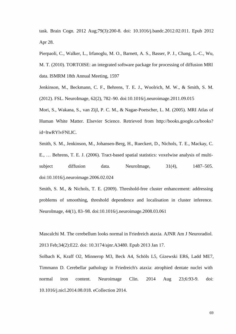
task. Brain Cogn. 2012 Aug;79(3):200-8. doi: 10.1016/j.bandc.2012.02.011. Epub 2012
Apr 28.
Pierpaoli, C., Walker, L., Irfanoglu, M. O., Barnett, A. S.,Basser, P. J., Chang, L.-C., Wu,
M. T. (2010). TORTOISE: an integrated software package for processing of diffusion MRI
data. ISMRM 18th Annual Meeting, 1597
Jenkinson, M., Beckmann, C. F., Behrens, T. E. J., Woolrich,M. W., & Smith, S. M.
(2012). FSL. NeuroImage, 62(2), 782–90. doi:10.1016/j.neuroimage.2011.09.015
Mori, S., Wakana, S., van Zijl, P. C. M., & Nagae-Poetscher, L. M. (2005). MRI Atlas of
Human White Matter. Elsevier Science. Retrieved from http://books.google.ca/books?
id=ltwRYlvFNLIC.
Smith, S. M., Jenkinson, M., Johansen-Berg, H., Rueckert, D., Nichols, T. E., Mackay, C.
E., … Behrens, T. E. J. (2006). Tract-based spatial statistics: voxelwise analysis of multi-
subject diffusion data. NeuroImage, 31(4), 1487–505.
doi:10.1016/j.neuroimage.2006.02.024
Smith, S. M., & Nichols, T. E. (2009). Threshold-free cluster enhancement: addressing
problems of smoothing, threshold dependence and localisation in cluster inference.
NeuroImage, 44(1), 83–98. doi:10.1016/j.neuroimage.2008.03.061
Mascalchi M. The cerebellum looks normal in Friedreich ataxia. AJNR Am J Neuroradiol.
2013 Feb;34(2):E22. doi: 10.3174/ajnr.A3480. Epub 2013 Jan 17.
Solbach K, Kraff O2, Minnerop M3, Beck A4, Schöls L5, Gizewski ER6, Ladd ME7,
Timmann D. Cerebellar pathology in Friedreich's ataxia: atrophied dentate nuclei with
normal iron content. Neuroimage Clin. 2014 Aug 23;6:93-9. doi:
10.1016/j.nicl.2014.08.018. eCollection 2014.
69
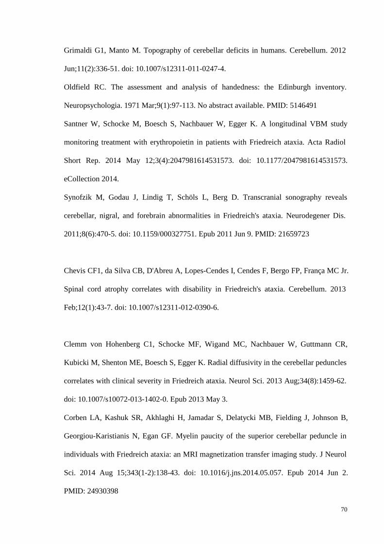
Grimaldi G1, Manto M. Topography of cerebellar deficits in humans. Cerebellum. 2012
Jun;11(2):336-51. doi: 10.1007/s12311-011-0247-4.
Oldfield RC. The assessment and analysis of handedness: theEdinburgh inventory.
Neuropsychologia. 1971 Mar;9(1):97-113. No abstract available. PMID: 5146491
Santner W, Schocke M, Boesch S, Nachbauer W, Egger K. A longitudinal VBM study
monitoring treatment with erythropoietin in patients withFriedreich ataxia. Acta Radiol
Short Rep. 2014 May 12;3(4):2047981614531573. doi: 10.1177/2047981614531573.
eCollection 2014.
Synofzik M, Godau J, Lindig T, Schöls L, Berg D. Transcranialsonography reveals
cerebellar, nigral, and forebrain abnormalities in Friedreich's ataxia. Neurodegener Dis.
2011;8(6):470-5. doi: 10.1159/000327751. Epub 2011 Jun 9. PMID: 21659723
Chevis CF1, da Silva CB, D'Abreu A, Lopes-Cendes I, Cendes F,Bergo FP, França MC Jr.
Spinal cord atrophy correlates with disability in Friedreich's ataxia. Cerebellum. 2013
Feb;12(1):43-7. doi: 10.1007/s12311-012-0390-6.
Clemm von Hohenberg C1, Schocke MF, Wigand MC, Nachbauer W, Guttmann CR,
Kubicki M, Shenton ME, Boesch S, Egger K. Radial diffusivityin the cerebellar peduncles
correlates with clinical severity in Friedreich ataxia. Neurol Sci. 2013 Aug;34(8):1459-62.
doi: 10.1007/s10072-013-1402-0. Epub 2013 May 3.
Corben LA, Kashuk SR, Akhlaghi H, Jamadar S, Delatycki MB, Fielding J, Johnson B,
Georgiou-Karistianis N, Egan GF. Myelin paucity of the superior cerebellar peduncle in
individuals with Friedreich ataxia: an MRI magnetization transfer imaging study. J Neurol
Sci. 2014 Aug 15;343(1-2):138-43. doi: 10.1016/j.jns.2014.05.057. Epub 2014 Jun 2.
PMID: 24930398
70
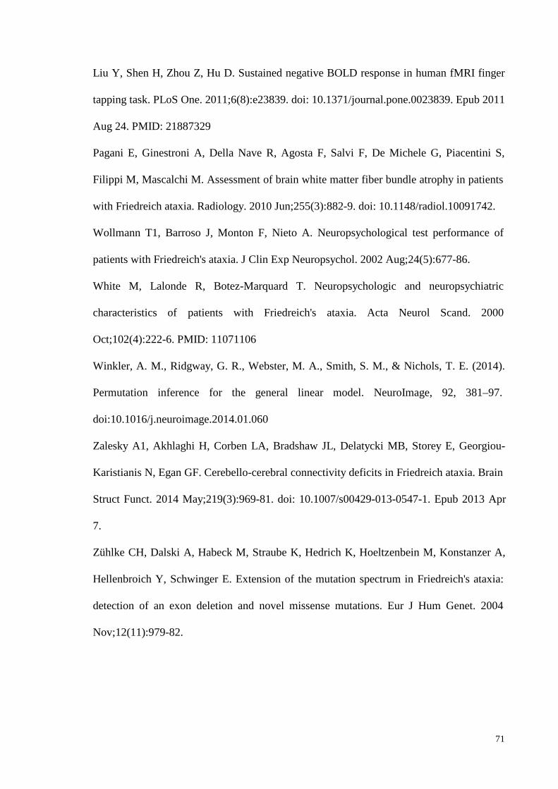
Liu Y, Shen H, Zhou Z, Hu D. Sustained negative BOLD response in human fMRI finger
tapping task. PLoS One. 2011;6(8):e23839. doi: 10.1371/journal.pone.0023839. Epub 2011
Aug 24. PMID: 21887329
Pagani E, Ginestroni A, Della Nave R, Agosta F, Salvi F, De Michele G, Piacentini S,
Filippi M, Mascalchi M. Assessment of brain white matter fiber bundle atrophy in patients
with Friedreich ataxia. Radiology. 2010 Jun;255(3):882-9. doi: 10.1148/radiol.10091742.
Wollmann T1, Barroso J, Monton F, Nieto A. Neuropsychological test performance of
patients with Friedreich's ataxia. J Clin Exp Neuropsychol. 2002 Aug;24(5):677-86.
White M, Lalonde R, Botez-Marquard T. Neuropsychologic andneuropsychiatric
characteristics of patients with Friedreich's ataxia. Acta Neurol Scand. 2000
Oct;102(4):222-6. PMID: 11071106
Winkler, A. M., Ridgway, G. R., Webster, M. A., Smith, S. M., &Nichols, T. E. (2014).
Permutation inference for the general linear model. NeuroImage, 92, 381–97.
doi:10.1016/j.neuroimage.2014.01.060
Zalesky A1, Akhlaghi H, Corben LA, Bradshaw JL, Delatycki MB, Storey E, Georgiou-
Karistianis N, Egan GF. Cerebello-cerebral connectivity deficits in Friedreich ataxia. Brain
Struct Funct. 2014 May;219(3):969-81. doi: 10.1007/s00429-013-0547-1. Epub 2013 Apr
7.
Zühlke CH, Dalski A, Habeck M, Straube K, Hedrich K, Hoeltzenbein M, Konstanzer A,
Hellenbroich Y, Schwinger E. Extension of the mutation spectrum in Friedreich's ataxia:
detection of an exon deletion and novel missense mutations.Eur J Hum Genet. 2004
Nov;12(11):979-82.
71
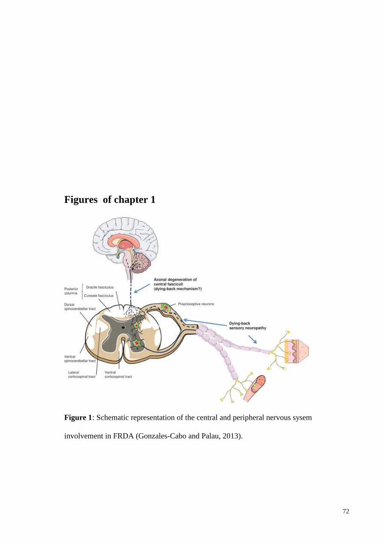
Figures of chapter 1
Figure 1: Schematic representation of the central and peripheral nervous sysem
involvement in FRDA (Gonzales-Cabo and Palau, 2013).
72
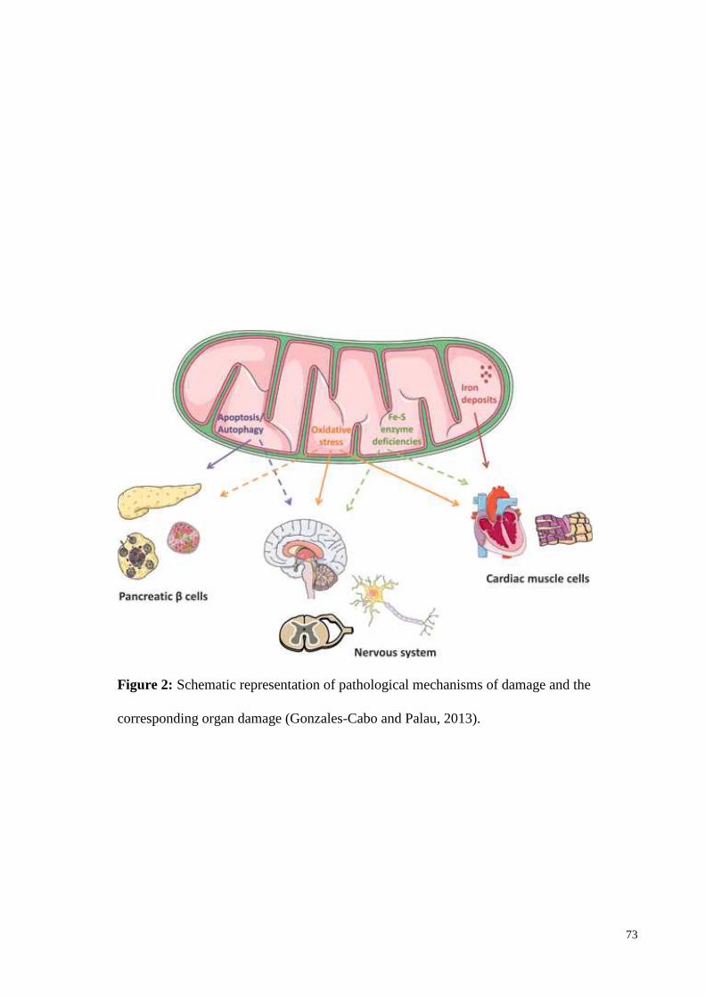
Figure 2: Schematic representation of pathological mechanisms of damage and the
corresponding organ damage (Gonzales-Cabo and Palau, 2013).
73
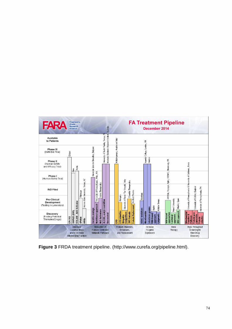
Figure 3 FRDA treatment pipeline. (http://www.curefa.org/pipeline.html).
74
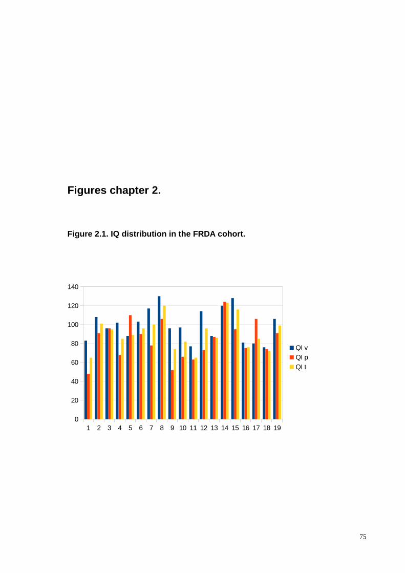
Figures chapter 2.
Figure 2.1. IQ distribution in the FRDA cohort.
75
1 2 3 4 5 6 7 8 9 10 11 12 13 14 15 16 17 18 19
0
20
40
60
80
100
120
140
QI v
QI p
QI t

Tables chapter 2.
Table 2.1: participants clinical and genetic featur es.
76

GAA: guanine adenosine adenosine, AAO: age at onset, AAV: age at the visit, DD disease
duration.
Table 2.2. Clinical data, onset symptoms and additional ones.
77

Legend: NA: non applicable, GI: Gastrointestinal, NY: nystagmus, DTR: deep tendon reflexes (- absent, + present, ++ brisk), DM: diabetes mellitus (years after onset), W-d-a: Wheelchair dependency age, Heart involvement: (+ hypertrophy), R: Right, L: left, AFO: ankle foot orthosis, V ventricle.
Table 2.2. (Continues)
78
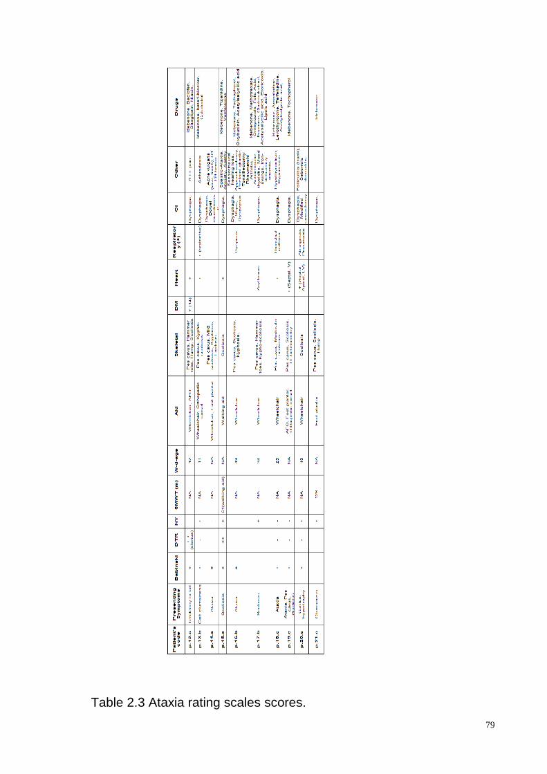
Table 2.3 Ataxia rating scales scores.
79
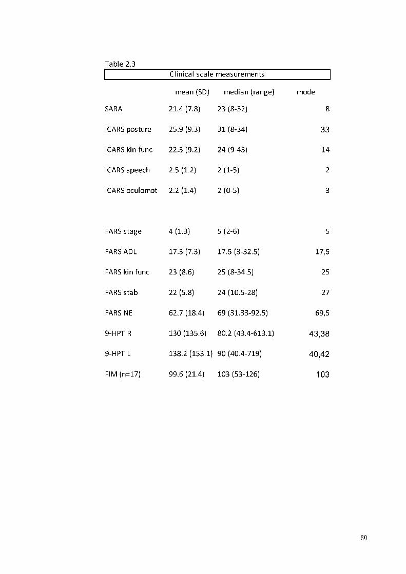
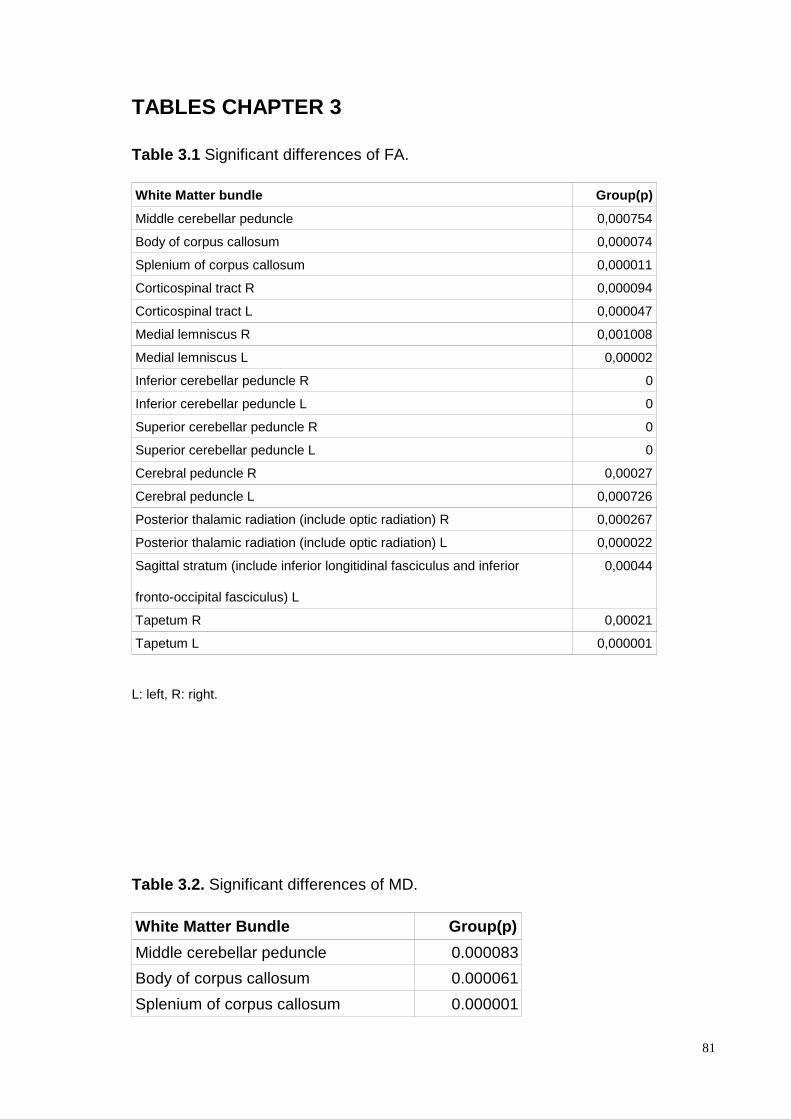
TABLES CHAPTER 3
Table 3.1 Significant differences of FA.
White Matter bundle Group(p)
Middle cerebellar peduncle 0,000754
Body of corpus callosum 0,000074
Splenium of corpus callosum 0,000011
Corticospinal tract R 0,000094
Corticospinal tract L 0,000047
Medial lemniscus R 0,001008
Medial lemniscus L 0,00002
Inferior cerebellar peduncle R 0
Inferior cerebellar peduncle L 0
Superior cerebellar peduncle R 0
Superior cerebellar peduncle L 0
Cerebral peduncle R 0,00027
Cerebral peduncle L 0,000726
Posterior thalamic radiation (include optic radiation) R 0,000267
Posterior thalamic radiation (include optic radiation) L 0,000022
Sagittal stratum (include inferior longitidinal fasciculus and inferior
fronto-occipital fasciculus) L
0,00044
Tapetum R 0,00021
Tapetum L 0,000001
L: left, R: right.
Table 3.2. Significant differences of MD.
White Matter Bundle Group(p)
Middle cerebellar peduncle 0.000083
Body of corpus callosum 0.000061
Splenium of corpus callosum 0.000001
81
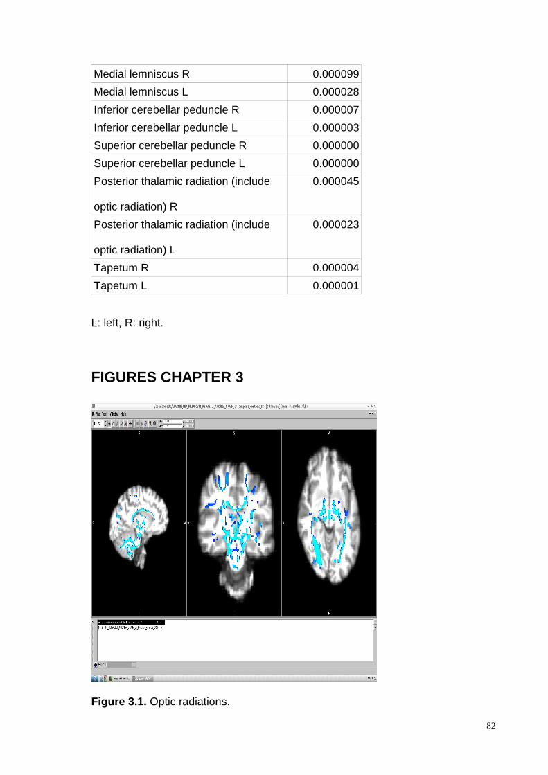
Medial lemniscus R 0.000099
Medial lemniscus L 0.000028
Inferior cerebellar peduncle R 0.000007
Inferior cerebellar peduncle L 0.000003
Superior cerebellar peduncle R 0.000000
Superior cerebellar peduncle L 0.000000
Posterior thalamic radiation (include
optic radiation) R
0.000045
Posterior thalamic radiation (include
optic radiation) L
0.000023
Tapetum R 0.000004
Tapetum L 0.000001
L: left, R: right.
FIGURES CHAPTER 3
Figure 3.1. Optic radiations.
82

Figure 3.2 . Corticospinal tracts.
Figure 3.3 Corticospinal tracts.
83
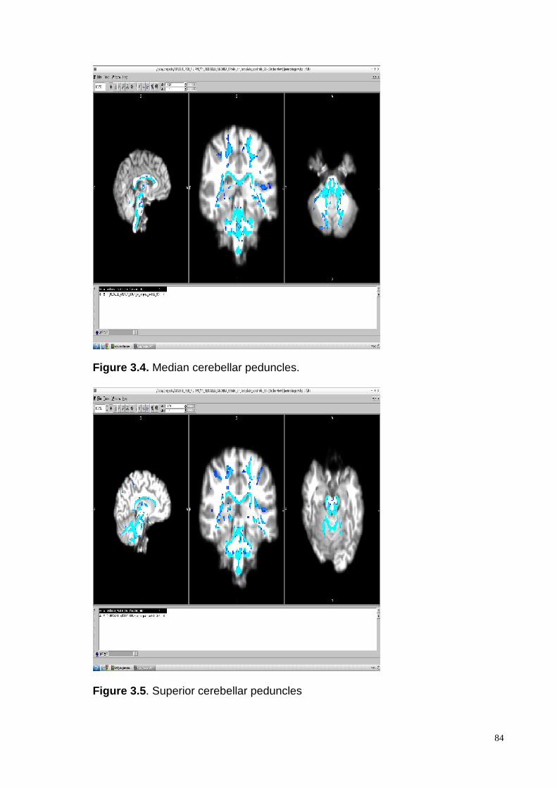
Figure 3.4. Median cerebellar peduncles.
Figure 3.5 . Superior cerebellar peduncles
84
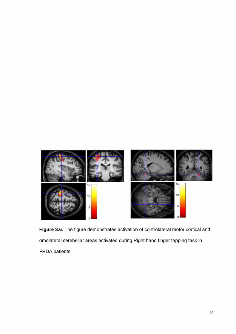
Figure 3.6 . The figure demonstrates activation of controlateral motor cortical and
omolateral cerebellar areas activated during Right hand finger tapping task in
FRDA patients.
85
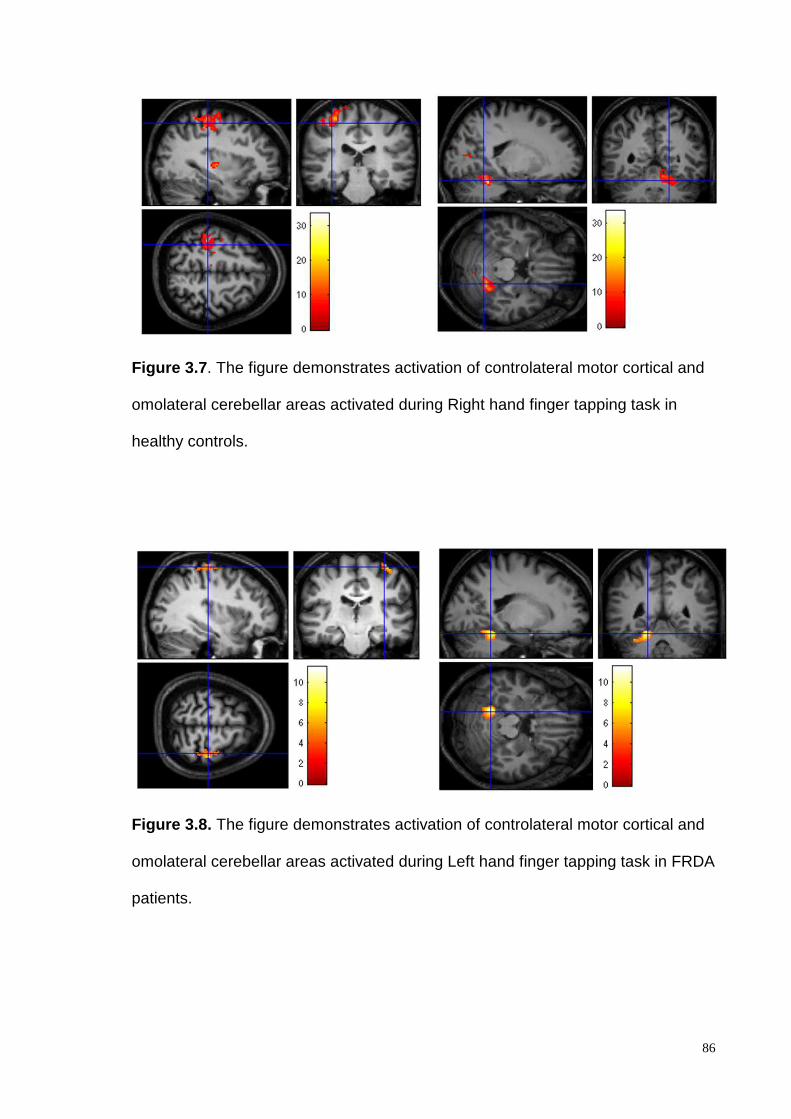
Figure 3.7 . The figure demonstrates activation of controlateral motor cortical and
omolateral cerebellar areas activated during Right hand finger tapping task in
healthy controls.
Figure 3.8. The figure demonstrates activation of controlateral motor cortical and
omolateral cerebellar areas activated during Left hand finger tapping task in FRDA
patients.
86
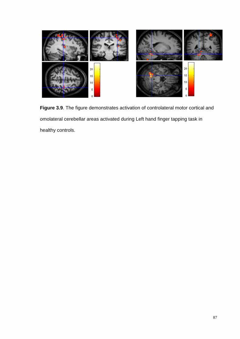
Figure 3.9 . The figure demonstrates activation of controlateral motor cortical and
omolateral cerebellar areas activated during Left hand finger tapping task in
healthy controls.
87
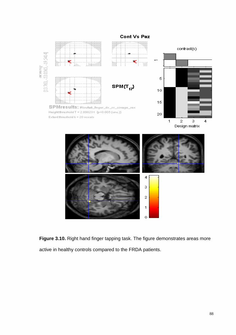
Figure 3.10. Right hand finger tapping task. The figure demonstrates areas more
active in healthy controls compared to the FRDA patients.
88
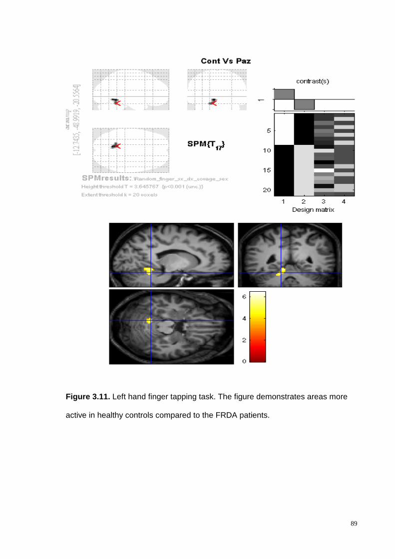
Figure 3.11. Left hand finger tapping task. The figure demonstrates areas more
active in healthy controls compared to the FRDA patients.
89