07 Moretti
6
190 ACT A oTorhinolAr yngologiCA iTAliCA 2013;33:190-195 Rib grafts in septorhinoplasty Innesti di costa nella settorinoplastica A. Moretti 1 , S. Sciuto 2 1 Dpamn f Mdal, oal and Bhnlgal Sns, eNt Sn, “G. d’Annnz” unvsy f ch- Psaa, ialy; 2 casa d ca Nsa Sgna dlla Md, rm, ialy SummAry Ats cata as a b cs t staa at ata cstct sptpast f a stcta sppt. i t stct t asa skt, ats cata ca b ast t asa spt, t ac t b, bt csta cata s cs t bst at ata patts q aj cstct. rb cata s a tsta ata cstct sptpast, spca s s a a ats tss a q. Ts ats ata as a at cpcats sc as spt, ct a xts cpa t ats a apastc pats. i t pst st, t ats aaz a scss t s ats b cata 54 patts t pa a s sptpast. its s s as sst cass c t s a t a a a at catas tss t b at asa ak cstct a spat ct pt. Key wordS: Reconstructiv e septhorhinoplasty • Grafts • Rib cartilage riASSunTo La cartilagine autologa viene generalmente consider ata come il materiale di prima scelta per gli innesti nella cosiddetta settorinoplastica ricostruttiva sia per quanto riguarda il sostegno strutturale del naso che per gli effetti di riempimento che da essa si possono ottenere. Nella ristrutturazione dell’architettura di sostegno nasale il tessuto cartilagineo può essere ricavato dal setto, dal padiglione auricolare o dalla costa. Quest’ultima sede rappresenta il sito donatore principale nelle ricostruzioni maggiori ed in particolare nella chirurgia di revisione da eseguire in pazienti con deplezione cartilaginea. La cartilagine costale autologa se confrontata con omoinnesti ed impianti alloplastici presenta una ba ssa percentuale di complicanze, soprattutto infezioni ed estrusione anche se non è scevra da possibile riassorbimento e distorsione. Nel presente studio gli Autori analizzano e discutono l’impiego della cartilagine costale autologa in 54 pazienti sottoposti a settorinoplastica primaria o di revisione e ne suggeriscono l’impiego in tutti quei casi in cui vi è la necessità di avere a disposizione una congrua quantità di tessuto da innestare per ristabilire la struttura di sostegno e migliorare la funzionalità nasale. PArole ChiAve: Settorinoplastica ricostruttiva • Innesti • Cartilagine costale Acta Otorhinola ryngol Ital 2013 ;33:190-1 95 Introduction Grats in septorhinoplasty can be obtained rom dierent autologous and homologous tissues or rom alloplastic materials. Autologous cartilage presents many advantag- es compared to other kinds o grats: it survives as a liv- ing tissue, seldom undergoes resorption, does not stimu- late an immune response, is ideal or all types o grating and presents only biological costs, but its use requires longer operation time. Septum, auricular concha and rib are the best cartilaginous donor sites or autologous na- sal grating. Rib cartilage is the grat material o choice or dorsal augmentation and reconstructive support when sucient septal cartilage is not available 1 . Autologous rib cartilage is oten overlooked in reconstructive sep- torhinoplasty because o potential donor-site morbidity and the warping eect 2-8 ; however, its use is indispensa- ble when large amounts o tissue and multiple grats are required, especially in patients already surgically treated, with cartilage depletion. Usually two teams work simul- taneously, but since most warping occurs within 15-60 min o harvesting (early warping), the same surgeon can harvest the rib beore modeling the grat and preparing the recipient site, preerably using an open approach, to veriy cartilaginous structure modications. The central portion o the 5 th to 8 th rib is preerred by some surgeons 9 ; however , the 11 th and 12 th ree-foating ribs are naturally straighter, require less carving and undergo less warp- ing 10 . To reduce the warping eect, Gunter suggested to reinorce larger grats (dorsal onlay grat and columellar strut) with a centrally placed Kirschner-wire to provide a more stable and predictable result 6 . The grat is pre- erably harvested rom the right side to prevent the pos- sibility o conounding any cardiac chest pain, although
Transcript of 07 Moretti
7/30/2019 07 Moretti
http://slidepdf.com/reader/full/07-moretti 1/6
190
ACTA oTorhinolAryngologiCA iTAliCA 2013;33:190-195
Rib grafts in septorhinoplasty
Innesti di costa nella settorinoplastica A. Moretti1, S. Sciuto2 1 Dpamn f Mdal, oal and Bhnlgal Sns, eNt Sn, “G. d’Annnz” unvsy f ch-Psaa, ialy; 2 casa d ca Nsa Sgna dlla Md, rm, ialy
SummAry
Ats cata as a b cs t staa at ata cstct sptpast
f a stcta sppt. i t stct t asa skt, ats cata ca b ast t asa spt, t
ac t b, bt csta cata s cs t bst at ata patts q aj cstct. rb cata s a
tsta ata cstct sptpast, spca s s a a ats tss a q. Ts
ats ata as a at cpcats sc as spt, ct a xts cpa t ats a apastc
pats. i t pst st, t ats aaz a scss t s ats b cata 54 patts t pa
a s sptpast. its s s as sst cass c t s a t a a a at catas tss t bat asa ak cstct a spat ct pt.
Key wordS: Reconstructive septhorhinoplasty • Grafts • Rib cartilage
riASSunTo
La cartilagine autologa viene generalmente considerata come il materiale di prima scelta per gli innesti nella cosiddetta settorinoplastica
ricostruttiva sia per quanto riguarda il sostegno strutturale del naso che per gli effetti di riempimento che da essa si possono ottenere. Nella
ristrutturazione dell’architettura di sostegno nasale il tessuto cartilagineo può essere ricavato dal setto, dal padiglione auricolare o dalla
costa. Quest’ultima sede rappresenta il sito donatore principale nelle ricostruzioni maggiori ed in particolare nella chirurgia di revisione
da eseguire in pazienti con deplezione cartilaginea. La cartilagine costale autologa se confrontata con omoinnesti ed impianti alloplastici
presenta una bassa percentuale di complicanze, soprattutto infezioni ed estrusione anche se non è scevra da possibile riassorbimento e
distorsione. Nel presente studio gli Autori analizzano e discutono l’impiego della cartilagine costale autologa in 54 pazienti sottoposti asettorinoplastica primaria o di revisione e ne suggeriscono l’impiego in tutti quei casi in cui vi è la necessità di avere a disposizione una
congrua quantità di tessuto da innestare per ristabilire la struttura di sostegno e migliorare la funzionalità nasale.
PArole ChiAve: Settorinoplastica ricostruttiva • Innesti • Cartilagine costale
Acta Otorhinolaryngol Ital 2013;33:190-195
Introduction
Grats in septorhinoplasty can be obtained rom dierent
autologous and homologous tissues or rom alloplastic
materials. Autologous cartilage presents many advantag-
es compared to other kinds o grats: it survives as a liv-
ing tissue, seldom undergoes resorption, does not stimu-
late an immune response, is ideal or all types o grating
and presents only biological costs, but its use requires
longer operation time. Septum, auricular concha and rib
are the best cartilaginous donor sites or autologous na-
sal grating. Rib cartilage is the grat material o choice
or dorsal augmentation and reconstructive support when
sucient septal cartilage is not available 1. Autologous
rib cartilage is oten overlooked in reconstructive sep-torhinoplasty because o potential donor-site morbidity
and the warping eect 2-8; however, its use is indispensa-
ble when large amounts o tissue and multiple grats are
required, especially in patients already surgically treated,
with cartilage depletion. Usually two teams work simul-taneously, but since most warping occurs within 15-60
min o harvesting (early warping), the same surgeon can
harvest the rib beore modeling the grat and preparing
the recipient site, preerably using an open approach, to
veriy cartilaginous structure modications. The central
portion o the 5th to 8th rib is preerred by some surgeons 9;
however, the 11th and 12th ree-foating ribs are naturally
straighter, require less carving and undergo less warp-
ing 10. To reduce the warping eect, Gunter suggested to
reinorce larger grats (dorsal onlay grat and columellar
strut) with a centrally placed Kirschner-wire to provide
a more stable and predictable result 6. The grat is pre-erably harvested rom the right side to prevent the pos-
sibility o conounding any cardiac chest pain, although
7/30/2019 07 Moretti
http://slidepdf.com/reader/full/07-moretti 2/6
Rib grafts in septorhinoplasty
191
in some cases, to acilitate a two-team approach, rib car-
tilage harvesting is perormed on the patient’s let side.
Rib cartilage can be harvested circumerentially, ater
superior and inerior perhicondrium elevation, taking
special precaution not to injure the inerior line neuro-
vascular bundle or closely adherent pleura on the medialsurace. Rib harvesting can be also limited to the outer
lamella preserving the internal costal arch. By preserv-
ing the inner lamella o the rib, postoperative morbidi-
ties, including pain, splinting and pneumothorax, are re-
duced 10. In revision rhinoplasty, the dorsum is requently
over-resected and the septal L-structure is weakened. In
these cases, dorsal spreader grats should be placed along
either side o the septum to provide a stable recipient site
or the dorsal onlay grat 11. The spreader grats should
be stabilized with horizontal mattress sutures at the same
level as the septum and extend rom the keystone area to
the septal angle in preparation or receiving the dorsal
onlay grat. Spreader grats are used to widen a narrow
dorsum when necessary to obtain better symmetry o the
middle third o the nose, but also to improve the acute
angle o the internal nasal valve when related respira-
tory insuciency is present. To restore eective L-strut
support, it is very important to prepare a precise pocket
between the medial crura or placement o the columellar
strut. To obtain a stable and strong medial ramework in
most severe depleted cartilaginous cases, it is necessary
to hinge the columellar strut with the dorsal grat 11. The
present study describes the authors’ experience in the useo autologous rib grats in primary and revision cases o
septorhinoplasty.
Methods
We retrospectively analyzed data rom 54 patients who
underwent septorhinoplasty using autologous rib cartilage
grats in the last 10 years. The study population consisted
o 33 male and 21 emale patients (mean age: 34 years; age
range: 16-64 years). All septorhinoplasties were perormed
under general anaesthesia in patients with depletion or de-
ormities o the osteocartilagineous nasal ramework usingan open approach. Twelve patients were primary cases: 8
with post-traumatic deormities and 4 presented congenital
abnormalities. Three patients showed stigmata o cocaine
abuse, 1 presented sequelae due to haematoma o the sep-
tum, and 38 were revision cases, 7 o which previously
treated with alloplastic implants. The patients had several
degrees o saddle nose deormity and complained o nasal
obstruction: in 42 cases, most o the septum was absent,
and 6 patients had previous aggressive septoplasty with
septal peroration in 2 cases. Eleven patients had internal
nasal valve insuciency. All patients showed tip deorm-
ity and alteration o the relationship between dorsal linesand columella-tip complex. In correction o the iatrogenic
nasal deormities such as stigmata o alloplastic implant,
saddle nose, septal perorations, valvular collapse and na-
sal airfow obstruction, the procedure included placement
o septal grats, alar, tip and dorsal onlay grats, spreader
grats, columellar struts and shield grats.
In male patients, an incision was made over the seventh
costal cartilage and in the women under the breast creaseto hide the scar. Ater skin incision, the external oblique
muscle was reached and the ascia over this muscle was
opened. The overlying muscles were spread, parallel o
the direction o their bres and appropriately retracted,
to reduce postoperative pain, until exposing the underly-
ing costal cartilage. The perichondrium was incised paral-
lel to the outer surace and circumerentially elevated to
perorm ull-thickness harvesting o the rib cartilage. In
some cases, when the harvesting was limited to the outer
lamella, the perichondrium was elevated only to the ex-
ternal surace. Once elevation was complete, the desired
section o the rib was perormed.
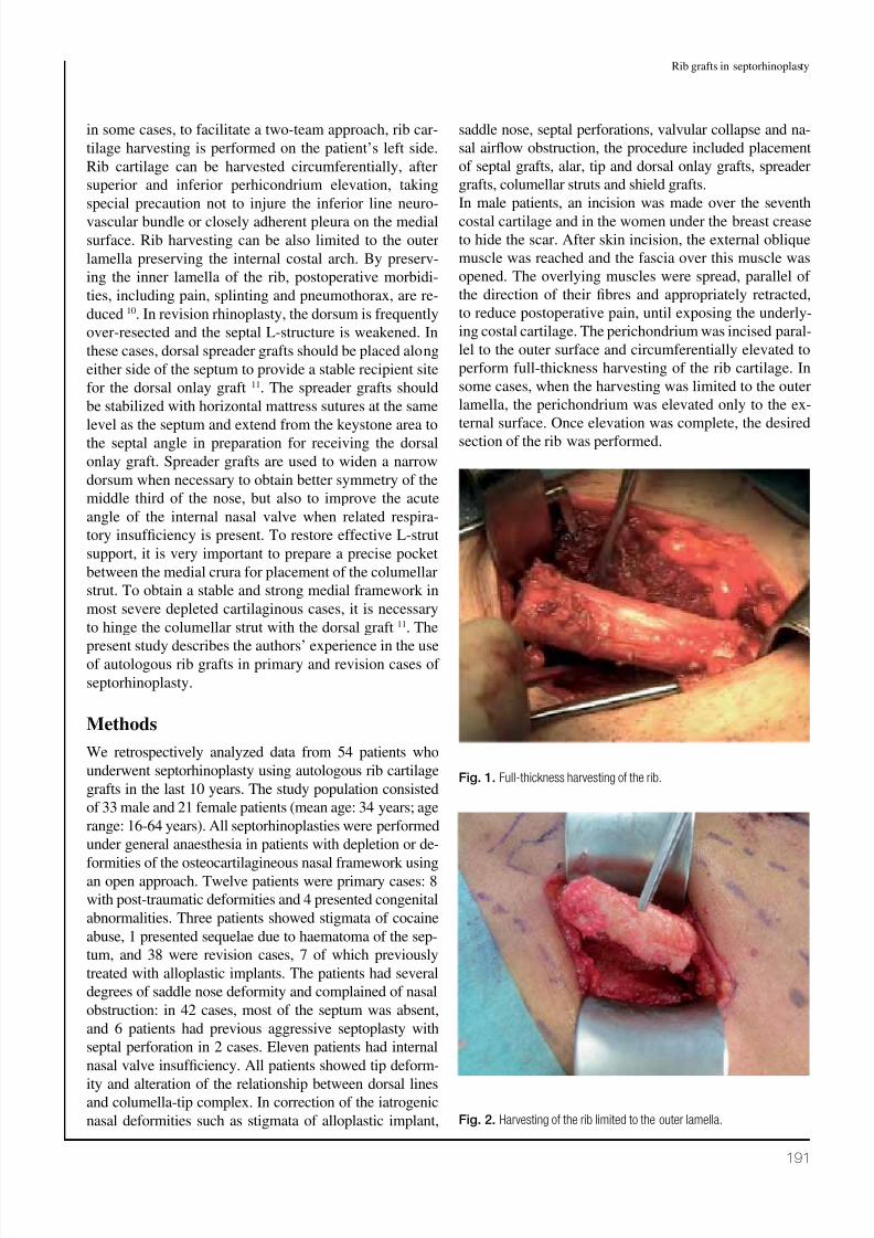
Fig. 1. Full-thickness harvesting o the rib.
Fig. 2. Harvesting o the rib limited to the outer lamella.

7/30/2019 07 Moretti
http://slidepdf.com/reader/full/07-moretti 3/6
A. Moretti, S. Sciuto
192
Rib cartilage can be removed ull-thickness (Fig. 1), tak-
ing special precaution to the neurovascular bundle or the
closely adherent pleura medially. The harvesting can be
also conducted only to the outer portion to maintain the
continuity o the internal costal arch (Fig. 2). By preserv-
ing the inner lamella o the rib, postoperative morbidities,including pain, splinting and pneumothorax, are reduced.
Ater the grat was removed, the donor site was closed in
layers without drains. The harvested costal cartilage was
shaped as a vertical strip except in patients with severe
saddle nose deormity when a large amount o cartilage is
required to recreate the new L-structure with a columellar
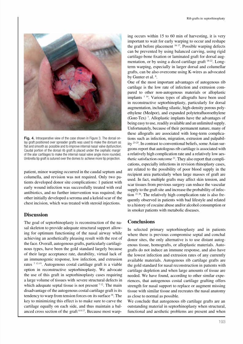
strut hinged with a large dorsal grat (Fig. 3). The central
portion was usually used in dorsal reshaping (dorsal and
spreader grat) and or septal reconstruction, while the pe-
ripheral portion was shaped in alar and tip replacement
grats ater approximately 30 min to allow most o the
warping to occur. All the grats were inserted using an
open approach that oers the best exposure o nasal struc-
tures by providing grat positioning and stabilization in
the desired locations without distorsion. The costal carti-
lage grats were used as dorsal, septal and columellar strut
in all cases (Fig. 4), while spreader grats were utilized in
17 patients to provide urther stability to the septal grat or
to improve nasal valve unction. In each case, the dorsum
was made as fat and smooth as possible beore the dorsal
onlay grat was placed (Fig. 5). Alar and tip onlay grats
(Fig. 6) had various combinations with the previous grats
in 48 patients to address structural deects. Only in threecases, with underprojected tip, was a shield grat used.
Results
The ollow-up ranged rom 6 to 36 months, with an av-
erage o 18 months. None o the patients had any intra-
operative complications. Oral analgesic was always ad-
equate or pain control and chest pain subsided within
1 to 6 weeks postoperatively. Cosmetic appearance and
nasal obstruction were improved in all cases. Regarding
complications, none o the 54 patients had grats extru-
sion. Four patients had inection in recipient sites (3 onthe columella and 1 on the dorsum) with signicant grats
resorption in 2 cases with a history o cocaine abuse.
Warping deect was noticed in 3 patients ater the oedema
subsided. In 2 o these, distorsion occurred in the dorsal
onlay grat and revision surgery was required. In the third
Fig. 3. Preoperative and2-years postoperative photo-graphs o a patient who had twoprevious aggressive septorhino-plasty. All the grats used wereharvested rom rib cartilage.
7/30/2019 07 Moretti
http://slidepdf.com/reader/full/07-moretti 4/6
Rib grafts in septorhinoplasty
193
patient, minor warping occurred in the caudal septum and
columella, and revision was not required. Only two pa-
tients developed donor site complications: 1 patient with
early wound inection was successully treated with oral
antibiotics, and no urther intervention was required; the
other initially developed a seroma and a keloid scar o the
chest incision, which was treated with steroid injections.
Discussion
The goal o septorhinoplasty is reconstruction o the na-
sal skeleton to provide adequate structural support allow-
ing or optimum unctioning o the nasal airway while
achieving an aesthetically pleasing result with the rest o
the ace. Overall, autogenous grats, particularly cartilagi-
nous types, have been the gold standard largely because
o their large acceptance rate, durability, virtual lack o
an immunogenic response, low inection, and extrusion
rates 7 12-15. Autogenous costal cartilage grat is a viable
option in reconstructive septorhinoplasty. We advocate
the use o this grat in septorhinoplasty cases requiring
a large volume o tissues with severe structural deects in
which adequate septal tissue is not present 7 12. The main
disadvantage o the autogenous costal cartilage grat is its
tendency to warp rom tension orces on its surace 16. The
key to minimizing this eect is to make sure to carve thecartilage equally on each side, and thus maintain a bal-
anced cross section o the grat 6 8 17. Because most warp-
ing occurs within 15 to 60 min o harvesting, it is very
important to wait or early warping to occur and reshape
the grat beore placement 16 17. Possible warping deects
can be prevented by using balanced carving, using rigid
cartilage-bone xation or laminated grat or dorsal aug-
mentation, or by using a diced cartilage grat 18-22. Long-term warping, especially in larger dorsal and columellar
grats, can be also overcome using K-wires as advocated
by Gunter et al. 6.
One o the most important advantages o autogenous rib
cartilage is the low rate o inection and extrusion com-
pared to other non-autogenous materials or alloplastic
implants 1 14. Various types o allograts have been used
in reconstructive septorhinoplasty, particularly or dorsal
augmentation, including silastic, high-density porous poly-
ethylene (Medpor), and expanded polytetrafuoroethylene
(Gore-Tex) 7. Alloplastic implants have the advantages o
being easy to use, readily available and an unlimited supply.
Unortunately, because o their permanent nature, many o
these allograts are associated with long-term complica-
tions such as inection, migration, extrusion and palpabil-
ity 23-25. In contrast to conventional belies, some Asian sur-
geons report that autologous rib cartilage is associated with
a relatively high complication rate and a relatively low aes-
thetic satisaction outcome 21. They also report that compli-
cations, especially inections in revision rhinoplasty cases,
are related to the possibility o poor blood supply in the
recipient area particularly when large masses o grat are
used. In act, multiple grats may aect skin tension, andscar tissues rom previous surgery can reduce the vascular
supply to the grat site and increase the probability o inec-
tion 21 26. The relatively high complication rate is also re-
quently observed in patients with bad liestyle and related
to a history o cocaine abuse and/or alcohol consumption or
in smoker patients with metabolic diseases.
Conclusions
In selected primary septorhinoplasty and in patients
where there is previous compromise septal and conchal
donor sites, the only alternative is to use distant autog-enous tissue, homograts, or alloplastic materials. Auto-
grats do not induce an immune response, and also have
the lowest inection and extrusion rates o any currently
available materials. Autogenous rib cartilage grats are
the gold standard or nasal reconstruction in patients with
cartilage depletion and when large amounts o tissue are
needed. We have ound, according to other similar expe-
riences, that autogenous costal cartilage grating oers
strength or nasal support to replace or augment missing
tissue with similar tissue and recreates the nasal anatomy
as close to normal as possible.
We conclude that autogenous rib cartilage grats are anoutstanding material in septorhinoplasty when structural,
unctional and aesthetic problems are present and when
Fig. 4. Intraoperative view o the case shown in Figure 3. The dorsal on-lay grat positioned over spreader grats was used to make the dorsum asat and smooth as possible and to improve internal nasal valve dysunction.Caudal portion o the dorsal rib grat is placed under the cephalic margino the alar cartilages to make the internal nasal valve angle more rounded. Umbrella tip grat is sutured over the domes to achieve more tip projection.

7/30/2019 07 Moretti
http://slidepdf.com/reader/full/07-moretti 5/6
A. Moretti, S. Sciuto
194
an eective volume lling and reconstruction o the nasal
structures are needed. Its use should however be adopted
keeping in mind the possibility o complications.
References
1 Porter JP. Grafts in rhinoplasty, alloplastic vs autogenous.Arch Otolaryngol Head Neck Surg 2000;126:558-61.
2 Brent B. The versatile cartilage autograft: current trends inclinical transplantation. Clin Plast Surg 1979;6:163-80.
3 Kim DW, Shah AR, Toriumi DM. Concentric and eccentriccarved costal cartilage. Arch Facial Plast Surg 2006;8:42-6.
4 Agaoglu G, Erol OO. In situ split costal cartilage graft har-vesting through a small incision using a gouge. Plast Recon-str Surg 2000;106:932-5.
5 Maas CS, Monhian N, Shah SB. Implants in rhinoplasty. Fa-cial Plast Surg 1997;13:279-90.
6 Gunter JP, Clark CP, Friedman RM. Internal stabilization of cartilaginous rib cartilage grafts in rhinoplasty: a barrier tocartilage warping. Plast Reconstr Surg 1997;100:161-9.
7 Moshaver A, Gantous A. The use of autogenous costal carti-lage graft in septorhinoplasty. Otolaryngol Head Neck Surg2007;137:862-7.
Fig. 5. Above: saddle nose asthe result o septoplasty withcomplications leading to almosttotal resorption o the septumand collapse o the cartilaginousvault. Below: the end result ob-tained by balancing the profle
through reconstruction o thecartilaginous dorsum and exci-sion o the osseous dorsum
Fig. 6. Intraoperative photograph o the case shown in Figure 5. Recon-struction o an L-shaped structure to support the dorsum completed withgrats to ensure the projection and defnition o the tip.
7/30/2019 07 Moretti
http://slidepdf.com/reader/full/07-moretti 6/6
Rib grafts in septorhinoplasty
195
8 Gibson T, Davis WB. The distortion of autogenous car-tilage grafts: its cause and prevention. Br J Plast Surg1958;10:257-73.
9 Lee M, Inman J, Ducic Y. Central segment harvest of costalcartilage in rhinoplasty. Laryngoscope 2011;121:2155-8.
10 Ansari K, Asaria J, Hilger P, et al. Grafts and implants in rhi-noplasty. Techniques and long-term results. Operative Tech-niques in Otolaryngol 2008;19:42-58.
11 Yilmaz M, Vayvada H, Menderes A, et al. Dorsal nasal aug-mentation with rib cartilage graft: long-term results and pa-tient satisfaction. J Cranioac Surg 2007;18:1457-62.
12 Araco A, Gravante G, Araco F, et al. Autologous cartilagegrafts rhinoplasties. Aesthetic Plast Surg 2006;30:169-74.
13 Vuyk HD, Adamson PA. Biomaterials in rhinoplasty. ClinOtolaryngol 1998;23:209-17.
14 Adamson PA. Grafts in rhinoplasty: autogenous grafts aresuperior to alloplastic. Arch Otolaryngol Head Neck Surg
2000;126:561-2.15 Toriumi DM. Autogenous grafts are worth the extra time.
Arch Otolaryngol Head Neck Surg 2000;126:562-4.
16 Adams WP, Rohrich RJ, Gunter JP, et al. The rate of warpingin irradiated and nonirradiated homograft rib cartilage: acontrolled comparison and clinical implications. Plast Re-constr Surg 1999;103:265-70.
17 Cakmak O, Ergin T. The versatile autogenous costal car-tilage graft in septorhinoplasty. Arch Facial Plast Surg2002;4:172-6.
18 Swanepoel PF, Fysh R. Laminated dorsal beam graft to elim-inate postoperative twisting complications. Arch Facial PlastSurg 2007;9:285-9.
19 Sherris DA, Kern EB. The versatile autogenous rib graft inseptorhinoplasty. Am J Rhinol 1998;12:221-7.
20 Daniel RK. Diced cartilage grafts in rhinoplasty surgery:current techniques and applications. Plast Reconstr Surg2008;122:1883-91.
21 Moon BJ, Lee HJ, Jang YJ. Outcomes following rhinoplastyusing autologous costal cartilage. Arch Facial Plast Surg2012;14:175-80.
22 Jang YJ, Min JY, Lau BC. A multilayer cartilaginoustip-grafting technique for improved nasal tip refine-ment in Asian rhinoplasty. Otolaryngol Head Neck Surg2011;145:217-22.
23 Davis PKB, Jones SM. The complications of silastic im- plants: experience with 137 consecutive cases. Br J PlastSurg 1971;24:405-11.
24 Godin MS, Waldman R, Johnson CM. The use of expanded polytetrafluoroethylene (Gore-Tex) in rhinoplasty. Arch Oto-laryngol Head Neck Surg 1995;121:1131-6.
25 Raghavan U, Jones NS, Romo R. Immediate autogenous car-tilage grafts in rhinoplasty after alloplastic implant rejec-tion. Arch Facial Plast Surg 2004;6:192-6.
26 Holt GR, Garner ET, McLarey D. Postoperative sequelaeand complications of rhinoplasty. Otolaryngol Clin NorthAm 1987;20:853-76.
Address or correspondence: Antonio Moretti, Department o Me-
dical, Oral and Biotechnological Sciences, ENT Section “G. d’An-
nunzio” University o Chieti-Pescara, via dei Vestini, 66100 Chieti,
Italy. Tel. +39 0871 3554070 . E-mail: [email protected]
Received: December 15, 2012 - Accepted: January 7, 2013